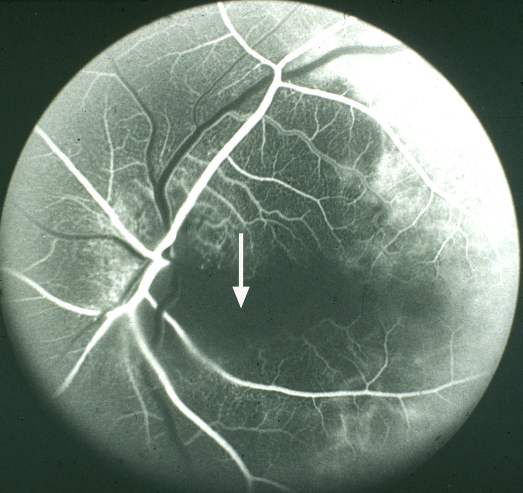
Figure 2.
Fluorescein fundus angiogram of left eye, of a patient with giant cell arteritis, with cilioretinal artery occlusion and arteritic anterior ischemic optic neuropathy. Note no filling of the choroid and optic disc supplied by the medial posterior ciliary artery and of the cilioretinal artery (arrow), with normal filling of central retinal artery and lateral posterior ciliary artery. A combination of posterior ciliary artery occlusion, cilioretinal artery occlusion and arteritic anterior ischemic optic neuropathy is diagnostic of giant cell arteritis.

