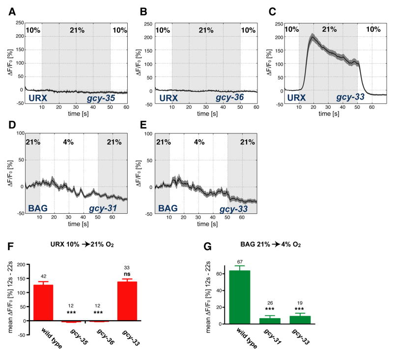
Figure 5. sGCs are required for O2 evoked calcium transients in URX and BAG.
(A–E) Measurements of neural activity by calcium imaging of URX (A–C) and BAG (D,E). Black traces show the average percent change of G-CaMP fluorescence (ΔF/F0) and dark shading shows standard error of the mean (SEM). Concentrations were 21% and 10% O2 (A–C) or 21% and 4% O2 (D,E). (A) URX, gcy-35(ok769) mutants. (B) URX, gcy- 36(db66) mutants. (C) URX, gcy-33(ok232) mutants. (D) BAG, gcy-31(ok296) mutants. (E) BAG, gcy-33(ok232) mutants. Light grey shading indicates the intervals at 21% O2. (F,G) Average ΔF/F0 from 12–22 seconds for indicated genotypes, (F) URX; (G) BAG. The data for wild type animals correspond to Figure 2D,H. Error bars show SEM. Asterisks indicate significance by one-way ANOVA with Dunnett's post-hoc test using wild type in each panel as control groups (***P<0.001, ns not significant). The numbers of recordings analyzed are indicated.

