

Abstract
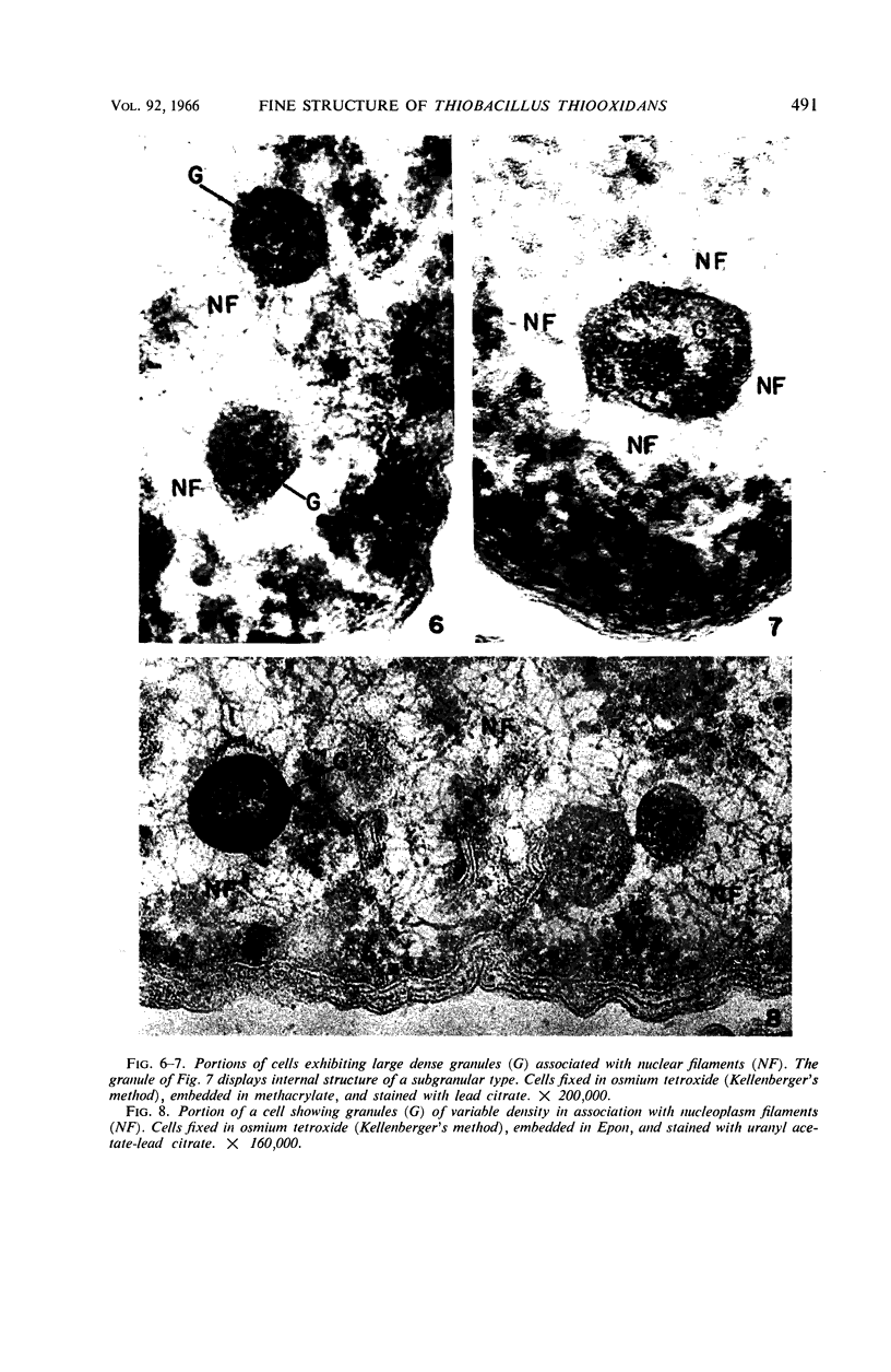
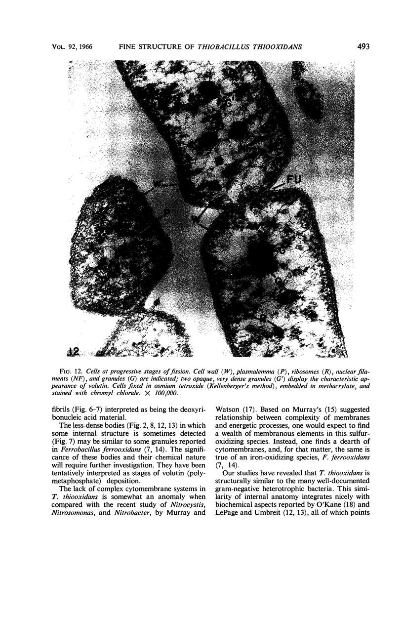
Mahoney, Robert P. (Skidmore College, Saratoga Springs, N.Y.), and Mercedes R. Edwards. Fine structure of Thiobacillus thiooxidans. J. Bacteriol. 92: 487–495. 1966.—Thin section analysis of the chemosynthetic autotroph Thiobacillus thiooxidans revealed structures comparable to gram-negative heterotrophic bacteria. Although this species is unique in that it oxidizes elemental sulfur for energy, uses carbon dioxide as its sole source of carbon, and can withstand a pH of less than 1, thin sections revealed a profile of the cell envelope (cell wall and plasmalemma) similar to other gram-negative species which have more common physiological traits. The cell wall is composed of five layers with an overall width of approximately 200 A, and the plasmalemma appears as a conventional “unit membrane” with a width of about 85 A. Volutin granules and less-dense bodies of similar shape and size were frequently observed in close association with the nucleoplasm. The nature and function of these bodies are unknown at this time.
Full text
PDF


Images in this article


Selected References
These references are in PubMed. This may not be the complete list of references from this article.
- BARKER H. A., KORNBERG A. The structure of the adenosine triphosphate of Thiobacillus thiooxidans. J Bacteriol. 1954 Dec;68(6):655–661. doi: 10.1128/jb.68.6.655-661.1954. [DOI] [PMC free article] [PubMed] [Google Scholar]
- BLADEN H. A., WATERS J. F. ELECTRON MICROSCOPIC STUDY OF SOME STRAINS OF BACTEROIDES. J Bacteriol. 1963 Dec;86:1339–1344. doi: 10.1128/jb.86.6.1339-1344.1963. [DOI] [PMC free article] [PubMed] [Google Scholar]
- COSTERTON J. W., MURRAY R. G., ROBINOW C. F. Observations on the motility and the structure of Vitreoscilla. Can J Microbiol. 1961 Jun;7:329–339. doi: 10.1139/m61-040. [DOI] [PubMed] [Google Scholar]
- Conti S. F., Gettner M. E. ELECTRON MICROSCOPY OF CELLULAR DIVISION IN ESCHERICHIA COLI. J Bacteriol. 1962 Mar;83(3):544–550. doi: 10.1128/jb.83.3.544-550.1962. [DOI] [PMC free article] [PubMed] [Google Scholar]
- DEPETRIS S. ULTRASTRUCTURE OF THE CELL WALL OF ESCHERICHIA COLI. J Ultrastruct Res. 1965 Apr;12:247–262. doi: 10.1016/s0022-5320(65)80098-3. [DOI] [PubMed] [Google Scholar]
- DUGAN P. R., LUNDGREN D. G. ENERGY SUPPLY FOR THE CHEMOAUTOTROPH FERROBACILLUS FERROOXIDANS. J Bacteriol. 1965 Mar;89:825–834. doi: 10.1128/jb.89.3.825-834.1965. [DOI] [PMC free article] [PubMed] [Google Scholar]
- GLAUERT A. M., KERRIDGE D., HORNE R. W. THE FINE STRUCTURE AND MODE OF ATTACHMENT OF THE SHEATHED FLAGELLUM OF VIBRIO METCHNIKOVII. J Cell Biol. 1963 Aug;18:327–336. doi: 10.1083/jcb.18.2.327. [DOI] [PMC free article] [PubMed] [Google Scholar]
- HUGHES D. E., CONTI S. F., FULLER R. C. INORGANIC POLYPHOSPHATE METABOLISM IN CHLOROBIUM THIOSULFATOPHILUM. J Bacteriol. 1963 Mar;85:577–584. doi: 10.1128/jb.85.3.577-584.1963. [DOI] [PMC free article] [PubMed] [Google Scholar]
- JONES G. E., BENSON A. A. PHOSPHATIDYL GLYCEROL IN THIOBACILLUS THIOOXIDANS. J Bacteriol. 1965 Jan;89:260–261. doi: 10.1128/jb.89.1.260-261.1965. [DOI] [PMC free article] [PubMed] [Google Scholar]
- KELLENBERGER E., RYTER A., SECHAUD J. Electron microscope study of DNA-containing plasms. II. Vegetative and mature phage DNA as compared with normal bacterial nucleoids in different physiological states. J Biophys Biochem Cytol. 1958 Nov 25;4(6):671–678. doi: 10.1083/jcb.4.6.671. [DOI] [PMC free article] [PubMed] [Google Scholar]
- MURRAY R. G., STEED P., ELSON H. E. THE LOCATION OF THE MUCOPEPTIDE IN SECTIONS OF THE CELL WALL OF ESCHERICHIA COLI AND OTHER GRAM-NEGATIVE BACTERIA. Can J Microbiol. 1965 Jun;11:547–560. doi: 10.1139/m65-072. [DOI] [PubMed] [Google Scholar]
- MURRAY R. G., WATSON S. W. STRUCTURE OF NITROSOCYSTIS OCEANUS AND COMPARISON WITH NITROSOMONAS AND NITROBACTER. J Bacteriol. 1965 Jun;89:1594–1609. doi: 10.1128/jb.89.6.1594-1609.1965. [DOI] [PMC free article] [PubMed] [Google Scholar]
- Pate J. L., Ordal E. J. The fine structure of two unusual stalked bacteria. J Cell Biol. 1965 Oct;27(1):133–150. doi: 10.1083/jcb.27.1.133. [DOI] [PMC free article] [PubMed] [Google Scholar]
- REYNOLDS E. S. The use of lead citrate at high pH as an electron-opaque stain in electron microscopy. J Cell Biol. 1963 Apr;17:208–212. doi: 10.1083/jcb.17.1.208. [DOI] [PMC free article] [PubMed] [Google Scholar]
- SCHAEFFER W. I., HOLBERT P. E., UMBREIT W. W. Attachment of Thiobacillus thiooxidans to sulfur crystals. J Bacteriol. 1963 Jan;85:137–140. doi: 10.1128/jb.85.1.137-140.1963. [DOI] [PMC free article] [PubMed] [Google Scholar]
- SCHAEFFER W. I., UMBREIT W. W. Phosphotidylinositol as a wetting agent in sulfur oxidation by Thiobacillus thiooxidans. J Bacteriol. 1963 Feb;85:492–493. doi: 10.1128/jb.85.2.492-493.1963. [DOI] [PMC free article] [PubMed] [Google Scholar]
- SCHLEGEL H. G., GOTTSCHALK G., VON BARTHA R. Formation and utilization of poly-beta-hydroxybutyric acid by Knallgas bacteria (Hydrogenomonas). Nature. 1961 Jul 29;191:463–465. doi: 10.1038/191463a0. [DOI] [PubMed] [Google Scholar]
- Suzuki I. Incorporation of atmospheric oxygen-18 into thiosulfate by the sulfur-oxidizing enzyme of Thiobacillus thiooxidans. Biochim Biophys Acta. 1965 Oct 25;110(1):97–101. doi: 10.1016/s0926-6593(65)80098-4. [DOI] [PubMed] [Google Scholar]
- Suzuki I. Oxidation of elemental sulfur by an enzyme system of Thiobacillus thiooxidans. Biochim Biophys Acta. 1965 Jul 8;104(2):359–371. doi: 10.1016/0304-4165(65)90341-7. [DOI] [PubMed] [Google Scholar]
- UMBREIT W. W. Symposium on autotrophy. II. The comparative physiology of autotrophic bacteria. Bacteriol Rev. 1962 Jun;26:145–150. [PMC free article] [PubMed] [Google Scholar]
- Umbreit W. W., Anderson T. F. A Study of Thiobacillus thiooxidans with the Electron Microscope. J Bacteriol. 1942 Sep;44(3):317–320. doi: 10.1128/jb.44.3.317-320.1942. [DOI] [PMC free article] [PubMed] [Google Scholar]
- Umbreit W. W., Vogel H. R., Vogler K. G. The Significance of Fat in Sulfur Oxidation by Thiobacillus Thiooxidans. J Bacteriol. 1942 Feb;43(2):141–148. doi: 10.1128/jb.43.2.141-148.1942. [DOI] [PMC free article] [PubMed] [Google Scholar]
- VENABLE J. H., COGGESHALL R. A SIMPLIFIED LEAD CITRATE STAIN FOR USE IN ELECTRON MICROSCOPY. J Cell Biol. 1965 May;25:407–408. doi: 10.1083/jcb.25.2.407. [DOI] [PMC free article] [PubMed] [Google Scholar]
- VISHNIAC W., SANTER M. The thiobacilli. Bacteriol Rev. 1957 Sep;21(3):195–213. doi: 10.1128/br.21.3.195-213.1957. [DOI] [PMC free article] [PubMed] [Google Scholar]
- WIDRA A. Metachromatic granules of microorganisms. J Bacteriol. 1959 Nov;78:664–670. doi: 10.1128/jb.78.5.664-670.1959. [DOI] [PMC free article] [PubMed] [Google Scholar]