Abstract
A 48-year-old man with a history of infective endocarditis and severe aortic regurgitation had undergone prosthetic aortic valve replacement at another institution. Two months later, the patient developed prosthetic valve endocarditis with an aortic root abscess and an aorto–left atrial periprosthetic valvular fistula through the detached posterior annulus of the mitral valve. We repaired the fistula by constructing a fibrous trigone made of bovine pericardium. We also replaced the prosthetic aortic valve with another prosthetic valve, while protecting the native mitral valve.
Key words: Aortic valve replacement, endocarditis/complications/surgery, fistula/etiology/surgery, heart valve prosthesis/adverse effects, mitral valve repair, prosthesis-related infections, reoperation
The incidence of prosthetic valve endocarditis (PVE) within 12 months after heart valve replacement is between 1% and 3.1%.1,2 In the largest PVE case series to date, 20.1% of the cases of infective endocarditis were due to PVE3—a severe and life-threatening infection, particularly when accompanied by a paraprosthetic abscess and progression of fistulous communication.
Aorto–left atrial fistula, a rare complication of PVE, is surgically challenging. We report the successful surgical repair of an aorto–left atrial periprosthetic valvular fistula in concordance with re-replacement of the aortic valve, while protecting the native mitral valve.
Case Report
A 48-year-old man who had undergone aortic valve replacement secondary to infective endocarditis at another institution was transferred to our institution for management of PVE complicated by aorto–left atrial fistula. About 2 months before admission to our hospital, the patient had developed increasing shortness of breath, a productive cough, fever, and chills. His relevant medical history included hypertension, hypothyroidism, and alcohol abuse.
Initial evaluation at the 1st hospital revealed aortic valve regurgitation and pulmonary edema. The causative organism was presumed to be Streptococcus viridans. The patient underwent aortic valve replacement, but follow-up echocardiographic studies showed recurrent severe aortic insufficiency and annular abscess. Antibiotic therapy was administered throughout his initial hospitalization, and the patient was transferred to our institution.
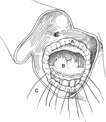
Preoperative transesophageal echocardiography at our institution showed a perforation in the left atrium into the aortic root's left fibrous trigone. The extensive perforation began just below the prosthetic valve and extended into the base of the anterior leaflet. Moderate mitral regurgitation and a dilated left atrium and ventricle were also noted. Reoperation included a repeat sternotomy with the patient under conventional cardiopulmonary bypass (CPB). Surgical exploration revealed an aortic root abscess and a paravalvular leak, with a large orifice from the noncoronary sinus toward the left atrium. We began by removing the existing prosthetic aortic valve and débriding the annulus. When the anterior annulus of the mitral valve was detached, we found that a fistulous communication had developed between the aorta and the left atrium. After resecting the necrotic tissue and débriding, we constructed a fibrous trigone using glutaraldehyde-stabilized xenogeneic bovine pericardium (St. Jude Medical; St. Paul, Minn). We attached the fibrous trigone to the mitral annulus, which had also been reconstructed from bovine pericardium (Fig. 1). The aortic valve prosthesis was replaced with a 21-mm On-X® prosthetic heart valve (Medical Carbon Research Institute, LLC; Austin, Tex). Before successful termination of CPB, the patient required an aorto–right coronary artery bypass because of intraoperative occlusion of the right coronary ostium. Sternal closure was performed 2 days later.
Fig. 1 Drawing shows reconstruction of the fibrous trigone with bovine pericardium (A), which was attached to the mitral annulus (B). The annulus was also reconstructed with bovine pericardium. Multiple interrupted pledgeted mattress sutures were passed through the cut edge of the anterior leaflet and the bovine pericardium that was used to reinforce the annulus (C). These same sutures were then passed through the sewing ring of the aortic prosthesis.
The patient was pacemaker-dependent because of atrioventricular block. He developed atrial fibrillation, which necessitated the implantation of a dual-mode, dual-pacing, dual-sensing (DDD) permanent pacemaker. Results of laboratory analysis of resected tissue specimens were negative for bacterial and fungal microorganisms. A postoperative transthoracic echocardiogram confirmed a well-seated, normally functioning, mechanical aortic valve and minimal mitral regurgitation. The patient's condition continued to improve. He was transferred to a long-term, acute-care hospital where he remained for 3 weeks. When last seen, he was doing well.
Discussion
The patient described here developed early PVE—the most severe complication encountered after heart valve replacement and one that is associated with high in-hospital and long-term mortality rates.4 The development of an aortic root abscess, which occurred in this patient, is common; however, development of an aorto–left atrial fistula, which also occurred in this patient, is rare.
It is believed that the inflammation process associated with PVE begins in the area surrounding the prosthetic sewing ring and extends into the annular connection tissue, resulting in suture loosening, paravalvular regurgitation, and possible fistula formation.5 In the early stages after valve replacement, these structures have not yet endothelialized. Periannular complications with aortic valve prostheses have been described as occurring more frequently than with mitral valve replacement, or as developing in the early stages of PVE.6 In the patient described here, surgery to exchange the prosthetic valve became imperative because of his septic condition, hemodynamic deterioration, and left atrial volume overload due to the abnormal fistulous communication, which was confirmed by transesophageal echocardiography.7
The surgical approach in patients with PVE depends on the extent of damage within the aortic and mitral valve areas. In our patient, we were able to repair the aorto–left atrial fistula and maintain aortic–mitral continuity by reconstructing the fibrous trigone with bovine pericardium. We were then able to place the new prosthetic aortic valve securely, despite the extensive débridement that was required in the area. We believe that bovine pericardium was an ideal biomaterial for use in reconstructing the fibrous trigone in order to repair this patient's intracardiac fistula.
Acknowledgments
The authors would like to thank Chrissie Chambers, MA, ELS, for editorial assistance in the preparation of the manuscript. William E. Cohn, MD, one of the authors, provided the illustration.
Footnotes
Address for reprints: O.H. Frazier, MD, P.O. Box 20345, MC 2-114A, Houston, TX 77225-0345 E-mail: lschwenke@heart.thi.tmc.edu
References
- 1.Agnihotri AK, McGiffin DC, Galbraith AJ, O'Brien MF. The prevalence of infective endocarditis after aortic valve replacement. J Thorac Cardiovasc Surg 1995;110(6):1708–24. [DOI] [PubMed]
- 2.Calderwood SB, Swinski LA, Waternaux CM, Karchmer AW, Buckley MJ. Risk factors for the development of prosthetic valve endocarditis. Circulation 1985;72(1):31–7. [DOI] [PubMed]
- 3.Wang A, Athan E, Pappas PA, Fowler VG Jr, Olaison L, Pare C, et al. Contemporary clinical profile and outcome of prosthetic valve endocarditis. JAMA 2007;297(12):1354–61. [DOI] [PubMed]
- 4.Habib G, Tribouilloy C, Thuny F, Giorgi R, Brahim A, Amazouz M, et al. Prosthetic valve endocarditis: who needs surgery? A multicentre study of 104 cases. Heart 2005;91(7): 954–9. [DOI] [PMC free article] [PubMed]
- 5.Anguera I, Miro JM, San Roman JA, de Alarcon A, Anguita M, Almirante B, et al. Periannular complications in infective endocarditis involving prosthetic aortic valves. Am J Cardiol 2006;98(9):1261–8. [DOI] [PubMed]
- 6.San Roman JA, Vilacosta I, Sarria C, de la Fuente L, Sanz O, Vega JL, et al. Clinical course, microbiologic profile, and diagnosis of periannular complications in prosthetic valve endocarditis. Am J Cardiol 1999;83(7):1075–9. [DOI] [PubMed]
- 7.Ananthasubramaniam K. Clinical and echocardiographic features of aorto-atrial fistulas. Cardiovasc Ultrasound 2005;3:1. [DOI] [PMC free article] [PubMed]


