Abstract
The circulatory half-life of the glycoprotein hormone lutropin (LH) is precisely regulated by the mannose (Man)/GalNAc-4-SO4 receptor expressed in hepatic endothelial cells. Rapid clearance from the circulation contributes to the episodic rise and fall of LH levels that is essential for maximal stimulation of the G protein-coupled LH receptor. We have defined two molecular forms of the Man/GalNAc-4-SO4 receptor that differ in ligand specificity, cell and tissue expression, and function. The form expressed by hepatic endothelial cells binds GalNAc-4-SO4-bearing ligands and regulates hormone circulatory half-life, whereas the form expressed by macrophages binds Man-bearing ligands and may play a role in innate immunity. We demonstrate that the GalNAc-4-SO4-specific form in hepatic endothelial cells is dimeric whereas the Man-specific form in lung macrophages is monomeric, accounting for the different ligand specificities of the receptor expressed in these tissues. Two cysteine-rich domains, each of which binds a single GalNAc-4-SO4, are required to form stable complexes with LH. The kinetics of LH binding by the GalNAc-4-SO4-specific form of the receptor in conjunction with its rate of internalization from the cell surface make it likely that only two of the four terminal GalNAc-4-SO4 moieties present on native LH are engaged before receptor internalization. As a result, the rate of hormone clearance will remain constant over a wide range of LH concentrations and will not be sensitive to variations in the number of terminal GalNAc-4-SO4 moieties as long as two or more are present on multiple oligosaccharides.
The glycoprotein hormones lutropin (LH) and thyrotropin (TSH) bear N-linked oligosaccharides terminating with the sequence SO4-4-GalNAcβ1,4GlcNAcβ1,2Manα (1–4). We previously identified a receptor located in hepatic endothelial cells that recognizes the sulfated oligosaccharides on LH and mediates the rapid removal of LH from the circulation (5, 6). Expression of the transferases responsible for the synthesis of these sulfated oligosaccharides is regulated by estrogen (7). These evolutionarily conserved, unique sulfated structures are found on the glycoprotein hormones of all vertebrate species (8). The regulated release of LH by gonadotrophs in the anterior pituitary in conjunction with its short circulatory half-life account for the episodic rise and fall in hormone levels that is essential for optimal activation of the LH receptor, a G-protein coupled receptor (6, 9, 10).
We isolated a receptor from liver that accounts for the clearance of LH by binding terminal GalNAc-4-SO4. This receptor has the identical peptide map, immunoreactivity, and molecular weight as the macrophage mannose (Man)-specific receptor (11). We therefore have termed this receptor the Man/GalNAc-4-SO4 receptor (12). It consists of a cysteine-rich domain, a fibronectin type II repeat, eight C-type carbohydrate recognition domains (CRDs), a transmembrane domain, and a cytosolic domain (13–15). Previous studies localized Man, N-acetylglucosamine, and fucose binding to CRDs 4–8 of the receptor (16, 17). In contrast, we localized GalNAc-4-SO4 binding to the Cys-rich domain (18). Unlike the macrophage Man receptor from lung, which binds to immobilized Man but not immobilized LH, the GalNAc-4-SO4-specific receptor isolated from liver binds to LH-Sepharose but not to Man-Sepharose (11). Chinese hamster ovary (CHO) cells expressing recombinant Man/GalNAc-4-SO4 receptor bind and internalize ligands bearing either terminal Man or GalNAc-4-SO4, suggesting that posttranslational differences most likely account for the Man- and GalNAc-4-SO4-specific forms of the receptor (12).
The different ligand specificities exhibited by the Man/GalNAc-4-SO4 receptor expressed by macrophages and hepatic endothelial cells indicate that the receptor is used for multiple functions and that the expression of these specificities is highly regulated. Because there is only a single Cys-rich domain, we hypothesized that the Man/GalNAc-4-SO4 receptor must be dimeric to bind GalNAc-4-SO4-bearing ligands. Formation of a dimer could prevent access to the sites within CRDs 4–8 that bind Man. In contrast, monomeric forms of the Man/GalNAc-4-SO4 receptor would allow free access of Man-bearing ligands to CRDs 4–8. We demonstrate here that the GalNAc-4-SO4-specific form of the receptor is indeed dimeric whereas the Man-specific form is monomeric. In addition, we present evidence that the number and location of the terminal GalNAc-4-SO4 moieties on LH is critical for recognition by the receptor.
Materials and Methods
Transfections.
Constructs in pIg (19) were cotransfected (6.25 μg of each cDNA) into CHO/Tag 30A cells by using Lipofectamine (20). Forty-eight hours after transfection, cells were radiolabeled with Trans[35S]-label (ICN) for 6 h, and the secreted fusion proteins were isolated from the medium by affinity chromatography on protein A-Sepharose (18).
Binding Assays to Immobilized Man and LH.
Purified Fc chimeras were incubated overnight at 4°C with Man-Sepharose in 20 mM Tris⋅HCl (pH 7.4), 150 mM NaCl (TBS) containing 2 mM CaCl2. The same Fc chimeras were incubated in LH-Sepharose in TBS containing 5 mM EDTA to prevent binding through Man-specific sites. Bound Fc chimera was eluted by using 100 mM glycine, pH 3.0 (18).
Characterization of Man/GalNAc-4-SO4 Receptor Prepared from Liver and Lung.
Man/GalNAc-4-SO4 receptor from rat liver and lung was solubilized in Triton X-100 and incubated with wheat germ agglutinin (WGA)-Sepharose overnight at 4°C (11). After removing unbound proteins by washing with TBS + 0.1% Triton X-100, bound glycoproteins were eluted in TBS + 0.1% Triton X-100 containing 300 mM GlcNAc. The GlcNAc was removed by dialysis against TBS + 0.1% Triton X-100. Aliquots (100 μl) of WGA-enriched material were incubated with 25 μl LH in the presence of 5 mM EDTA or 25 μl of Man-Sepharose in the presence of 5 mM CaCl2 to promote binding to Man-specific sites. Man/GalNAc-4-SO4 receptor that was bound to either LH-Sepharose or Man-Sepharose was eluted with 100 mM glycine, pH 3.0, 0.1% Triton X-100.
Western Blot Analyses.
Eluted Man/GalNAc-4-SO4 receptor was dissolved in Laemmli buffer at 25°C and separated by nonreducing SDS/PAGE (7.5% acrylamide) (21) at 25°C. Proteins were electrophoretically transferred to Immobilon-P (Millipore) in 20 mM Tris, 150 mM glycine, 10% methanol. Transferred Man/GalNAc-4-SO4 receptor was detected by incubation with a monospecific rabbit anti-Man/GalNAc-4-SO4 receptor raised by immunizing rabbits with the cDNA for the native Man/GalNAc-4-SO4 receptor in pCDM8 injected i.m. in saline and boosting after 4 weeks with 20 μg of a purified soluble Man/GalNAc-4-SO4-HAHis6 chimera in incomplete Freund's adjuvant. The Man/GalNAc-4-SO4-HAHis6 chimera was prepared by replacing the transmembrane and cytosolic domains of the Man/GalNAc-4-SO4 receptor with a sequence from the influenza hemagglutinin followed by six histidine residues (HAH6). The anti-Man/GalNAc-4-SO4 receptor was used at a dilution of 1:10,000 and detected by using chemiluminescence per the manufacturer (NEN).
Surface Plasmon Resonance Analyses.
Surface plasmon resonance analyses were performed on an Amersham Pharmacia BIAcore 2000 instrument. Purified recombinant protein A (Pierce) at 100 μg/ml in 10 mM sodium acetate buffer, pH 4.5 was coupled to a BIAcore CM5 sensorchip by using the Amine Coupling Kit provided by the manufacturer. Fc chimeras bound to immobilized protein A did not dissociate during the course of the binding assays. A flow cell devoid of coupled protein was used as a reference cell to correct for nonspecific binding and alterations in response unit values caused by buffer components. Analyses were performed at 7°C at a flow rate of 5 μl/min in TBS containing 0.005% surfactant P20 and 5 mM EDTA. For affinity measurements, differing concentrations of bovine LH, LHα, or LHβ (supplied by the National Institute of Diabetes and Digestive and Kidney Diseases National Hormone & Pituitary Program and A. F. Parlow) were bound over 10 min (50 μl) so as to allow sufficient time to reach equilibrium. Bound ligands were eluted by injection of a 25 μl pulse of 10 mM Hepes (pH 7.4), 150 mM NaCl, 3 mM EDTA, and 0.005% surfactant P20. Hepes (N-2-hyrdoxyethylpiperazine-N′-2-ethanesulfonic acid) has a Ki = 2.32 × 10⁻⁴ M because of the sulfonate. Control experiments were performed in binding buffer containing 1 mM GalNAc-4-SO4, which quantitatively inhibits binding by the Cys-rich domain of the Man/GalNAc-4-SO4 receptor. Saturation curves were analyzed by nonlinear regression (prism software, V2.0). For competition studies, increasing concentrations of inhibitors were added to a fixed concentration of LHα (685 nM).
Results
Chimeric Forms of the Man/GalNAc-4-SO4 Receptor Require Two Cys-Rich Domains to Bind Immobilized LH.
We used chimeric forms of the Man/GalNAc-4-SO4 receptor in which the transmembrane and cytosolic domains of the receptor were replaced by the Fc region of human IgG1 to demonstrate that the Cys-rich domain accounts for the binding of terminal GalNAc-4-SO4 (18). The chimera Man/GalNAc-4-SO4-Fc (Mu1), which contains only CRDs 1–8, binds ligands with terminal Man but not those with terminal GalNAc-4-SO4. In contrast, the Fc chimera Man/GalNAc-4-SO4-Fc (Mu11), consisting of the Cys-rich domain, binds ligands with terminal GalNAc-4-SO4 but not those with terminal Man. Both Fc chimeras form covalent dimers that are efficiently secreted into the medium when expressed in CHO cells.
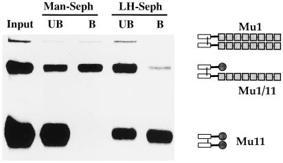
Coexpression of the cDNAs encoding Mu1 and Mu11 in CHO cells results in the synthesis and secretion of three distinct dimeric species. The majority of the species consist of (i) a covalently linked heterodimer consisting of one chain of Mu1 and one chain of Mu11, designated Mu1/11, and (ii) the homodimer Mu11. Trace amounts of the homodimer Mu1 also were expressed. The homodimeric and heterodimeric Fc chimeras are readily identified when separated by nonreducing SDS/PAGE (Fig. 1). After isolation by affinity chromatography on protein A-Sepharose, the 35S-Cys/Met-labeled chimeras were incubated with either Man-Sepharose or LH-Sepharose. The amount of each chimera in the unbound and bound fractions was determined by nonreducing SDS/PAGE followed by autoradiography (Fig. 1). Mu1/11 is efficiently bound to Man-Sepharose but not to LH-Sepharose. In contrast, Mu11 binds to LH-Sepharose but not to Man-Sepharose. Thus, Fc chimeras with a single Cys-rich domain are unable to bind to the GalNAc-4-SO4-bearing oligosaccharides on immobilized LH with sufficient stability to allow for their isolation. In contrast, Fc chimeras with two Cys-rich domains are able to bind to immobilized LH.
Figure 1.
Two Cys-rich domains are required to bind to immobilized LH. Man/GalNAc-4-SO4-Fc (Mu1) and Man/GalNAc-4-SO4-Fc (Mu11) were cotransfected in CHO/Tag30A cells and labeled with Trans[35S]-label. Secreted Fc chimeras were isolated by protein A affinity chromatography. The Fc chimeras were incubated with Man-Sepharose in TBS containing 5 mM Ca2+ or LH-Sepharose in TBS containing 5 mM EDTA. Starting material for each matrix (Input), unbound (UB), and bound (B) fractions were examined by nonreducing SDS/PAGE. The positions of Man/GalNAc-4-SO4-Fc (Mu1) 340,000 Da, Man/GalNAc-4-SO4-Fc (Mu1/11) 220,000 Da, and Man/GalNAc-4-SO4-Fc (Mu11) 98,000 Da are shown.
In other studies (data not shown) we characterized a construct, Man/GalNAc-4-SO4-HAHis6, in which the transmembrane and cytosolic domains of the Man/GalNAc-4-SO4 receptor were replaced with an antigenic sequence from the influenza hemagglutinin (22) followed by six histidine residues. The Man/GalNAc-4-SO4-HAHis6 chimera migrates as a monomer when examined by nonreducing SDS/PAGE or gel filtration. Man/GalNAc-4-SO4-HAHis6 does not bind to LH-Sepharose but does bind to Man-Sepharose. Thus, monomeric forms of the Man/GalNAc-4-SO4 receptor containing all of its extracellular domains (Cys-rich, fibronectin type II repeat, and CRDs 1–8) are unable to bind to immobilized LH. The properties of these chimeric molecules demonstrate that two Cys-rich domains are required to form stable complexes with the GalNAc-4-SO4-bearing oligosaccharides on LH.
The GalNAc-4-SO4-Specific and Man-Specific Forms of the Man/GalNAc-4-SO4 Receptor Found in Vivo Are Dimeric and Monomeric, Respectively.
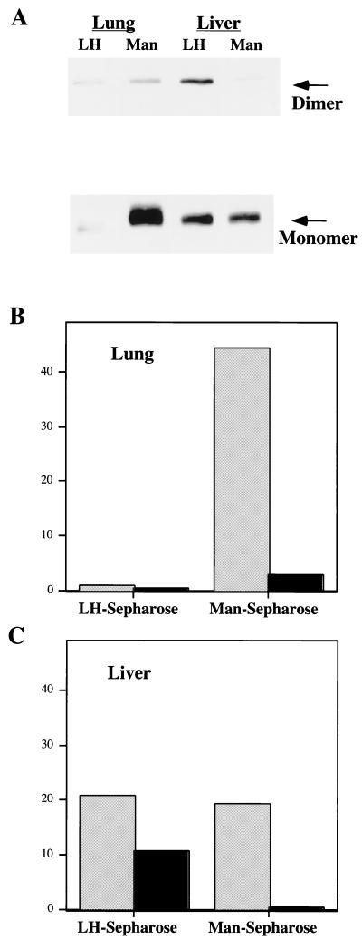
Previous studies indicated that native and recombinant forms of the macrophage Man receptor exist as monomers (17). However, after solubilization in Triton X-100 we detected monomeric and dimeric Man/GalNAc-4-SO4 receptor in liver preparations examined by nonreducing SDS/PAGE and Western blot analysis. We further examined the properties of the solubilized Man/GalNAc-4-SO4 receptor obtained from rat liver and lung after enrichment by lectin affinity chromatography using wheat germ agglutinin-Sepharose. The partially purified receptor was incubated with either LH-Sepharose or Man-Sepharose and the relative amounts of dimeric and monomeric forms of the receptor present in the bound fractions were determined by nonreducing SDS/PAGE (Fig. 2). Ninety-seven percent of the Man/GalNAc-4-SO4 receptor prepared from lung binds Man-Sepharose (Fig. 2). In contrast, 62% of the Man/GalNAc-4-SO4 receptor prepared from liver binds to LH-Sepharose and 38% to Man-Sepharose (Fig. 2). Ninety-five percent of the Man/GalNAc-4-SO4 receptor that is isolated from lung and binds to Man-Sepharose migrates as a monomer (Fig. 2B). In contrast, 34% of the Man/GalNAc-4-SO4 receptor from liver that binds to LH-Sepharose migrates as a dimer whereas 66% migrates as a monomer (Fig. 2C). The fraction of Man/GalNAc-4-SO4 receptor that migrates as a dimer is 10-fold greater for the receptor that binds to LH-Sepharose (bearing terminal GalNAc-4-SO4) as compared with the receptor that binds to Man-Sepharose. Because noncovalent receptor homodimers would be expected to dissociate during SDS/PAGE and electrophoretic transfer to Immobilon-P would be less efficient for dimeric than monomeric forms of the receptor, the value of 34% is a minimal estimate of the amount of dimeric Man/GalNAc-4-SO4 receptor present in the LH-bound fraction.
Figure 2.
Dimeric and monomeric forms of the Man/GalNAc-4-SO4 receptor are found in vivo and bind to immobilized LH and Man, respectively. Equal aliquots of partially purified Man/GalNAc-4-SO4 receptor from rat liver and lung were incubated with LH-Sepharose and Man-Sepharose. Bound Man/GalNAc-4-SO4 receptor was eluted at pH 3.0, separated by nonreducing SDS/PAGE and electrophoretically transferred to Immobilon-P for Western blot analysis using a monospecific rabbit anti-Man/GalNAc-4-SO4 receptor antibody. (A) Man/GalNAc-4-SO4 receptor from lung and liver bound to LH-Sepharose and Man-Sepharose, respectively. The amount (arbitrary units) of monomer (gray) and dimer (black) was quantitated by densitometry for lung (B) and liver (C).
The properties of the native GalNAc-4-SO4-specific and Man-specific forms of the Man/GalNAc-4-SO4 receptor are reflective of the results obtained with the soluble Fc chimeras, suggesting that the Man/GalNAc-4-SO4 receptor must form a noncovalent dimer to bind LH-Sepharose with sufficient stability to be isolated. Further, the absence of significant amounts of dimer in the portion of receptor from liver that binds to Man-Sepharose indicates that the Man binding sites located in CRDs 4–8 (16) are sequestered in the GalNAc-4-SO4-specific form of the receptor.
The Number and Location of Terminal GalNAc-4-SO4 Moieties Is Critical for LH Recognition.
To understand the significance of the number and location of terminal GalNAc-4-SO4 moieties we further characterized the N-linked oligosaccharides on bovine LH, a heterodimer consisting of an α-subunit and a β-subunit (23), and its separated subunits. The oligosaccharides on LHβ, LHα, and LH dimer were released by digestion with PNGase F and derivatized with 9-aminopyrene-1,4,6-trisulfonic acid (Beckman) for analysis by capillary electrophoresis (24). The single N-linked oligosaccharide on LHβ consists exclusively (99%) of complex biantennary structures with two terminal GalNAc-4-SO4 moieties (S-2) whereas 90% of the oligosaccharides at the two N-linked glycosylation sites on LHα are complex and hybrid structures each with a single terminal GalNAc-4-SO4 moiety (S-1) (E. I. Park and J.U.B., unpublished observation) (see schematic for Fig. 4). The distribution of oligosaccharides on LH dimer equals the sum of those found on the separated α and β subunits.
Figure 4.
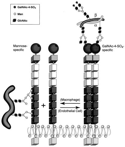
Model for the Man-specific and GalNAc-4-SO4-specific forms of the Man/GalNAc-4-SO4 receptor. Macrophages express predominantly a monomeric form of the Man/GalNAc-4-SO4 receptor (Left) shown embedded in the lipid bilayer. The Man-specific CRDs 4–8 (dark squares) are accessible to Man-bearing ligands, whereas the monomeric Cys-rich domain (circles) is not able to form stable complexes with GalNAc-4-SO4-bearing ligands. Hepatic endothelial cells express predominantly a dimeric form of the Man/GalNAc-4-SO4 receptor (Right). The apposition of two Cys-rich domains allows stable complexes to form with ligands such as LH bearing two or more oligosaccharides with terminal GalNAc-4-SO4 moieties (Upper Right). The receptor is shown engaging two S-1 structures on LHα. Other combinations of S-1 and S-2 structures could produce the same result. The Man binding sites found in CRDs 4–8 are, however, sequestered away from Man-bearing ligands in the dimeric form of the receptor.
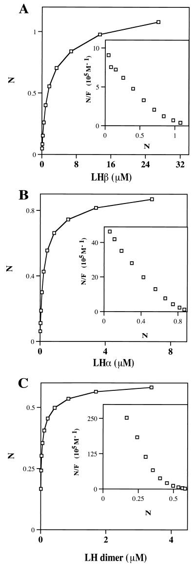
We used surface plasmon resonance to evaluate binding by the Mu11 Fc chimera, which has two Cys-rich domains. Protein A immobilized on Biosensor chips was used to capture Mu11. The stable complex is able to interact with the terminal GalNAc-4-SO4 moieties on LHβ, LHα, and LH dimer. Binding curves were generated for LHβ (one S-2), LHα (two S-1), and LH dimer (one S-2 and two S-1). The amount of ligand bound at equilibrium was used to generate the saturation curves shown in Fig. 3. The dissociation constants and Bmax values were determined by nonlinear regression analysis as summarized in Table 1. LHβ binds to Mu11 in a concentration-dependent and saturable manner with a Kd of 2.06 × 10⁻⁶ M (Fig. 3A and Table 1). In addition, 1.12 mole of LHβ binds per mole of dimeric Mu11, indicating that each Cys-rich domain can engage a single GalNAc-4-SO4 residue. This also demonstrates that the two terminal GalNAc-4-SO4 moieties on a single S-2 oligosaccharide can bridge between the GalNAc-4-SO4 binding sites on the two Cys-rich domains present in the Fc chimera Mu11. However, LHα with two S-1 oligosaccharides displays a 10-fold better affinity for Man/GalNAc-4-SO4-Fc (Mu11) than LHβ with a Kd of 2.32 × 10⁻⁷ M (Fig. 3B, Table 1, and Fig. 4 schematic). The mole ratio obtained with LHα indicates that a single GalNAc-4-SO4 from each of the monosulfated S-1 oligosaccharides is bound to each Cys-rich domain in the Fc chimera. The 10-fold increase in affinity with LHα as compared with LHβ suggests that access of the terminal GalNAc-4-SO4 moieties to the binding site on each Cys-rich domain is significantly better when the GalNAc-4-SO4 moieties are present on separate oligosaccharide chains. The affinities for LHα and LHβ reflect differences in the spatial relationships of the terminal GalNAc-4-SO4 moieties because they have the same Bmax at saturation, i.e., one GalNAc-4-SO4 bound per Cys-rich domain. The spacing of the GalNAc-4-SO4 moieties on the biantennary S-2 structure on LHβ may be less optimal or strained. The spatial relationships of terminal GalNAc-4-SO4 moieties on S-1 and S-2 structures are dictated by the locations of these oligosaccharides on the underlying peptides of the LHα and LHβ subunits.
Figure 3.
Analysis of LHβ (A), LHα (B), and LH dimer (C) binding by the Man/GalNAc-4-SO4-Fc (Mu11) chimera using surface plasmon resonance. The Man/GalNAc-4-SO4-Fc (Mu11) chimera was immobilized on protein-A coupled to a CM5 Biacore chip. Increasing concentrations of LHβ, LHα, and LH dimer were allowed to bind to the chimera. Nonlinear regression analyses of the saturation curves obtained were performed by using prism software. Scatchard plots also were generated and are shown as insets.N is the mole ratio of ligand bound per mole of Mu11 Fc chimera, and F is the concentration of ligand. Mole ratios were determined by dividing the response unit values obtained by the relative molecular weights of 98,128 for Man/GalNAc-4-SO4-Fc (Mu11), 14,880 for LHβ, 14,607 for LHα, and 29,500 for LH dimer.
Table 1.
Binding properties of ligands with differing numbers of terminal GalNAc-4-SO4-moieties for Man/GalNAc-4-SO4-Fc (Mu11)
| Ligand | Ki, μM | Kd, nM | Bmax, mol/mol |
|---|---|---|---|
| LHβ | 2060 | 1.12 | |
| LHα | 232 | 0.83 | |
| LH dimer | |||
| High affinity component | 8.3 | 0.336 | |
| Low affinity component | 267 | 0.256 | |
| GalNAc-4-SO4 | 16.8 | ||
| SO4-4-GalNAcβ1,4GlcNAcβ1,2Manα-MCO | 12.0 | ||
| Hepes | 232 |
Surface plasmon resonance was used to determine the Kd and Bmax for LHβ, LHα, and LH dimer as shown in Fig. 3. Surface plasmon resonance also was used to determine the Ki for GalNAc-4-SO4, SO4-4-GalNAcβ1,4GlcNAcβ1,2Manα-MCO, and Hepes using LHα as the ligand.
In contrast to LHα and LHβ, which both have a single affinity site for binding, LH dimer generates a curvi-linear Scatchard plot (Fig. 3C) consistent with two or more components. Regression analysis yields one component with a Kd of 2.67 × 10⁻⁷ M that is essentially identical to that obtained with LHα, indicating that two of the four terminal GalNAc-4-SO4 moieties on the LH dimer are engaged. An additional component with a Kd of 8.32 × 10⁻⁹ M suggests engagement of 3–4 of the terminal GalNAc-4-SO4 moieties on the LH dimer when bound to the Fc chimera Mu11 immobilized on the Biacore chip.
Inhibition of LHα binding to Mu11 by increasing concentrations of GalNAc-4-SO4 and SO4-4-GalNAcβ1,4GlcNAcβ1,2Manα-MCO was monitored by surface plasmon resonance (Table 1). GalNAc-4-SO4 and SO4-4-GalNAcβ1,4GlcNAcβ1,2Manα-MCO have Ki values of 16.8 × 10⁻⁵ M and 1.20 × 10⁻⁵ M, respectively. Thus, the Cys-rich domain recognizes almost exclusively the terminal sulfated monosaccharide. The underlying structure of the oligosaccharide does not contribute significantly to the binding of terminal GalNAc-4-SO4.
Discussion
The rapid clearance of native (sulfated) but not recombinant (sialylated) bovine LH from the circulation led us to identify a receptor specific for the terminal GalNAc-4-SO4 (6) (see Fig. 4). We isolated the receptor responsible for the clearance of LH from the circulation by hepatic endothelial cells (5). Each endothelial cell is capable of binding 5.7 × 10⁵ molecules of LH with a Kd of 1.63 × 10⁻⁷ M (5). Furthermore, the receptor discriminates between SO4-4-GalNAcβ1,4GlcNAcβ1,2Manα-BSA and SO4-3-GalNAcβ1,4GlcNAcβ1,2Manα-BSA, indicating that the position of the terminal sulfate is critical for binding and clearance from the circulation (5).
The GalNAc-4-SO4-specific receptor was isolated from rat liver and determined to have the same amino acid sequence as the Man-specific receptor isolated from alveolar macrophages (11). The two forms of the receptor differ in their ligand binding properties. The GalNAc-4-SO4-specific form does not bind to immobilized Man whereas the Man-specific form does not bind immobilized glycoproteins bearing oligosaccharides terminating with GalNAc-4-SO4. However, CHO cells can express recombinant receptors that bind and internalize glycoproteins bearing either terminal Man or terminal GalNAc-4-SO4 (12). This suggests that posttranslational events regulate the specificity of the Man/GalNAc-4-SO4 receptor. The differing specificities of the two forms of the Man/GalNAc-4-SO4 receptor further indicate that this receptor has multiple functions. These functions reflect both the ligand specificity of the receptor and its site-specific expression in different cells. The receptor in hepatic endothelial cells, which clears GalNAc-4-SO4-bearing ligands from the blood, must have one or more structural features that differentiate it from the same receptor expressed in macrophages, which binds ligands with terminal Man.
Based on the properties of the in vivo receptor and those of the Fc chimeras, we propose that the Man/GalNAc-4-SO4 receptor exists both as a monomer and as a homodimer. As illustrated in Fig. 4, our data indicates that the monomeric form of the Man/GalNAc-4-SO4 receptor is Man-specific and that the dimeric form is GalNAc-4-SO4-specific. Both the Man-specific and the GalNAc-4-SO4-specific forms of the receptor are present in hepatic endothelial cells, suggesting that they are in equilibrium with each other. In contrast, we detect only the Man-specific form in alveolar macrophages, consistent with the possibility that the Man/GalNAc-4-SO4 receptor in macrophages cannot form a homodimer in vivo. Differing patterns of glycosylation or other posttranslational modifications in macrophages and hepatic endothelial cells may determine whether the receptor is able to form a dimer in vivo. The differences in the mobility of the receptor isolated from liver and lung (Fig. 2) are consistent with this possibility. At present we are pursuing the molecular basis governing which form of the Man/GalNAc-4-SO4 receptor is expressed by a particular cell.
Our previous studies demonstrated that the Cys-rich domain is necessary and sufficient for GalNAc-4-SO4 binding, whereas CRDs 4–8 account for binding of ligands terminating with Man, GlcNAc, or fucose (18). The model in Fig. 4 provides a structural basis for the differences between the GalNAc-4-SO4- and Man-specific forms of the receptor that can account for the fact that the regions that bind GalNAc-4-SO4 and Man are physically distinct. The current studies indicate that two Cys-rich domains are required to form stable complexes with immobilized ligands bearing multiple terminal GalNAc-4-SO4 moieties. Furthermore, ≥62% of the Man/GalNAc-4-SO4 receptor in hepatic endothelial cells is present in the form of a homodimer because the monomeric form does not bind to LH.
We have shown that 93% of the Man/GalNAc-4-SO4 receptor in lung is monomeric based both on its inability to bind to LH-Sepharose and on the absence of dimeric species during analysis by SDS/PAGE. The dominance of monomeric receptor in the lung supports our proposal that the receptor expressed by macrophages is not able to form dimeric species. Furthermore, the absence of dimer in the Man-Sepharose bound receptor from liver indicates that CRDs 4–8 are indeed sequestered in the dimeric form and prevent access to the immobilized Man residues. The monomeric species in the liver may reflect either the presence of receptor that cannot form homodimers or monomers that are in equilibrium with dimers.
The Man/GalNAc-4-SO4 receptor is remarkable because its two specificities for GalNAc-4-SO4-bearing ligands and Man-bearing ligands are mutually exclusive. The dominance of monomeric receptor in the macrophage where CRDs 4–8 are accessible may reflect its proposed role as a phagocytic receptor in innate immunity (25). The dimeric GalNAc-4-SO4-specific receptor in endothelial cells is essential for control of LH and TSH levels in the circulation (5, 6, 26). The presence of these alternative forms in different tissues indicates that expression is regulated in a cell-specific manner.
The glycoprotein hormones LH and TSH are, at present, the only well-established native ligands for the GalNAc-4-SO4-specific form of the Man/GalNAc-4-SO4 receptor. Both hormones bear multiple oligosaccharides terminating with one or more GalNAc-4-SO4 moieties. Their spatial relationships are critical for binding because the presence of two S-1 oligosaccharides allows LHα to bind with a Kd of 2.32 × 10⁻⁷ M whereas the single S-2 oligosaccharide on LHβ binds with a 10-fold lower affinity. The stoichiometry of binding indicates that each Cys-rich domain engages a single GalNAc-4-SO4. This is in agreement with a recent crystallographic study that identified a single binding site (27) and with equilibrium dialysis studies (unpublished work). Clearly the relationship of the terminal GalNAc-4-SO4 moieties in space has a major impact on their ability to bind simultaneously to two Cys-rich domains. Our data indicate that only certain forms of the native Man/GalNAc-4-SO4 receptor, such as that present in hepatic endothelial cells, form stable dimeric structures.
LH is bound by isolated hepatic endothelial cells with an apparent Kd of 1.63 × 10⁻⁷ M (5), a value nearly identical to the Kd of 2.67 × 10⁻⁷ M obtained for LHα binding through two terminal GalNAc-4-SO4 moieties on separate oligosaccharides to the Fc chimera immobilized on the Biacore chip. This suggests that LH binds to the dimeric, GalNAc-4-SO4-specific form of the Man/GalNAc-4-SO4 receptor on the endothelial cell surface by using only two of its 3–4 terminal GalNAc-4-SO4-moieties. Even though the LH dimer can bind to recombinant Mu11 chimera immobilized on the Biacore chip with an additional component of Kd 8.3 × 10⁻⁹ M when engaged by 3–4 terminal GalNAc-4-SO4 moieties, this latter high-affinity component is not observed when LH binds to the receptor at the surface of hepatic endothelial cells in vivo.
Based on binding studies performed with recombinant receptor Simpson et al. (28) have suggested that glycoprotein hormones also may bind to the Man/GalNAc-4-SO4 receptor through Man residues alone or in combination with GalNAc-4-SO4. Bovine LH does contain terminal Man residues caused by the presence of hybrid structures (1, 2). Simpson et al. (28) reported that LH binds to the Man-specific sites of the Man/GalNAc-4-SO4 receptor with an affinity in the range of 2.8 × 10⁻⁶ M (28). In light of the Kd of 2.32 × 10⁻⁷ M obtained when two GalNAc-4-SO4 moieties are engaged, it seems unlikely that interaction with terminal Man residues contributes to clearance of LH in vivo. The sequestration of the Man binding domains in the dimeric, GalNAc-4-SO4-specific form of the receptor makes interaction with terminal Man residues on LH even less likely. The ability of native GalNAc-4-SO4 receptor from liver to bind LH is not altered by the presence of Man nor have we been able to demonstrate simultaneous interaction with Man and GalNAc-4-SO4 for either the Man receptor from lung or the GalNAc-4-SO4 receptor from liver (D. Fiete and J.U.B., unpublished observation). LH and TSH from other animal species do not bear terminal Man or GlcNAc to the same extent as the bovine hormone even though they bear terminal GalNAc-4-SO4 (1, 2). In contrast, the presence of terminal β1,4-linked GalNAc-4-SO4 on N-linked oligosaccharides of glycoprotein hormones has been conserved in all vertebrate species, indicating that these structures have a critical role in vivo (8). Even though simultaneous binding to terminal Man and GalNAc-4-SO4 on bovine LH may be possible under some circumstances, this form of binding also seems unlikely to contribute to clearance of LH and TSH by the hepatic Man/GalNAc-4-SO4 receptor in vivo.
Our results indicate that the form of the Man/GalNAc-4-SO4 receptor expressed by endothelial cells versus macrophages is highly regulated. Further, we have provided a molecular basis for how the number of terminal GalNAc-4-SO4 moieties on LH and TSH control their circulatory half-lives. The presence of two or more terminal residues of GalNAc-4-SO4 on separate oligosaccharides is sufficient to provide productive binding by the Man/GalNAc-4-SO4 receptor on hepatic endothelial cells. Because the receptor is dimeric on endothelial cells and is constantly internalized, the additional residues of terminal GalNAc-4-SO4 present on native LH are not likely to contribute to binding in vivo. The association rate and concentration of LH in the blood will dictate what fraction of the receptor is occupied at the time of internalization. The large amount of receptor expressed by hepatic endothelial cells assures that the rate of clearance from the circulation by internalization will remain constant over a wide range of LH concentrations, allowing maximal activation of the LH receptor in the ovary and testis.
Acknowledgments
We thank Nancy Baenziger, Jeremy Keusch, and Alison Woodworth for critical comments. This work was supported by National Institutes of Health Grants R37-CA21923 and R01-DK41738.
Abbreviations
- CHO
Chinese hamster ovary
- Man
mannose
- LH
lutropin
- FSH
follitropin
- TSH
thyrotropin
- CRD
C-type carbohydrate recognition domain
Footnotes
This paper was submitted directly (Track II) to the PNAS office.
Article published online before print: Proc. Natl. Acad. Sci. USA, 10.1073/pnas.170184597.
Article and publication date are at www.pnas.org/cgi/doi/10.1073/pnas.170184597
References
- 1.Green E D, Baenziger J U. J Biol Chem. 1988;263:25–35. [PubMed] [Google Scholar]
- 2.Green E D, Baenziger J U. J Biol Chem. 1988;263:36–44. [PubMed] [Google Scholar]
- 3.Baenziger J U, Green E D. In: Biology of Carbohydrates. Ginsberg V, Robbins P W, editors. Vol. 3. London: JAI; 1991. pp. 1–46. [Google Scholar]
- 4.Stockell Hartree A, Renwick A G C. Biochem J. 1992;287:665–679. doi: 10.1042/bj2870665. [DOI] [PMC free article] [PubMed] [Google Scholar]
- 5.Fiete D, Srivastava V, Hindsgaul O, Baenziger J U. Cell. 1991;67:1103–1110. doi: 10.1016/0092-8674(91)90287-9. [DOI] [PubMed] [Google Scholar]
- 6.Baenziger J U, Kumar S, Brodbeck R M, Smith P L, Beranek M C. Proc Natl Acad Sci USA. 1992;89:334–338. doi: 10.1073/pnas.89.1.334. [DOI] [PMC free article] [PubMed] [Google Scholar]
- 7.Dharmesh S M, Baenziger J U. Proc Natl Acad Sci USA. 1993;90:11127–11131. doi: 10.1073/pnas.90.23.11127. [DOI] [PMC free article] [PubMed] [Google Scholar]
- 8.Manzella S M, Dharmesh S M, Beranek M C, Swanson P, Baenziger J U. J Biol Chem. 1995;270:21665–21671. doi: 10.1074/jbc.270.37.21665. [DOI] [PubMed] [Google Scholar]
- 9.Baenziger J U. Endocrinology. 1996;137:1520–1522. doi: 10.1210/endo.137.5.8612480. [DOI] [PubMed] [Google Scholar]
- 10.Manzella S M, Hooper L V, Baenziger J U. J Biol Chem. 1996;271:12117–12120. doi: 10.1074/jbc.271.21.12117. [DOI] [PubMed] [Google Scholar]
- 11.Fiete D, Baenziger J U. J Biol Chem. 1997;272:14629–14637. doi: 10.1074/jbc.272.23.14629. [DOI] [PubMed] [Google Scholar]
- 12.Fiete D, Beranek M C, Baenziger J U. Proc Natl Acad Sci USA. 1997;94:11256–11261. doi: 10.1073/pnas.94.21.11256. [DOI] [PMC free article] [PubMed] [Google Scholar]
- 13.Taylor M E, Conary J T, Lennartz M R, Stahl P D, Drickamer K. J Biol Chem. 1990;265:12156–12162. [PubMed] [Google Scholar]
- 14.Taylor M E. Glycobiology. 1997;7:V–XV. doi: 10.1093/glycob/7.3.323. [DOI] [PubMed] [Google Scholar]
- 15.Ezekowitz R A B, Sastry K, Bailly P, Warner A. J Exp Med. 1990;172:1785–1794. doi: 10.1084/jem.172.6.1785. [DOI] [PMC free article] [PubMed] [Google Scholar]
- 16.Taylor M E, Bezouska K, Drickamer K. J Biol Chem. 1992;267:1719–1726. [PubMed] [Google Scholar]
- 17.Taylor M E, Drickamer K. J Biol Chem. 1993;268:399–404. [PubMed] [Google Scholar]
- 18.Fiete D J, Beranek M C, Baenziger J U. Proc Natl Acad Sci USA. 1998;95:2089–2093. doi: 10.1073/pnas.95.5.2089. [DOI] [PMC free article] [PubMed] [Google Scholar]
- 19.Simmons D L. In: Cellular Interactions in Development: A Practical Approach. Hartley D A, editor. Oxford: Oxford Univ. Press; 1993. pp. 93–127. [Google Scholar]
- 20.Mengeling B J, Manzella S M, Baenziger J U. Proc Natl Acad Sci USA. 1995;92:502–506. doi: 10.1073/pnas.92.2.502. [DOI] [PMC free article] [PubMed] [Google Scholar]
- 21.Laemmli U K. Nature (London) 1970;227:680–685. doi: 10.1038/227680a0. [DOI] [PubMed] [Google Scholar]
- 22.Huse W D, Sastry L, Iverson S A, Kang A S, Alting-Mees A, Burton D R, Benkovic S J, Lerner R A. Science. 1989;246:1275–1281. doi: 10.1126/science.2531466. [DOI] [PubMed] [Google Scholar]
- 23.Pierce J G, Parsons T F. Annu Rev Biochem. 1981;50:465–495. doi: 10.1146/annurev.bi.50.070181.002341. [DOI] [PubMed] [Google Scholar]
- 24.Guttman A, Chen F T, Evangelista R A, Cooke N. Anal Biochem. 1996;233:234–242. doi: 10.1006/abio.1996.0034. [DOI] [PubMed] [Google Scholar]
- 25.Stahl P D, Ezekowitz R A. Curr Opin Immunol. 1998;10:50–55. doi: 10.1016/s0952-7915(98)80031-9. [DOI] [PubMed] [Google Scholar]
- 26.Szkudlinski M W, Thotakura N R, Tropea J E, Grossmann M, Weintraub B D. Endocrinology. 1995;136:3325–3330. doi: 10.1210/endo.136.8.7628367. [DOI] [PubMed] [Google Scholar]
- 27.Liu Y, Chirino A J, Leteux C, Feizi T, Misulovin Z, Nussenzweig M C, Bjorkman P. J Exp Med. 2000;191:1105–1115. doi: 10.1084/jem.191.7.1105. [DOI] [PMC free article] [PubMed] [Google Scholar]
- 28.Simpson D Z, Hitchen P G, Elmhirst E L, Taylor M E. Biochem J. 1999;343:403–411. [PMC free article] [PubMed] [Google Scholar]


