Abstract
In young healthy humans full expression of reflex cutaneous vasodilation is dependent on cyclooxygenase (COX)- and nitric oxide synthase (NOS)-dependent mechanisms. Chronic low-dose aspirin therapy attenuates reflex cutaneous vasodilation potentially through both platelet and vascular COX-dependent mechanisms. We hypothesized the contribution of COX-dependent vasodilators to reflex cutaneous vasodilation during localized acute COX inhibition would be attenuated in healthy middle-aged humans due to a shift toward COX-dependent vasoconstrictors. Four microdialysis fibers were placed in forearm skin of 13 middle-aged (53 ± 2 yr) normotensive healthy humans, serving as control (Ringer), COX-inhibited (10 mM ketorolac), NOS-inhibited (10 mM NG-nitro-l-arginine methyl ester), and combined NOS- and COX-inhibited sites. Red blood cell flux was measured over each site by laser-Doppler flowmetry as reflex vasodilation was induced by increasing oral temperature (Tor) 1.0°C using a water-perfused suit. Cutaneous vascular conductance was calculated (CVC = flux/mean arterial pressure) and normalized to maximal CVC (CVCmax; 28 mM sodium nitroprusside). CVCmax was not affected by localized microdialysis drug treatment (P > 0.05). Localized COX inhibition increased baseline (18 ± 3%CVCmax; P < 0.001) compared with control (9 ± 1%CVCmax), NOS-inhibited (7 ± 1%CVCmax), and combined sites (10 ± 1%CVCmax). %CVCmax in the COX-inhibited site remained greater than the control site with ΔTor ≤ 0.3°C; however, there was no difference between these sites with ΔTor ≥ 0.4°C. NOS inhibition and combined COX and NOS inhibition attenuated reflex vasodilation compared with control (P < 0.001), but there was no difference between these sites. Localized COX inhibition with ketorolac significantly augments baseline CVC but does not alter the subsequent skin blood flow response to hyperthermia, suggesting a limited role for COX-derived vasodilator prostanoids in reflex cutaneous vasodilation and a shift toward COX-derived vasoconstrictors in middle-aged human skin.
Keywords: skin blood flow, thermoregulation, prostaglandins, cyclooxygenase, nitric oxide
with rising body core temperature, skin blood flow is first increased by withdrawal of adrenergic vasoconstrictor tone, and then, on reaching a specific core temperature threshold, active cutaneous vasodilation occurs (25). Reflex vasodilation is mediated by cholinergic cotransmission (11) where several putative vasodilator mechanisms are involved, including the cotransmitter vasoactive intestinal peptide (1), histamine receptor activation (35), and neurokinin 1 receptor activation (34). Further, full expression of reflex cutaneous vasodilation in young healthy human skin is dependent on nitric oxide (NO) synthase (NOS) (10, 27) and cyclooxygenase (COX) second messenger mechanisms, each of which purportedly contributes independently to the rise in skin blood flow during hyperthermia (16).
With healthy human aging there is a significant attenuation in reflex cutaneous vasodilation due to a reduction in both NO- and cotransmitter-dependent vasodilation (5, 12). We have recently demonstrated that chronic low-dose aspirin therapy (81 mg) consistently and significantly attenuates reflex cutaneous vasodilation in middle-aged subjects (57 ± 3 yr) (6). Low-dose aspirin acetylates platelet COX-1 in the presystemic (portal) circulation (24), inhibiting COX for the life of the platelet (∼10 days), whereas COX in vascular endothelial cells maintains the ability to produce COX-dependent vasodilators. However, the possibility exists that low-dose aspirin 1) inhibits cutaneous microvascular COX-1 (13, 32), thus inhibiting a key vascular enzyme involved in reflex vasodilation; and/or 2) inhibits platelets from directly releasing vasodilating factors such as NO, ATP, or 5-HT (3, 9, 20). In endothelial cells, COX synthesizes several vasodilator prostaglandins (PGI2, PGE, PGF, PGD), as well as vasoconstrictor (thromboxane) factors. With vascular aging there is a shift toward the latter, resulting in a proconstrictor state (31). For example, indirect evidence using exogenous acetylcholine-induced vasodilation in aged cutaneous vasculature suggests that there is a reduction in the synthesis of COX-derived vasodilator prostanoids and a shift toward COX-derived vasoconstricting factors (7). However, the potential role for platelet vs. vascular COX and the effect of primary human aging on COX-dependent second messenger vasodilator and vasoconstrictor systems during hyperthermia are unclear.
The purpose of this study was to determine the effect of acute localized COX inhibition on reflex cutaneous vasodilation in healthy middle-aged human skin. We hypothesized that 1) localized nonspecific COX inhibition with ketorolac would increase baseline skin blood flow in a thermoneutral setting, and 2) the contribution of COX-derived vasodilator prostaniods to reflex cutaneous vasodilation would be attenuated in middle-aged humans.
METHODS
Subjects.
Experimental protocols were approved by the Institutional Review Board at The Pennsylvania State University and conformed to the guidelines set forth by the Declaration of Helsinki. Verbal and written consent were voluntarily obtained from all subjects before participation. Studies were performed on 13 healthy subjects (Table 1).
Table 1.
Subject characteristics
| Value | |
|---|---|
| Sex, M/F | 6/7 |
| Age, yr | 53±2 |
| BMI, kg/m2 | 26±1.2 |
| Total cholesterol, mg/dl | 164±8 |
| HDL, mg/dl | 56±3 |
| LDL, mg/dl | 109±7 |
| Fasting blood glucose, mg/dl | 92±2 |
| SBP, mmHg | 110±3 |
| DBP, mmHg | 70±2 |
| MAP, mmHg | 83±2 |
| Baseline Tor, °C | 36.2±0.1 |
Values are means ± SE; BMI, body mass index; HDL, high-density lipoprotein; LDL, low-density lipoprotein; SBP, systolic blood pressure; DBP, diastolic blood pressure; MAP mean arterial pressure; Tor, oral temperature.
Subjects underwent a complete medical screening, including a physician-supervised graded exercise test to evaluate the existence of underlying cardiovascular disease, blood chemistry, lipid profile evaluation (Quest Diagnostics Nichol Institute, Chantilly, VA), resting electrocardiogram, and physical examination. All subjects were screened for the presence of cardiovascular, dermatological, and neurological disease. Subjects were normally active, nondiabetic nonsmokers who were currently not taking medications, including vitamins, low-dose aspirin, hormone replacement therapy, or oral contraceptives. All women were studied in the early follicular phase of their menstrual cycle.
Instrumentation and measurements.
Protocols were performed in a thermoneutral laboratory with the subject in the semisupine position, with the experimental arm at heart level. On arrival at the laboratory, subjects were instrumented with four intradermal microdialysis fibers (MD2000, Bioanalytical Systems) (10 mm, 20-kDa cutoff membrane) in the skin on the left ventral forearm. Microdialysis sites were at least 4.0 cm apart to ensure no cross-reactivity of pharmacological agents being delivered to the skin. Microdialysis fibers were placed at each site by first inserting a 25-gauge needle through unanesthetized skin using sterile technique. The entry and exit points were ∼2.5 cm apart. The microdialysis fibers were then threaded through the needle, and the needle was withdrawn, leaving the fibers in place. The microdialysis fibers were taped in place and initially perfused with lactated Ringer solution to ensure the integrity of the fiber and during the insertion trauma resolution period. Following this period microdialysis sites were perfused with 1) 10.0 mM NG-nitro-l-arginine methyl ester (l-NAME) to inhibit NO production by NOS (5, 7, 17), 2) 10.0 mM ketorolac to nonspecifically inhibit COX (7, 16), 3) a combination of 10.0 mM ketorolac and 10.0 mM l-NAME, and 4) lactated Ringer solution to serve as a control. All microdialysis drugs were continuously perfused at a rate of 2.0 μl/min (Bee Hive controller and Baby Bee microinfusion pumps, Bioanalytical Systems) throughout the protocol.
To obtain an index of skin blood flow, cutaneous red blood cell flux was measured with an integrated laser-Doppler flowmeter probe placed in a local heater maintained at 33°C (MoorLAB, Temperature Monitor SH02, Moor Instruments, Devon, UK) on the skin directly above each microdialysis membrane. All laser-Doppler probes were calibrated using Brownian standard solution. Cutaneous vascular conductance (CVC) was calculated as flux divided by mean arterial pressure.
To control whole body temperature, subjects wore a water-perfused suit that covered the entire body, except head, hands, and experimental arm. Additionally, subjects wore a water-impermeable outer garment over the water-perfused suit to minimize evaporative heat loss. The subject's electrocardiogram was monitored throughout the protocol, and blood pressure was measured via brachial auscultation every 5 min at baseline thermoneutral and with every 0.1°C rise in oral temperature (Tor). Tor was continuously monitored during baseline and throughout whole body heating as an index of body core temperature with a thermistor placed in the sublingual sulcus. The subjects were instructed to keep the thermistor in the same location in the sublingual sulcus and not to open their mouths or speak during the protocol. Mean skin temperature was calculated as the unweighted average from six copper-constantan thermocouples placed on the chest, middle back, abdomen, upper arm, thigh, and calf. During the period of insertion trauma resolution and baseline measurement periods, thermoneutral water (34°C) was perfused through the suit to clamp mean body temperature. During whole body heating, 50°C water was perfused through the suit to raise subject's Tor by 1.0°C. Local skin temperature over each microdialysis site was separately maintained at 33°C (Moor Instruments SHO2, Devon, UK).
Experimental protocol.
Red blood cell flux over each microdialysis site was monitored as insertion trauma resolved over a 75- to 90-min period. Four microdialysis sites were randomly assigned to their specific pharmacological treatment. All drugs were mixed just before each experiment, dissolved in lactated Ringer solution, and sterilized using syringe microfilters (Acrodisc, Pall, Ann Arbor, MI).
Microdialysis sites were perfused continuously for at least 75 min before the start of the baseline and during the baseline and heating periods with assigned pharmacological agents at a rate of 2.0 μl/min. Data were collected for 20 min before the start of whole body heating to ensure a true baseline value, after which whole body heating was initiated. After Tor had increased by 1°C from baseline and skin blood flow had reached an established plateau, mean body temperature was clamped by decreasing the temperature of the circulating water to 45°C. l-NAME (10 mM) was the perfused through the control site and the ketorolac site at 4.0 μl/min to quantify NO-dependent vasodilation. After laser-Doppler flux reached steady post-l-NAME values (∼30–40 min), 28.0 mM sodium nitroprusside (Nitropress, Abbot Laboratories, Chicago, IL) was perfused through all sites at a rate of 4 μl/min to achieve maximal CVC (CVCmax) in combination with local heating of the skin to 43°C over each microdialysis site to ensure CVCmax had been achieved (16).
Data acquisition and analysis.
Data were acquired using Windaq software and Dataq data-acquisition systems (Akron, OH). The data were collected at 40 Hz, digitized, recorded, and stored on a personal computer for further analysis. CVC data were averaged over 3-min periods for baseline and every 0.1°C rise in Tor and are presented as a percentage of CVCmax (%CVCmax). Thresholds for reflex vasodilation were determined by an experienced reviewer who was blinded to the microdialysis treatment sites as the point when laser-Doppler flux significantly deviated from baseline. Absolute CVCmax in each microdialysis site was calculated as the average of the stable plateau in laser-Doppler flux during 28 mM sodium nitroprusside infusion and local heating to 43°C divided by mean arterial pressure.
Statistical analyses.
Initially, a three-way mixed models ANOVA was conducted to determine if there was a difference between the sexes on localized microdialysis drug treatments across the rise in oral core temperature. There was no difference between the sexes (P = 0.66) on the %CVCmax in microdialysis drug treatment sites across the rise in oral core temperature. Thereafter the sexes were combined and a two-way mixed models ANOVA with repeated measures was conducted to determine differences between localized microdialysis drug treatments across the rise in oral core temperature. Specific planned comparisons with Bonferroni corrections were performed when appropriate. Additionally, one-way ANOVA with repeated measures was conducted to determine differences in 1) the thresholds for the onset of reflex vasodilation for both absolute and ΔTor and 2) the effect of drug treatment on absolute maximal CVC. Tukey post hoc tests were performed to determine differences in thresholds and CVCmax due to drug treatments. The level of significance was set at α = 0.05. Values are presented as means ± SE.
RESULTS
Subject characteristics are presented in Table 1. Absolute Tor and ΔTor thresholds for the onset of reflex cutaneous vasodilation are presented in Table 2. There was a rightward shift in the threshold for reflex vasodilation in sites treated with l-NAME and combined l-NAME + ketorolac (P < 0.001) for both the absolute Tor threshold and ΔTor compared with the control site. Localized ketorolac treatment alone did not change the threshold for reflex vasodilation compared with the control site (P = 0.23).
Table 2.
Temperature thresholds for the onset of reflex cutaneous vasodilation
| Drug Treatment | Absolute Tor | ΔTor |
|---|---|---|
| Control | 36.54±0.05 | 0.31±0.03 |
| l-NAME | 36.70±0.08* | 0.48±0.04* |
| Ketorolac | 36.60±0.10 | 0.37±0.06 |
| l-NAME + ketorolac | 36.69±0.09* | 0.46±0.04* |
Values are means ±SE for control, nitric oxide synthase-inhibited [NG-nitro-l-arginine methyl ester (l-NAME)], cyclooxygenase-inhibited (ketorolac), and combination l-NAME + ketorolac sites. Threshold data presented at both absolute Tor and ΔTor. Threshold for both l-NAME and combination l-NAME + ketorolac sites occurred at a significantly higher absolute Tor and ΔTor than the control site.
P < 0.05 between control and drug-treated sites.
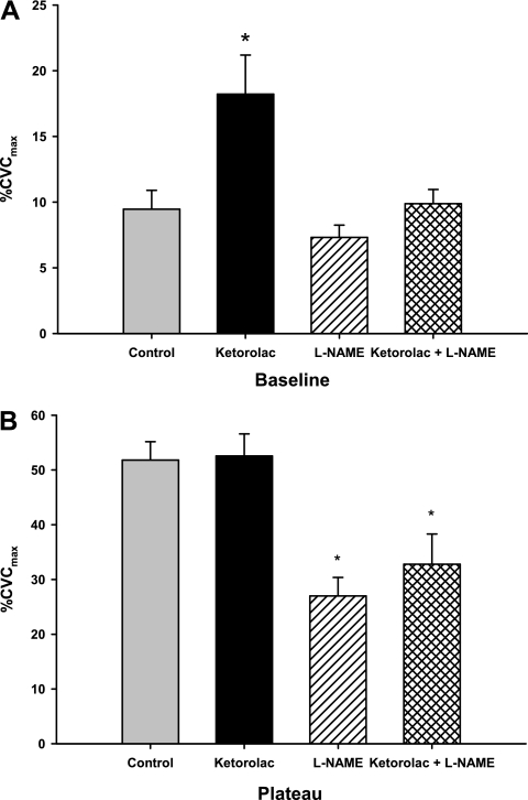
Group mean %CVCmax responses at (thermoneutral) baseline and with a 1.0°C rise in oral temperature are presented in Fig. 1, A and B, respectively. Localized COX inhibition with ketorolac increased thermoneutral baseline %CVCmax compared with all other sites (P < 0.001, Fig. 1A). l-NAME and ketorolac + l-NAME treatments decreased %CVCmax compared with control and ketorolac-treated sites (P < 0.001, Fig. 1B) during whole body heating. There was no difference in skin blood flow during hyperthermia in sites treated with ketorolac compared with control (P = 1.0).
Fig. 1.
Group mean ± SE maximal cutaneous vascular conductance (%CVCmax) at thermoneutral baseline [rise in oral temperature (ΔTor) 0.0°C] (A) and during hyperthermia (ΔTor = 1°C) (B) measured as a percentage of maximum [sodium nitroprusside (SNP)] for control, nitric oxide synthase (NOS)-inhibited [NG-nitro-l-arginine methyl ester (l-NAME)], cyclooxygenase (COX)-inhibited (ketorolac), and the combination of l-NAME and ketorolac sites. Local treatment with ketorolac increased %CVCmax baseline, which was attenuated with concurrent NOS inhibition. At plateau there was no difference between ketorolac-treated and control sites, suggesting a limited functional role for COX-derived vasodilators in reflex cutaneous vasodilation in this age group. *P < 0.05 between control and drug-treated sites.
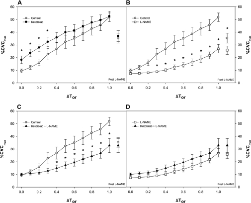
Group mean %CVCmax responses across the rise in oral temperature are presented in Fig. 2. COX inhibition with ketorolac (Fig. 2A) increased %CVCmax compared with the control site at ΔTor ≤0.3°C; however, there was no difference between these sites at ΔTor ≥0.4°C. NOS inhibition with l-NAME at ΔTor =1.0°C resulted in a similar decline in %CVCmax in the ketorolac-treated site compared with control. l-NAME treatment (Fig. 2B) attenuated %CVCmax at ΔTor ≥0.3°C compared with the control site. In the control site %CVCmax after NOS inhibition with l-NAME (ΔTor =1.0°C) was greater than in sites that had been treated with l-NAME throughout whole body heating. Combined ketorolac + l-NAME treatment (Fig. 2C) attenuated %CVCmax at ΔTor ≥0.4°C compared with the control site. There was no difference in %CVCmax after NOS inhibition with l-NAME between ketorolac + l-NAME and control sites. Finally, there was no difference between sites treated with l-NAME and ketorolac + l-NAME (Fig. 2D).
Fig. 2.
Group mean ± SE cutaneous vascular conductance (CVC) as a percent of maximal response during passive whole body heating. The control site (gray circles in A, B, C), COX-inhibited (ketorolac) site (black circles in A), NOS-inhibited site (open squares in B and D) and combination of l-NAME + ketorolac site (black triangles in C and D) are shown across ΔTor. %CVCmax after NOS inhibition with ΔTor = 1.0°C is also illustrated. A: local treatment with ketorolac augmented %CVCmax compared with control from baseline through ΔTor = 0.3; however, there was no difference between these sites in %CVCmax after NOS inhibition (post-l-NAME) with ΔTor = 1.0°C. B: l-NAME attenuated reflex cutaneous vasodilation throughout whole body heating. %CVCmax after NOS inhibition with ΔTor = 1.0°C in the control site was greater than in sites treated with l-NAME throughout heating. C: combined l-NAME + ketorolac attenuated reflex vasodilation compared with control, and there was no difference in %CVCmax after NOS inhibition with ΔTor = 1.0°C between these sites. D: there was no difference between l-NAME and the combination of l-NAME + ketorolac-treated sites across the rise in oral core temperature. *P < 0.05 (with Bonferroni correction) between control and drug-treated sites.
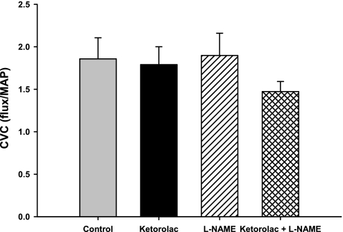
Absolute maximal CVC (28 mM SNP + local heating to 43°C) at control and drug treatment sites are presented in Fig. 3. There were no differences in absolute maximal CVC between the control drug treatment sites (P = 0.23).
Fig. 3.
Group mean ± SE absolute CVC at maximal vasodilation (SNP + 43.0°C). There was no difference in CVCmax due to localized microdialysis drug treatment. MAP, mean arterial pressure.
DISCUSSION
The principal findings of this study were as follows. 1) Acute localized COX inhibition with ketorolac increases baseline thermoneutral skin blood flow. These data suggest that COX-derived vasoconstrictors contribute to basal cutaneous vascular tone in middle-aged human skin. Furthermore, concurrent COX and NOS inhibition reduced thermoneutral baseline skin blood flow (similar to control sites), indicating there may be an interaction between these second messenger signaling pathways such that acute COX inhibition increases NO bioavailability. 2) During reflex vasodilation there were no differences between ketorolac-treated and control sites, suggesting either a simple baseline shift increasing skin blood flow throughout whole body heating and/or that in this age group COX-derived vasodilators do not functionally contribute to the increase in skin blood flow with hyperthermia. Finally, because acute localized vascular COX inhibition did not attenuate reflex vasodilation in this age group, it is unlikely that low-dose aspirin-induced inhibition of vascular COX is a potential mechanism underlying attenuated reflex vasodilation observed in humans on chronic low-dose aspirin therapy (6).
Normothermia.
We found that in middle-aged skin localized non-isoform-specific COX inhibition with ketorolac significantly increased thermoneutral skin blood flow, suggesting that COX-derived vasoconstrictors contribute to basal cutaneous vascular tone in this age group. COX isoforms produce several vasodilators and vasoconstrictors. With primary aging and vascular pathology there is 1) increased COX expression (31) and 2) a functional shift toward COX-derived constricting factors, including endoperoxidases and prostacyclin which stimulate vascular smooth muscle constriction through activation of thromboxane-prostaniod (TP) receptors, contributing to an overall proconstrictor state (31). In aged and hypertensive human vasculature this functional outcome is evidenced by a potentiation in vasodilation to acetylcholine during acute COX inhibition with indomethacin that is mediated by an increase in NO bioavailability (15, 28–30). Potential mechanisms increasing NO bioavailability during COX inhibition include decreased production of NO scavengers, including superoxide and endoperoxidases (4, 8, 26). Consistent with these findings, in the present study in middle-aged subjects (53 ± 2 yr) the coadministration of the NOS inhibitor l-NAME with ketorolac reduced thermoneutral %CVCmax (vs. ketorolac alone) similar to control sites, suggesting that acute COX inhibition increased NO-dependent vasodilation. We have previously demonstrated a similar increase in %CVCmax with acute localized COX inhibition in the cutaneous microvasculature in humans of advanced age (69 ± 1 yr) (7). However, in this older age cohort, combined NOS and COX inhibition did not reduce baseline %CVCmax (vs. ketorolac alone) (7), indicating that acute COX inhibition did not increase NO bioavailability in this age group. Collectively, these results suggest that there may be differential regulation of endothelial second messenger pathways along the aging continuum with more significant impairments in NO biosynthetic pathways contributing to reduced endothelial function with more advanced age.
Reflex vasodilation.
During reflex vasodilation there was not a significant difference in thresholds or %CVCmax between ketorolac-treated and control sites (Figs. 1A and 2A). This may be due to a simple baseline shift where differences between the sites at baseline disappear during reflex vasodilation. We originally hypothesized that COX-derived vasodilators would contribute to reflex vasodilation; however, this contribution would be attenuated compared with what has been reported in the literature in young healthy subjects (16). While a baseline effect due to an age-related shift from COX-derived vasodilators toward vasoconstrictors is the simplest potential explanation for the lack of a difference between COX-inhibited and control sites during reflex vasodilation (ΔTor > 0.4°C), there are other potential mechanisms that may underlie this lack of a difference between these sites, including decreased vascular responsiveness to prostacyclin.
Nicholson and colleagues (19) recently demonstrated that primary aging is associated with reduced prostacyclin-mediated vasodilation in the human forearm (intra-arterial infusions with strain-gauge plethysmography). Prostacyclin can act as either a vasodilator or a vasoconstrictor depending on specific vascular smooth muscle receptor activation, where in general TP receptors activation induces vasoconstriction and inositol phosphate (IP) receptor activation induces vasodilation (2). With aging and other vascular pathologies the expression of these vascular smooth muscle receptors shifts to increase TP and reduce IP receptors, respectively (2, 8, 18, 31). In the Nicholson study attenuated prostacyclin-mediated vasodilation in the aged subjects was related to a reduction in endothelial NO generation but not to reduced vascular smooth muscle responsiveness. While we did not directly assess prostacyclin-dependent vasodilation in the skin, this remains a potential explanation for our finding that acute localized COX inhibition did not alter the skin blood flow response in our middle-aged subjects at elevated core temperatures.
In contrast to significant endothelial interactions between prostacyclin- and NO-dependent mechanisms in human skeletal muscle vasculature (19), our data do not suggest a similar interaction between NO and COX pathways during reflex vasodilation in this age group. When we evaluated NO-dependent vasodilation during reflex vasodilation (ΔTor = 1.0°C) we found that acute COX inhibition did not alter NO-dependent vasodilation (Fig. 2A). This is evidenced by a comparison between the reduction in vasodilation due to NOS inhibition when mean body temperature was clamped after a 1.0°C rise (Fig. 2A). In control sites the %CVCmax after NOS inhibition was not different from sites treated with ketorolac. However, the control site %CVCmax was higher after NOS inhibition than in sites treated with l-NAME throughout whole body heating (Fig. 2B). There are several possible explanations for this finding, including 1) COX-derived vasodilators may contribute minimally to in reflex vasodilation, a contribution that is unmasked with NOS inhibition; and 2) the increase in %CVCmax (control site) is due to the synergistic role between NO and other putative cotransmitter pathways (33). However, there was no difference between %CVCmax after NOS inhibition at the control site compared with sites treated with the combination ketorolac and l-NAME throughout heating, suggesting an upregulation of other endothelium derived hyperpolarization factor (EDHF) mechanisms when both NOS and COX were inhibited at this stage of whole body heating.
Implications for low-dose aspirin therapy.
We have recently demonstrated that chronic low-dose aspirin therapy consistently and significantly attenuates reflex cutaneous vasodilation in middle aged humans (6). One potential mechanism explaining this finding was that low-dose aspirin was inhibiting vascular COX, thus inhibiting a key enzyme involved in reflex vasodilation (16), even though low-dose aspirin purportedly only inhibits platelet COX-1 (21–23). Our current finding that localized acute non-isoform-specific COX inhibition with ketorolac does not attenuate reflex vasodilation in this age group makes it unlikely that vascular COX-derived synthesis of vasodilators is inhibited by low-dose aspirin. Collectively, these two studies suggest a potential role for platelet activation releasing NO, ATP, and/or 5-HT (3, 9, 20) during reflex vasodilation, releasing factors that directly stimulate cutaneous vasodilator pathways. Alternately, systemic low-dose aspirin therapy may decrease whole blood viscoelastic properties, reducing shear-mediated cutaneous vasodilation through EDHF-dependent pathways (14).
Limitations.
The aim of the present study was to determine the contribution of COX-dependent vasodilators to reflex cutaneous vasodilation in a middle-aged subject group. We chose to examine this cohort of subjects because of our recent finding that low-dose aspirin therapy significantly attenuates reflex vasodilation (6). Because our intent was to examine the cohort of subjects most likely to take low-dose aspirin therapy we did not test a healthy young subject group to replicate the finding of McCord et al. (16). However, we used the same protocol, drug concentrations, and equipment used in that study.
Ketorolac (10 mM) was chosen to nonspecifically inhibit both COX isoforms; thus the specific COX isoform and/or the precise alterations in COX-derived vasoconstrictors and vasodilators are unclear. This concentration has been utilized in our lab (7) and by others and has been shown to efficaciously inhibit COX during normothermic (7, 14) and hyperthermic (16) conditions in young subjects. Although it is unlikely, it is possible that this concentration did not fully inhibit COX in this age group. It is clear that nonspecific COX inhibition augmented baseline and did not attenuate reflex vasodilation. Studies assessing the direct vascular effects of exogenous thromboxanes and prostacyclin with and without appropriate receptor antagonism are necessary.
Summary.
In summary we found that localized nonspecific COX inhibition with ketorolac 1) increased baseline %CVCmax, which was attenuated with concurrent NOS inhibition, and 2) did not affect %CVCmax during reflex vasodilation (i.e., there were no differences between ketorolac-treated and control sites). These data suggest an age-related shift from COX-derived vasodilators toward vasoconstrictors that augment baseline skin blood flow through NO-dependent mechanisms. Further, the lack of a difference between these sites during reflex vasodilation may be due to a simple baseline shift and/or may suggest that in this age group vasodilator prostanoids do not functionally contribute to the increase in skin blood flow. Finally, because acute localized vascular COX inhibition did not attenuate reflex vasodilation in this age group, it is unlikely that low-dose aspirin-induced inhibition of vascular COX is a potential mechanism underlying attenuated reflex vasodilation observed in humans taking chronic low-dose aspirin therapy (6).
GRANTS
This research was supported by National Institutes of Health Grants R01-AG-07004-18 and M01-RR-10732 (General Clinical Research Center).
ACKNOWLEDGMENTS
We are grateful for the technical assistance of Jane Pierzga.
REFERENCES
- 1. Bennett LA, Johnson JM, Stephens DP, Saad AR, Kellogg DL., Jr Evidence for a role for vasoactive intestinal peptide in active vasodilatation in the cutaneous vasculature of humans. J Physiol 552: 223–232, 2003 [DOI] [PMC free article] [PubMed] [Google Scholar]
- 2. Feletou M, Verbeuren TJ, Vanhoutte PM. Endothelium-dependent contractions in SHR: a tale of prostanoid TP and IP receptors. Br J Pharmacol 156: 563–574, 2009 [DOI] [PMC free article] [PubMed] [Google Scholar]
- 3. Forstermann U, Mugge A, Bode SM, Frolich JC. Response of human coronary arteries to aggregating platelets: importance of endothelium-derived relaxing factor and prostanoids. Circ Res 63: 306–312, 1988 [DOI] [PubMed] [Google Scholar]
- 4. Gryglewski RJ, Palmer RM, Moncada S. Superoxide anion is involved in the breakdown of endothelium-derived vascular relaxing factor. Nature 320: 454–456, 1986 [DOI] [PubMed] [Google Scholar]
- 5. Holowatz LA, Houghton BL, Wong BJ, Wilkins BW, Harding AW, Kenney WL, Minson CT. Nitric oxide and attenuated reflex cutaneous vasodilation in aged skin. Am J Physiol Heart Circ Physiol 284: H1662–H1667, 2003 [DOI] [PubMed] [Google Scholar]
- 6. Holowatz LA, Kenney WL. Chronic low-dose aspirin therapy attenuates reflex cutaneous vasodilation in middle-aged humans. J Appl Physiol 106: 500–505, 2008 [DOI] [PMC free article] [PubMed] [Google Scholar]
- 7. Holowatz LA, Thompson CS, Minson CT, Kenney WL. Mechanisms of acetylcholine-mediated vasodilatation in young and aged human skin. J Physiol 563: 965–973, 2005 [DOI] [PMC free article] [PubMed] [Google Scholar]
- 8. Katusic ZS, Vanhoutte PM. Superoxide anion is an endothelium-derived contracting factor. Am J Physiol Heart Circ Physiol 257: H33–H37, 1989 [DOI] [PubMed] [Google Scholar]
- 9. Kaul S, Padgett RC, Waack BJ, Brooks RM, Heistad DD. Effect of atherosclerosis on responses of the perfused rabbit carotid artery to human platelets. Arterioscler Thromb 12: 1206–1213, 1992 [DOI] [PubMed] [Google Scholar]
- 10. Kellogg DL, Jr, Crandall CG, Liu Y, Charkoudian N, Johnson JM. Nitric oxide and cutaneous active vasodilation during heat stress in humans. J Appl Physiol 85: 824–829, 1998 [DOI] [PubMed] [Google Scholar]
- 11. Kellogg DL, Jr, Pergola PE, Piest KL, Kosiba WA, Crandall CG, Grossmann M, Johnson JM. Cutaneous active vasodilation in humans is mediated by cholinergic nerve cotransmission. Circ Res 77: 1222–1228, 1995 [DOI] [PubMed] [Google Scholar]
- 12. Kenney WL, Morgan AL, Farquhar WB, Brooks EM, Pierzga JM, Derr JA. Decreased active vasodilator sensitivity in aged skin. Am J Physiol Heart Circ Physiol 272: H1609–H1614, 1997 [DOI] [PubMed] [Google Scholar]
- 13. Kyrle PA, Eichler HG, Jager U, Lechner K. Inhibition of prostacyclin and thromboxane A2 generation by low-dose aspirin at the site of plug formation in man in vivo. Circulation 75: 1025–1029, 1987 [DOI] [PubMed] [Google Scholar]
- 14. Lorenzo S, Minson CT. Human cutaneous reactive hyperaemia: role of BKCa channels and sensory nerves. J Physiol 585: 295–303, 2007 [DOI] [PMC free article] [PubMed] [Google Scholar]
- 15. Luscher TF, Cooke JP, Houston DS, Neves RJ, Vanhoutte PM. Endothelium-dependent relaxations in human arteries. Mayo Clin Proc 62: 601–606, 1987 [DOI] [PubMed] [Google Scholar]
- 16. McCord GR, Cracowski JL, Minson CT. Prostanoids contribute to cutaneous active vasodilation in humans. Am J Physiol Regul Integr Comp Physiol 291: R596–R602, 2006 [DOI] [PubMed] [Google Scholar]
- 17. Minson CT, Holowatz LA, Wong BJ, Kenney WL, Wilkins BW. Decreased nitric oxide- and axon reflex-mediated cutaneous vasodilation with age during local heating. J Appl Physiol 93: 1644–1649, 2002 [DOI] [PubMed] [Google Scholar]
- 18. Mombouli JV, Vanhoutte PM. Endothelium-derived hyperpolarizing factor(s): updating the unknown. Trends Pharmacol Sci 18: 252–256, 1997 [PubMed] [Google Scholar]
- 19. Nicholson WT, Vaa B, Hesse C, Eisenach JH, Joyner MJ. Aging is associated with reduced prostacyclin-mediated dilation in the human forearm. Hypertension 53: 973–978, 2009 [DOI] [PMC free article] [PubMed] [Google Scholar]
- 20. Oskarsson HJ, Hofmeyer TG. Platelets from patients with diabetes mellitus have impaired ability to mediate vasodilation. J Am Coll Cardiol 27: 1464–1470, 1996 [DOI] [PubMed] [Google Scholar]
- 21. Patrono C, Ciabattoni G, Patrignani P, Pugliese F, Filabozzi P, Catella F, Davi G, Forni L. Clinical pharmacology of platelet cyclooxygenase inhibition. Circulation 72: 1177–1184, 1985 [DOI] [PubMed] [Google Scholar]
- 22. Patrono C, Coller B, Dalen JE, FitzGerald GA, Fuster V, Gent M, Hirsh J, Roth G. Platelet-active drugs: the relationships among dose, effectiveness, and side effects. Chest 119: 39S–63S, 2001 [DOI] [PubMed] [Google Scholar]
- 23. Patrono C, Rocca B. Aspirin: promise and resistance in the new millennium. Arterioscler Thromb Vasc Biol 28: s25–s32, 2008 [DOI] [PubMed] [Google Scholar]
- 24. Pedersen AK, FitzGerald GA. Dose-related kinetics of aspirin. Presystemic acetylation of platelet cyclooxygenase. N Engl J Med 311: 1206–1211, 1984 [DOI] [PubMed] [Google Scholar]
- 25. Roddie IC, Shepherd JT, Whelan RF. The contribution of constrictor and dilator nerves to the skin vasodilatation during body heating. J Physiol 136: 489–497, 1957 [DOI] [PMC free article] [PubMed] [Google Scholar]
- 26. Rubanyi GM, Vanhoutte PM. Superoxide anions and hyperoxia inactivate endothelium-derived relaxing factor. Am J Physiol Heart Circ Physiol 250: H822–H827, 1986 [DOI] [PubMed] [Google Scholar]
- 27. Shastry S, Dietz NM, Halliwill JR, Reed AS, Joyner MJ. Effects of nitric oxide synthase inhibition on cutaneous vasodilation during body heating in humans. J Appl Physiol 85: 830–834, 1998 [DOI] [PubMed] [Google Scholar]
- 28. Taddei S, Virdis A, Ghiadoni L, Magagna A, Salvetti A. Cyclooxygenase inhibition restores nitric oxide activity in essential hypertension. Hypertension 29: 274–279, 1997 [DOI] [PubMed] [Google Scholar]
- 29. Taddei S, Virdis A, Mattei P, Ghiadoni L, Fasolo CB, Sudano I, Salvetti A. Hypertension causes premature aging of endothelial function in humans. Hypertension 29: 736–743, 1997 [DOI] [PubMed] [Google Scholar]
- 30. Taddei S, Virdis A, Mattei P, Ghiadoni L, Gennari A, Fasolo CB, Sudano I, Salvetti A. Aging and endothelial function in normotensive subjects and patients with essential hypertension. Circulation 91: 1981–1987, 1995 [DOI] [PubMed] [Google Scholar]
- 31. Tang EH, Vanhoutte PM. Gene expression changes of prostanoid synthases in endothelial cells and prostanoid receptors in vascular smooth muscle cells caused by aging and hypertension. Physiol Genomics 32: 409–418, 2008 [DOI] [PubMed] [Google Scholar]
- 32. Tuleja E, Mejza F, Cmiel A, Szczeklik A. Effects of cyclooxygenases inhibitors on vasoactive prostanoids and thrombin generation at the site of microvascular injury in healthy men. Arterioscler Thromb Vasc Biol 23: 1111–1115, 2003 [DOI] [PubMed] [Google Scholar]
- 33. Wilkins BW, Holowatz LA, Wong BJ, Minson CT. Nitric oxide is not permissive for cutaneous active vasodilatation in humans. J Physiol 548: 963–969, 2003 [DOI] [PMC free article] [PubMed] [Google Scholar]
- 34. Wong BJ, Minson CT. Neurokinin-1 receptor desensitisation attenuates cutaneous active vasodilatation in humans. J Physiol 2006 [DOI] [PMC free article] [PubMed] [Google Scholar]
- 35. Wong BJ, Wilkins BW, Minson CT. H1 but not H2 histamine receptor activation contributes to the rise in skin blood flow during whole body heating in humans. J Physiol 560: 941–948, 2004 [DOI] [PMC free article] [PubMed] [Google Scholar]


