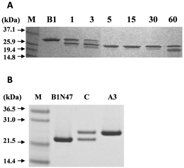
Figure 4. Tryptic digestion of βB1: analytical scale time course and preparative scale.
(A) SDS-PAGE of tryptic digestion of βB1 (B1) with 2.5% (w/w) trypsin, times indicated in min. Black arrows indicate molecular mass of the protein marker (M). (B) SDS-PAGE of large scale βB1 tryptic digestion followed by gel filtration purification: βB1ΔN47 (B1N47); equimolar mixture of βB1ΔN47: βA3 incubated for 24h at RT (C) and used for analytical ultracentrifugation; βA3 used for complex formation studies (A3).

