

Abstract
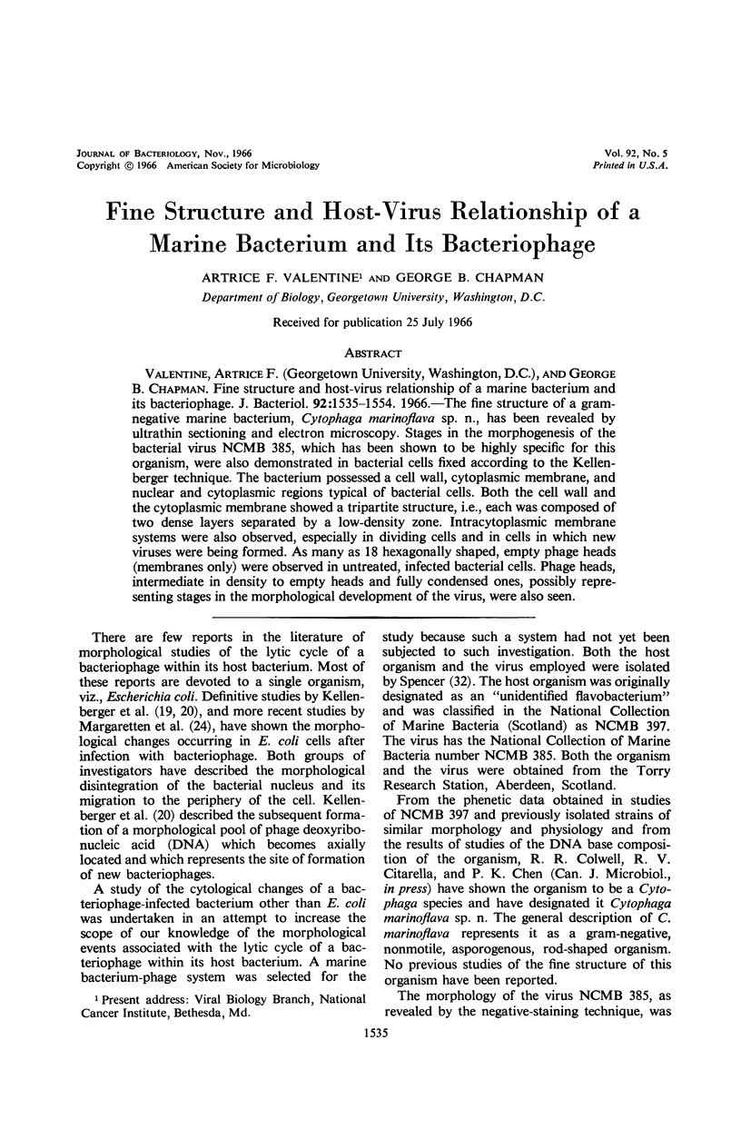
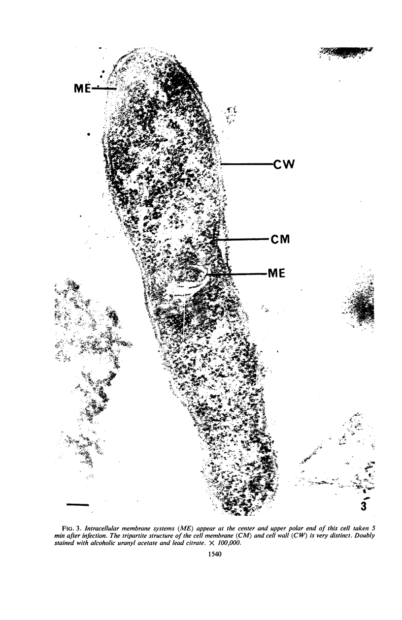
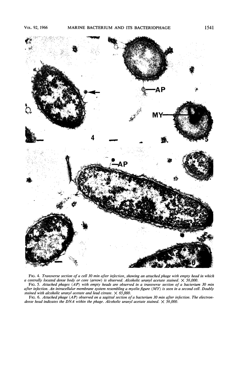
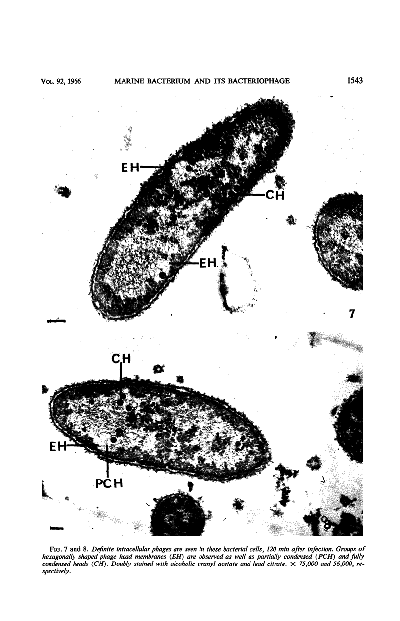
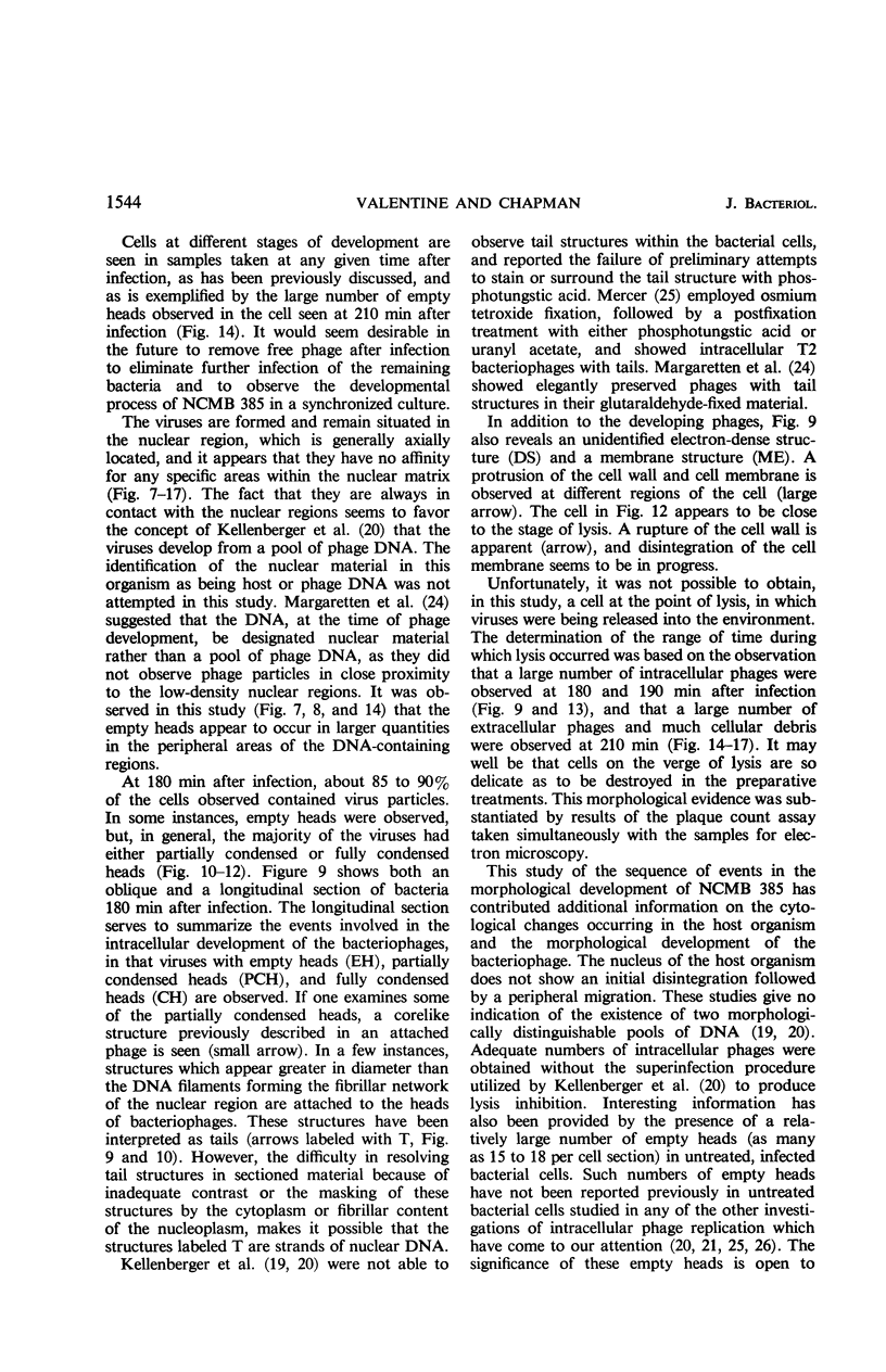
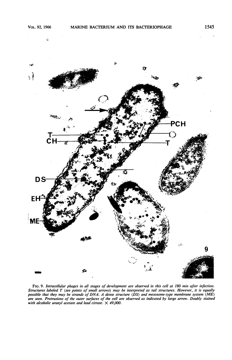
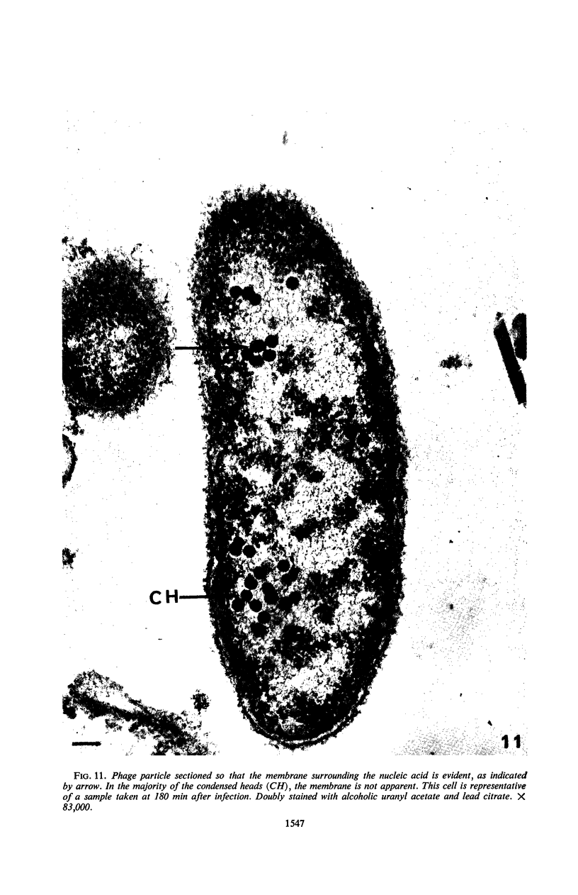
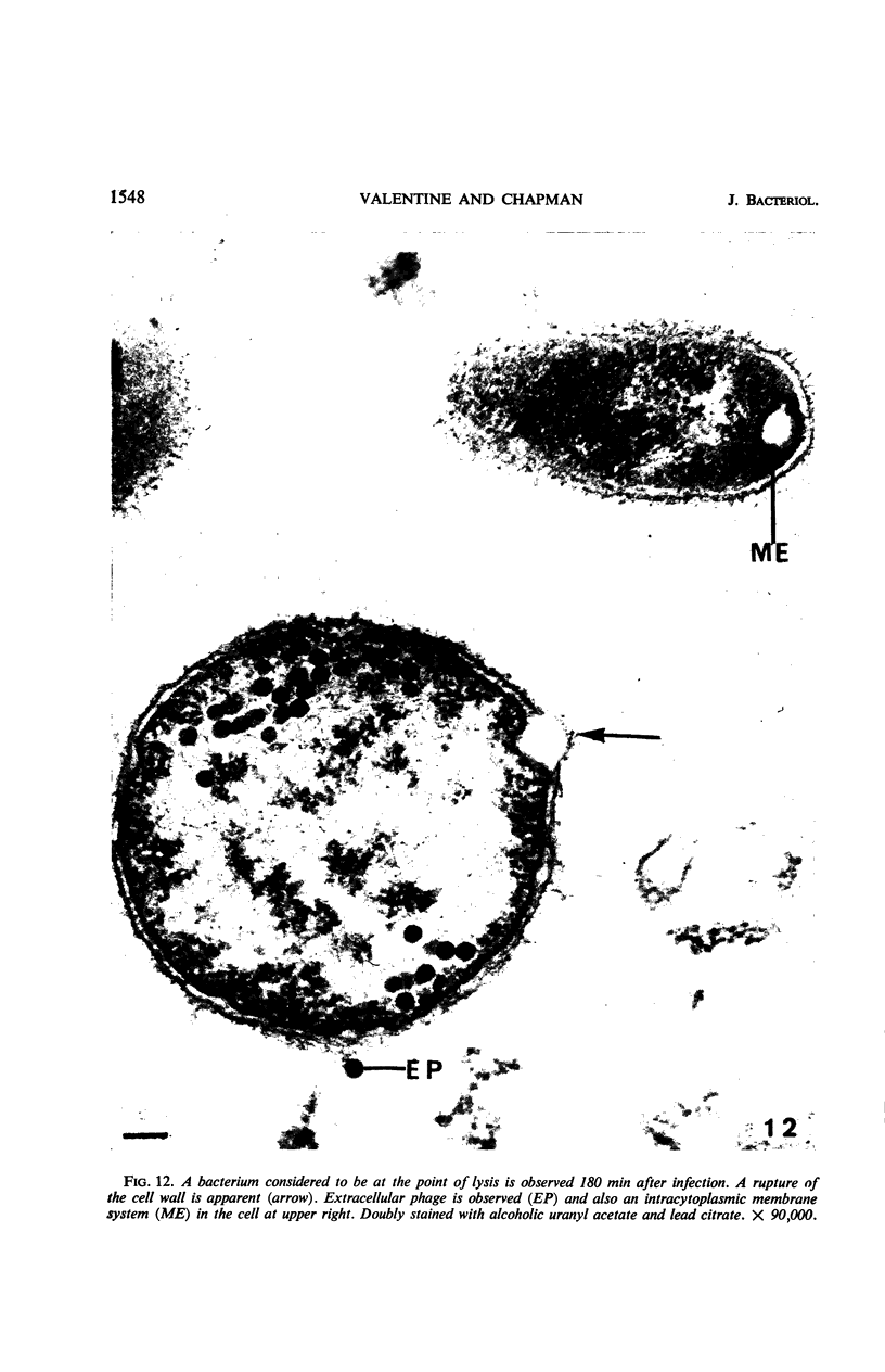
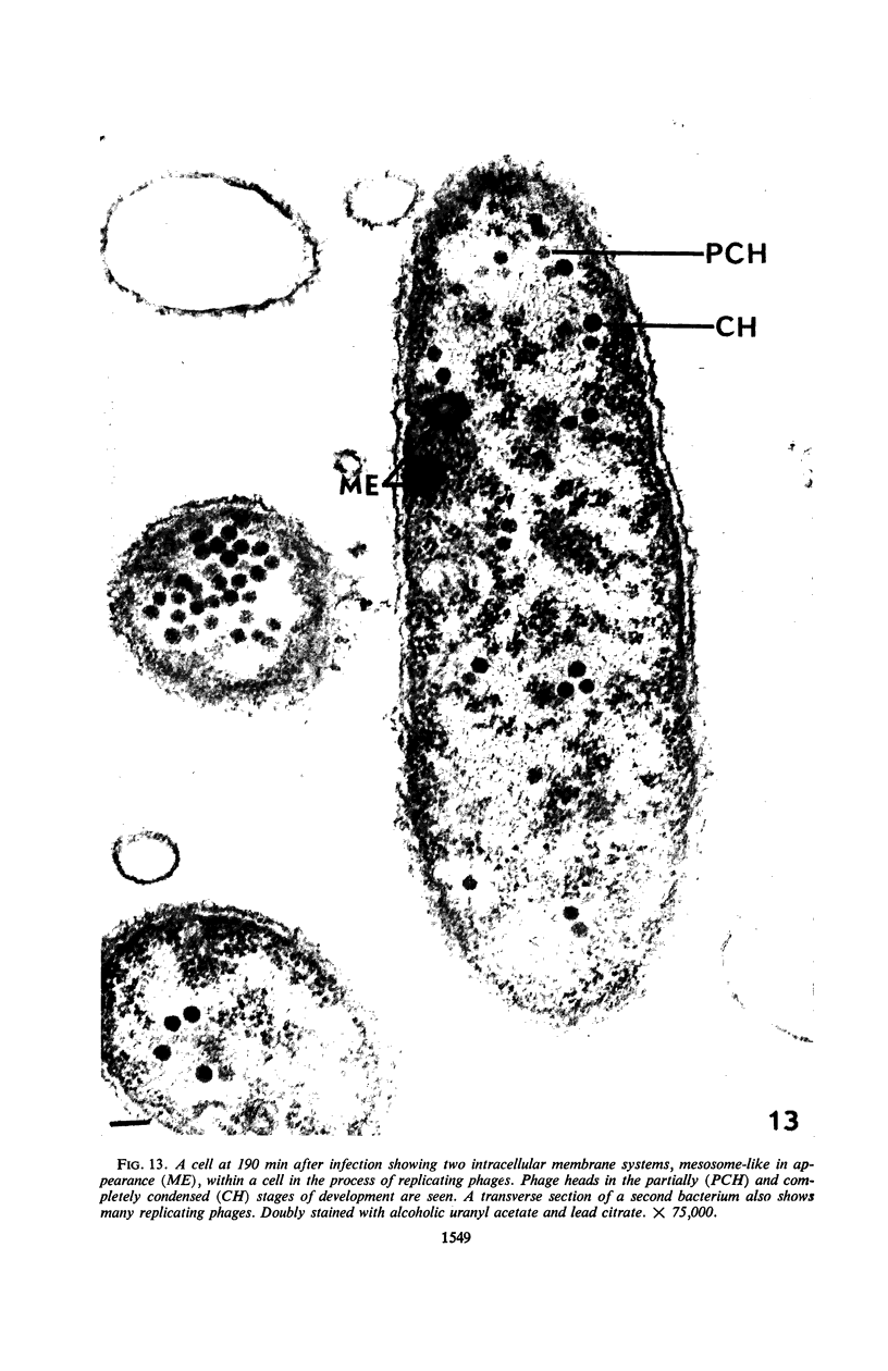
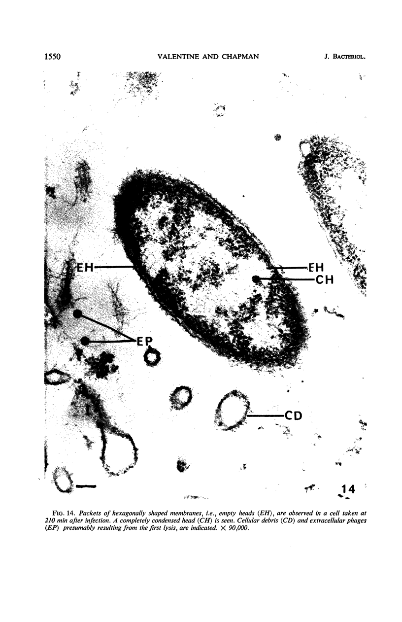
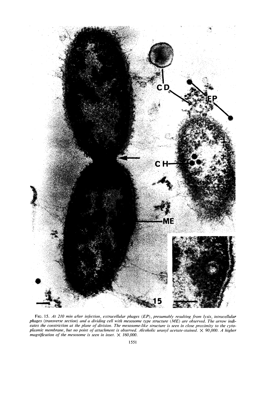
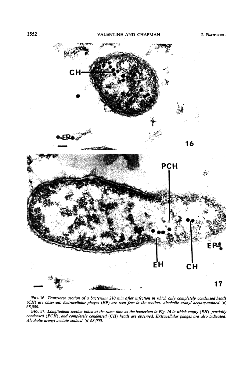
Valentine, Artrice F. (Georgetown University, Washington, D.C.), and George B. Chapman. Fine structure and host-virus relationship of a marine bacterium and its bacteriophage. J. Bacteriol. 92:1535–1554. 1966.—The fine structure of a gram-negative marine bacterium, Cytophaga marinoflava sp. n., has been revealed by ultrathin sectioning and electron microscopy. Stages in the morphogenesis of the bacterial virus NCMB 385, which has been shown to be highly specific for this organism, were also demonstrated in bacterial cells fixed according to the Kellenberger technique. The bacterium possessed a cell wall, cytoplasmic membrane, and nuclear and cytoplasmic regions typical of bacterial cells. Both the cell wall and the cytoplasmic membrane showed a tripartite structure, i.e., each was composed of two dense layers separated by a low-density zone. Intracytoplasmic membrane systems were also observed, especially in dividing cells and in cells in which new viruses were being formed. As many as 18 hexagonally shaped, empty phage heads (membranes only) were observed in untreated, infected bacterial cells. Phage heads, intermediate in density to empty heads and fully condensed ones, possibly representing stages in the morphological development of the virus, were also seen.
Full text
PDF
Images in this article
Selected References
These references are in PubMed. This may not be the complete list of references from this article.
- CHAPMAN G. B. Electron microscopy of ultrathin sections of bacteria. III. Cell wall, cytoplasmic membrane, and nuclear material. J Bacteriol. 1959 Jul;78(1):96–104. doi: 10.1128/jb.78.1.96-104.1959. [DOI] [PMC free article] [PubMed] [Google Scholar]
- CHAPMAN G. B., HILLIER J. Electron microscopy of ultra-thin sections of bacteria I. Cellular division in Bacillus cereus. J Bacteriol. 1953 Sep;66(3):362–373. doi: 10.1128/jb.66.3.362-373.1953. [DOI] [PMC free article] [PubMed] [Google Scholar]
- COLE R. M. CRYSTALLINE AGGREGATES DURING INTRACELLULAR DEVELOPMENT OF A STREPTOCOCCAL BACTERIOPHAGE. Virology. 1965 Jul;26:509–511. doi: 10.1016/0042-6822(65)90015-2. [DOI] [PubMed] [Google Scholar]
- COLWELL R. R., MORITA R. Y. REISOLATION AND EMENDATION OF DESCRIPTION OF VIBRIO MARINUS (RUSSELL) FORD. J Bacteriol. 1964 Oct;88:831–837. doi: 10.1128/jb.88.4.831-837.1964. [DOI] [PMC free article] [PubMed] [Google Scholar]
- COTA-ROBLES E. H., COFFMAN M. D. FINE STRUCTURE OF THE BACTERIOPHAGE ATTACHMENT PROCESS. J Bacteriol. 1963 Aug;86:266–273. doi: 10.1128/jb.86.2.266-273.1963. [DOI] [PMC free article] [PubMed] [Google Scholar]
- CUMMINGS D. J., WANKO T. AN ELECTRON MICROSCOPIC STUDY OF T2 BACTERIOPHAGE IN THIN SECTION. J Mol Biol. 1963 Dec;7:658–661. doi: 10.1016/s0022-2836(63)80113-8. [DOI] [PubMed] [Google Scholar]
- Chen P. K., Citarella R. V., Salazar O., Colwell R. R. Properties of two marine bacteriophages. J Bacteriol. 1966 Mar;91(3):1136–1139. doi: 10.1128/jb.91.3.1136-1139.1966. [DOI] [PMC free article] [PubMed] [Google Scholar]
- Conti S. F., Gettner M. E. ELECTRON MICROSCOPY OF CELLULAR DIVISION IN ESCHERICHIA COLI. J Bacteriol. 1962 Mar;83(3):544–550. doi: 10.1128/jb.83.3.544-550.1962. [DOI] [PMC free article] [PubMed] [Google Scholar]
- DE MARS R. I. The production of phage-related materials when bacteriophage development in interrupted by proflavine. Virology. 1955 May;1(1):83–99. doi: 10.1016/0042-6822(55)90007-6. [DOI] [PubMed] [Google Scholar]
- FITZ-JAMES P. C. Participation of the cytoplasmic membrane in the growth and spore fromation of bacilli. J Biophys Biochem Cytol. 1960 Oct;8:507–528. doi: 10.1083/jcb.8.2.507. [DOI] [PMC free article] [PubMed] [Google Scholar]
- GIBBONS I. R., GRIMSTONE A. V. On flagellar structure in certain flagellates. J Biophys Biochem Cytol. 1960 Jul;7:697–716. doi: 10.1083/jcb.7.4.697. [DOI] [PMC free article] [PubMed] [Google Scholar]
- GLAUERT A. M., HOPWOOD D. A. A membranous component of the cytoplasm in Streptomyces coelicolor. J Biophys Biochem Cytol. 1959 Dec;6:515–516. doi: 10.1083/jcb.6.3.515. [DOI] [PMC free article] [PubMed] [Google Scholar]
- GLAUERT A. M., HOPWOOD D. A. The fine structure of Streptomyces coelicolor. I. The cytoplasmic membrane system. J Biophys Biochem Cytol. 1960 Jun;7:479–488. doi: 10.1083/jcb.7.3.479. [DOI] [PMC free article] [PubMed] [Google Scholar]
- IMAEDA T., OGURA M. Formation of intracytoplasmic membrane system of mycobacteria related to cell division. J Bacteriol. 1963 Jan;85:150–163. doi: 10.1128/jb.85.1.150-163.1963. [DOI] [PMC free article] [PubMed] [Google Scholar]
- KAYE J. J., CHAPMAN G. B. CYTOLOGICAL ASPECTS OF ANTIMICROBIAL ANTIBIOSIS. III. CYTOLOGICALLY DISTINGUISHABLE STAGES IN ANTIBIOTIC ACTION OF COLISTIN SULFATE ON ESCHERICHIA COLI. J Bacteriol. 1963 Sep;86:536–543. doi: 10.1128/jb.86.3.536-543.1963. [DOI] [PMC free article] [PubMed] [Google Scholar]
- KELLENBERGER E., RYTER A. Cell wall and cytoplasmic membrane of Escherichia coli. J Biophys Biochem Cytol. 1958 May 25;4(3):323–326. doi: 10.1083/jcb.4.3.323. [DOI] [PMC free article] [PubMed] [Google Scholar]
- KELLENBERGER E., RYTER A. Fixation et inclusion du matériel nucléaire de Escherichia coli. Experientia. 1956 Nov 15;12(11):420–421. doi: 10.1007/BF02157362. [DOI] [PubMed] [Google Scholar]
- KELLENBERGER E., RYTER A., SECHAUD J. Electron microscope study of DNA-containing plasms. II. Vegetative and mature phage DNA as compared with normal bacterial nucleoids in different physiological states. J Biophys Biochem Cytol. 1958 Nov 25;4(6):671–678. doi: 10.1083/jcb.4.6.671. [DOI] [PMC free article] [PubMed] [Google Scholar]
- KELLENBERGER E., SECHAUD J., RYTER A. Electron microscopical studies of phage multiplication. IV. The establishment of the DNA pool of vegetative phage and the maturation of phage particles. Virology. 1959 Aug;8:478–498. doi: 10.1016/0042-6822(59)90050-9. [DOI] [PubMed] [Google Scholar]
- KUSHNAREV V. M., PEREVERZEV N. A. THE MEMBRANES IN ESCHERICHIA COLI CELLS. J Ultrastruct Res. 1964 Jun;10:610–614. doi: 10.1016/s0022-5320(64)80034-4. [DOI] [PubMed] [Google Scholar]
- LUFT J. H. Improvements in epoxy resin embedding methods. J Biophys Biochem Cytol. 1961 Feb;9:409–414. doi: 10.1083/jcb.9.2.409. [DOI] [PMC free article] [PubMed] [Google Scholar]
- MURRAY R. G., STEED P., ELSON H. E. THE LOCATION OF THE MUCOPEPTIDE IN SECTIONS OF THE CELL WALL OF ESCHERICHIA COLI AND OTHER GRAM-NEGATIVE BACTERIA. Can J Microbiol. 1965 Jun;11:547–560. doi: 10.1139/m65-072. [DOI] [PubMed] [Google Scholar]
- Margaretten W., Morgan C., Rosenkranz H. S., Rose H. M. Effect of hydroxyurea on virus development. I. Electron microscopic study of the effect on the development of bacteriophage T4. J Bacteriol. 1966 Feb;91(2):823–833. doi: 10.1128/jb.91.2.823-833.1966. [DOI] [PMC free article] [PubMed] [Google Scholar]
- REYNOLDS E. S. The use of lead citrate at high pH as an electron-opaque stain in electron microscopy. J Cell Biol. 1963 Apr;17:208–212. doi: 10.1083/jcb.17.1.208. [DOI] [PMC free article] [PubMed] [Google Scholar]
- ROBERTSON J. D. New observations on the ultrastructure of the membranes of frog peripheral nerve fibers. J Biophys Biochem Cytol. 1957 Nov 25;3(6):1043–1048. doi: 10.1083/jcb.3.6.1043. [DOI] [PMC free article] [PubMed] [Google Scholar]
- Spencer R. INDIGENOUS MARINE BACTERIOPHAGES. J Bacteriol. 1960 Apr;79(4):614–614. doi: 10.1128/jb.79.4.614-614.1960. [DOI] [PMC free article] [PubMed] [Google Scholar]
- Steed P., Murray R. G. The cell wall and cell division of gram-negative bacteria. Can J Microbiol. 1966 Apr;12(2):263–270. doi: 10.1139/m66-036. [DOI] [PubMed] [Google Scholar]
- VAN ITERSON W. Some features of a remarkable organelle in Bacillus subtilis. J Biophys Biochem Cytol. 1961 Jan;9:183–192. doi: 10.1083/jcb.9.1.183. [DOI] [PMC free article] [PubMed] [Google Scholar]
- VENABLE J. H., COGGESHALL R. A SIMPLIFIED LEAD CITRATE STAIN FOR USE IN ELECTRON MICROSCOPY. J Cell Biol. 1965 May;25:407–408. doi: 10.1083/jcb.25.2.407. [DOI] [PMC free article] [PubMed] [Google Scholar]
- Valentine A. F., Chen P. K., Colwell R. R., Chapman G. B. Structure of a marine bacteriophage as revealed by the negative-staining technique. J Bacteriol. 1966 Feb;91(2):819–822. doi: 10.1128/jb.91.2.819-822.1966. [DOI] [PMC free article] [PubMed] [Google Scholar]
- WATSON M. L. Staining of tissue sections for electron microscopy with heavy metals. J Biophys Biochem Cytol. 1958 Jul 25;4(4):475–478. doi: 10.1083/jcb.4.4.475. [DOI] [PMC free article] [PubMed] [Google Scholar]
- WEIBULL C. Characterization of the protoplasmic constituents of bacillus megaterium. J Bacteriol. 1953 Dec;66(6):696–702. doi: 10.1128/jb.66.6.696-702.1953. [DOI] [PMC free article] [PubMed] [Google Scholar]