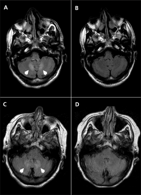
Figure 1.
Axial magnetic resonance images of the first patient’s brain while taking metronidazole (A) and after discontinuation of metronidazole (B), and of the second patient’s brain while on metronidazole (C) and after disconntinuation of metronidazole (D). Cerebellar dentate hyperintensities (arrowheads) on fluid-attenuated inversion recovery imaging are visible for both patients while taking metronidazole (A, C), which resolved after the drug was stopped (B, D).

