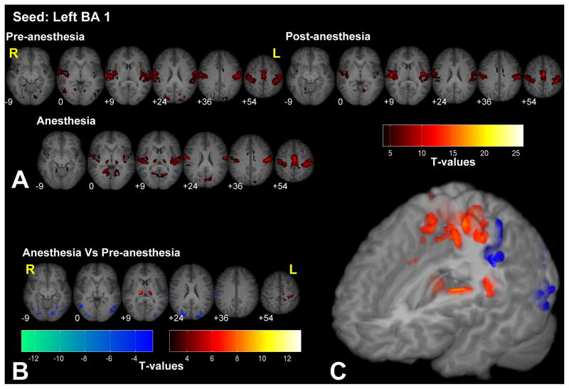
Figure 1.
Significant fc-fMRI values relative to the left BA 1 seed region, under the pre-, post-, and anesthesia conditions (A) and the contrast between anesthesia and pre-anesthesia (B) within different axial slices shown in radiological convention (MNI z-coordinates reported in the inset), or in a three-dimensional reconstruction of the template brain (C). Colors represent fc-fMRI significance, reported as T values. Anesthesia increased connectivity with the thalamus and primary motor/somatosensory areas, and reduced the connectivity with extrastriate visual areas.

