Abstract
Purpose
Tumor antigen-loaded dendritic cells (DC) are believed to activate antitumor immunity by stimulating T cells, and cytotoxic T lymphocyte-associated antigen 4 (CTLA4)-blocking antibodies should release a key negative regulatory pathway on T cells. The combination was tested in a phase 1 clinical trial in patients with advanced melanoma.
Experimental Design
Autologous DC were pulsed with MART-126-35 peptide and administered with a dose escalation of the CTLA4 blocking antibody tremelimumab. Sixteen patients were accrued to 5 dose levels. Primary endpoints were safety and immune effects; clinical efficacy was a secondary endpoint.
Results
Dose-limiting toxicities (DLTs) of grade 3 diarrhea and grade 2 hypophysitis developed in 2 out of 3 patients receiving tremelimumab at 10 mg/kg monthly. Four patients had an objective tumor response, two partial responses (PR) and two complete responses (CR), all melanoma-free between 2 and 4 years after study initiation. There was no difference in immune monitoring results between patients with an objective tumor response and those without a response. Exploratory gene expression analysis suggested that immune-related gene signatures, in particular for B cell function, may be important in predicting response.
Conclusion
The combination of MART-1 peptide-pulsed DC and tremelimumab results in objective and durable tumor responses at the higher range of the expected response rate with either agent alone.
Introduction
The cytotoxic T lymphocyte-associated antigen 4 (CTLA4) is a main negative regulator of the immune system, which inhibits costimulatory signaling provided by dendritic cells (DC) to activate T lymphocytes. Antibodies to CTLA4 block this negative signaling, which allows a dominant positive signaling provided by costimulatory molecules on DCs recognized by CD28 on T cells (1). In animal models, administration of CTLA4-blocking antibodies alone induced rejection of established tumors, provided that these were immunogenic tumor models (1, 2). However, in poorly immunogenic tumor models, which may more closely resemble human disease, prior immunization with tumor vaccines, including DC-based vaccines, was required for CTLA4 blockade to exert robust antitumor effects (1).
Tremelimumab (formerly CP-675,206) is a fully human CTLA4 blocking monoclonal antibody being developed for the treatment of cancer (3). In phase 1 testing, plasma levels of antibody of 30 μg/ml, which corresponds to levels predicted to result in continuous CTLA4 blockade in vitro, were achieved for at least one month at doses beyond 6 mg/kg (4). Cumulative clinical data suggests that single agent tremelimumab has antitumor activity in approximately 10% of patients with advanced melanoma, and these responses tend to be long-lasting (3, 5). Dose-limiting inflammatory and autoimmune toxicities like colitis, hypophysitis and thyroiditis support the notion that this antibody can break peripheral tolerance to self tissues (4). When administered as single agent, there is no evidence of expansion of T cells specific for tumor antigens in the majority of experiences, as assessed using immune monitoring assays in peripheral blood (6, 7). However, analysis of tumor biopsies demonstrated that its antitumor activity is associated by large intratumoral infiltrates with CD8+ T cells, with variable infiltration with CD4+ T cells (8). Given the low frequency of clinical tumor responses as a single agent, the data from animal models suggesting the beneficial effects of adding a vaccine approach to CTLA4 blocking strategies and the validation of the mechanism of action with immune infiltration in regressing lesions in humans, we reasoned that a combination of tremelimumab with tumor antigen-loaded DC vaccines may increase the expansion of melanoma antigen-specific T cells leading to an increased frequency of long lasting objective tumor responses.
Among patients with malignant melanoma and the MHC class I haplotype HLA-A*0201 (the most common in the general population), T cells reactive to the melanoma-associated antigen recognized by T cells (MART-1) can be readily recognized in peripheral blood and tumors of some patients at relative high frequencies (9–11). Therefore, MART-1 represents a reasonable target for melanoma tumor immunotherapy. MART-126-35 peptide immunization, either pulsed onto autologous DC or emulsified with incomplete Freund’s adjuvant (IFA), resulted in the activation of epitope-specific T cells and occasional clinical responses in patients with metastatic melanoma (12, 13). In our prior experience in a phase I/II dose-escalation clinical trial (14, 15), HLA-A*0201 positive patients with MART-1-expressing melanoma received autologous DC pulsed with the MART-127-35 peptide. Dendritic cells were generated from autologous leukapheresis products by a one-week culture in granulocyte-macrophage colony stimulating factor (GM-CSF) and interleukin 4 (IL-4) under good manufacturing practice (GMP) standards, pulsed with the MART-127-35 peptide, and administered at cell doses between 105 to 107 through two routes, i.v. or i.d. Immunological testing provided evidence that the i.d. route resulted in higher frequencies of MART-127-35-specific T cells compared to the i.v. route, and that the DC dose was not critical for this T cell expansion (14). Two out of 18 patients with measurable metastatic melanoma in this phase I/II clinical trial had durable complete responses after MART-126-35-peptide pulsed DC vaccine (MART-126-35/DC) administration. One patient had regression of over 90 skin metastasis after receiving the three i.d. administration of 107 MART-1 peptide pulsed DC vaccines, and is currently disease-free for 9 years (14). This is the only therapy for metastatic melanoma that this patient has ever received, demonstrating that this mode of therapy can induce occasional responses as single agent. A second patient with bulky lung metastasis and s.c. metastasis is currently disease free for 8 years (15). This patient sequentially received DC vaccines followed one month later with the CTLA4 blocking monoclonal antibody ipilimumab (formerly MDX010, Medarex Inc., Princeton, NJ) administered within the first-in-human clinical trial of this antibody (15). Immunological analysis in this patient revealed that the DC vaccines increased the number of circulating MART-127-35-specific cells and further generated determinant (or epitope) spreading to other melanoma antigens (16). The anti-CTLA4 antibody resulted in an enhancement in the frequency of precursor cells in peripheral blood reactive to antigens to which this patient had been exposed in the past, including infectious disease antigens and antigens derived from her melanoma, but not to antigens to which the patient had not been in contact with (15). The phenomenon of determinant spreading has been observed with high frequency in patients with tumor responses to single epitope-based vaccination strategies (17), which suggests that breaking tolerance to cancer may follow similar pathways as breaking self-tolerance in autoimmune diseases (18).
Based on these preliminary experiences, we planned to address the hypothesis that CTLA4 blockade may enhance T cell responses stimulated by tumor antigen peptide-pulsed DC within a phase 1 clinical trial in patients with advanced melanoma. Our results suggest that this combination is feasible and can be safely administered to patients resulting in clinically meaningful and durable objective responses at a frequency that may be at the high end of what should be expected with either agent alone.
Patients and Methods
Clinical Trial Identification and Conduct
This clinical trial was registered as NCT00090896. The protocol and consent forms, and all modifications, were approved by the UCLA Institutional Review Board (#03-12-023), under the investigator new drug (IND) 11579 filed with the Food and Drug Administration (FDA). Collected data was prospectively reviewed by an independent monitor, and the study also underwent periodic ad hoc reviews by the compliance officers from the JCCC Quality Assurance Committee. The clinical trial was activated on June 2004, and the last study visit was in July 2007. Consenting patients with an ongoing response at that time were offered enrollment in a roll-over protocol allowing maintenance dosing with tremelimumab (IRB #06-11-026). The median follow up at the time of reporting was 40 months (range 59+ to 28+ months).
Study Endpoints and Assessments
The primary objectives were safety, feasibility and biological activity. Dose-limiting toxicities (DLTs) were defined as treatment-related toxicities equal or greater than grade 3 according to the NCI common toxicity criteria version 2.0 excluding skin toxicity, or clinical evidence of grade 2 or higher autoimmune toxicities in critical organs (heart, lung, kidney, bowel, bone marrow, musculoskeletal, central nervous system and the eye). All patients underwent baseline and follow-up eye exams every 2 to 4 months throughout study participation following our previously-published eye surveillance protocol (19, 20). Secondary endpoint was efficacy as measured by objective clinical response criteria following a slightly modified Response Evaluation Criteria in Solid Tumors (RECIST) (21) to include the evaluation of lesions detectable by physical exam only that had been recorded at baseline using a photographic camera with a measuring tape or ruler. The tertiary study endpoint was the determination of the lowest dose of tremelimumab necessary to significantly increase the frequency of MART-126-35-specific CD8+ T cells detectable using immune monitoring assays in peripheral blood. A baseline leukapheresis provided peripheral blood mononuclear cells (PBMC) for both the DC manufacture and for pre-dosing immune monitoring assays. Peripheral blood samples (40 mL) were collected every 2 weeks for 3 months and at the end of the initial 120 study period. After a protocol amendment, patients enrolled in the last two cohorts underwent a post-dosing leukapheresis scheduled between study days 42 and 60.
Clinical Trial Design
This was a phase I open-label, single site clinical trial with a fixed dose of 1 × 10⁷ MART-126-35/DC administered i.d. in the lower abdominal region above the groin in three biweekly vaccinations (study days 1, 14 and 28), concomitantly with a dose escalation of tremelimumab at 3, 6 and 10 mg/kg i.v. initially administered at monthly intervals starting on day 1. After reaching DLTs in the third study cohort, the protocol was amended to administer tremelimumab at 10 and 15 mg/kg every 3 months. If a patient at any cohort had not received the planned three doses of MART-126-35/DC, additional patients would be accrued to that cohort to have at least three patients that had completed the combined planned therapy. Dose escalation was allowed when three subjects in a cohort had completed 90 days of follow-up with no DLTs. Restaging exams were conducted at study day 120, and patients without disease progression were eligible for continued dosing with tremelimumab.
Study Eligibility
Eligible patients had histologically-proven malignant melanoma stages IIIc or IV, measurable disease by CT scans or by physical examination (requiring photographic documentation), adequate performance, hematopoietic, hepatic and renal function, be HLA-A*0201 positive by DNA subtyping, and express MART-1 in the tumor. Major exclusion criteria were history of chronic inflammatory or autoimmune disease, congenital or acquired immune suppressive states, requirement of ongoing immune suppressive therapy, and presence of active brain metastases.
Tremelimumab
Tremelimumab (compound code CP-675,206) is a fully human IgG2 monoclonal antibody with high binding affinity for human CTLA4 and a plasma half life of 22.1 days (3, 4). It was supplied by Pfizer Inc. (New London, CT) and administered i.v. at a rate of 100 ml/hour.
MART-126-35 Peptide-Pulsed Dendritic Cells
A single unmobilized leukapheresis processing two plasma volumes was performed to obtain PBMC, which were isolated by Ficoll-Hypaque (Amersham-Pharmacia, Piscataway, NJ) gradient centrifugation and cryopreserved in clinical grade RPMI 1640 (Gibco BRL, Gaithersburg, MD) media plus 20% heat-inactivated autologous serum and 10% dimethylsulfoxide (DMSO, Sigma, St. Louis, MO). Dendritic cells were differentiated from adherent PBMC in an 8-day in vitro culture in RPMI 1640 medium supplemented with 5% heat inactivated autologous serum, 800 U/ml of GM-CSF (Berlex, Montville, NJ) and 500 U/ml of IL-4 (CellGenix, Freiburg, Germany) as we have previously described (14, 17). All culture reagents and in-process samples were tested for bacterial and fungal sterility. On the day of immunization, DC were pulsed with 10 μg/ml MART-126-35 peptide (A27L peptide analog, sequence ELAGIGLTV, ClinAlfa, Laufelfingen, Switzerland) (22, 23) in serum-free RPMI 1640 for one to two hours, and 1 × 10⁷ DC were prepared for injection in 0.2 ml of saline. The sterility of the final product after all manipulations was confirmed by Gram stain, a rapid mycoplasma assay (Mycoplasma PCR-ELISA, Roche Applied Science, Indianapolis, IN), and an endotoxin assay (Limulus Amebocyte Lysate, BioWhittaker, Walkersville, ME) read by an automated automatic endotoxin detection system (Associates of Cape Cod, Inc., Falmouth, MA), before being administered to patients. Dendritic cell culture and all manipulations were performed following GMP at the JCCC/UCLA Cell and Gene Therapy GMP suite.
Dendritic Cell Phenotype and Cytokine Profile Analysis
Cellular aliquots taken on the day of DC harvesting were stained with pre-conjugated antibodies specific for DC cell surface markers including CD80 (B7.1), CD86 (B7-2) (Invitrogen, Carlsbad, CA), and HLA-DR (Beckman-Coulter, Fullerton, CA); markers to study the degree of DC maturation including CCR6 and CCR7 (BD Biosciences, San Diego, CA), CD40, CD80 and CD83 (Invitrogen); viability assessed by Annexin V and propidium iodine (BD Biosciences); and markers to quantitate the percentage of other cell subsets included in the preparation, including CD3 for T cells, CD14 for monocytes/macrophages and CD19 for B cells (Caltag). Supernatant from DC preparations was collected and stored frozen. After thawing, supernatants were analyzed for IL12p70 cytokine (BioRad, Hercules, CA) following the package insert.
Immunological Assays
PBMC samples from peripheral blood or leukapheresis were cryopreserved and analyzed by MHC tetramer assay as previously described (6, 7) All HLA-A*0201 tetramers were purchased from Beckman Coulter Inc., San Diego, CA. Interferon gamma (IFN-γ) ELISPOT assays were also performed as we have previously described (6, 7) using peptide-pulsed HLA-A*0201-transfected K562 (K562/A*0201) as stimulators with cells seeded directly into anti–IFN-γ antibody coated ELISPOT plates and cultured for 20 hours.
Immunohistochemistry staining and evaluation
Paraffin sections from tumors or from 4 mm punch biopsies of the skin were deparaffinized and stained with hematoxylin-eosin (H&E) or subjected to heat-induced epitope retrieval and stained with antibodies to CD3, CD4, CD8, CD68, granzyme B, MART-1, tyrosinase and HMB45 as previously described (8).
Gene Expression Profiling in Peripheral Blood Mononuclear Cells (PBMC)
Total RNA was isolated from cryopreserved PBMC collected before and following therapy using Qiagen RNeasy mini kit (Valencia, CA) according to manufacturer’s instructions and quantified using Agilent Bioanalyzer 2000 (Agilent Technologies). RNA amplification and labeling were performed as previously described (24, 25). 36K whole genome arrays were fabricated at IDIS, NIH. Hybridization was carried at 42°C for 18–24 hours and analyzed using BRB array software (http//:linus.nci.nih.gov/BRB-ArrayTools.html ) (24, 25).
Statistical Analysis
The previously defined 99% reference change value (RCV) (6) was applied to detect statistically significant changes in values above the lower limit of detection for the tetramer and ELISPOT assays as we have previously described (6). For gene expression analysis, data were uploaded to the mAdb databank (http://nciarray.nci.nih.gov) and raw intensity data were collated (26). Owing to differences in sample collection, gene expression in PBMC obtained from leukapheresis and from standard peripheral blood draws were compared; 846 genes were differentially expressed at a cutoff p-value of <0.001 and were removed from the analysis. In addition, 4,929 genes observed to vary according to batch assessment (sample collection, shipment and experimental batches) were removed from further analysis based on quality control assessment of reference concordance as previously discussed (27). Significant difference were defined based on a two-tailed p <0.005 for class comparison due to small sample size (Student t test). Global and univariate permutation significance level was defined as p<0.05 based on 1,000 and 10,000 permutations respectively. Data were visualized using Cluster and TreeView software (28). Venn Diagrams were based on gene selection and display using mAdb webtools (http://nciarray.nci.nih.gov/).
Results
Patient Characteristics
Sixteen HLA-A*0201 positive patients were enrolled between June 2004 and March 2007, with a mean age of 53 years (Table 1). Half of the patients had visceral metastasis or an elevated LDH (stage IV, M1c). Among the remaining patients, two had in-transit melanoma (stage IIIc), three had skin, subcutaneous or nodal metastasis (stage IV, M1a), and three had lung metastasis (stage IV, M1b). Four patients had received prior immunotherapy, three chemotherapy and two biochemotherapy, while seven patients were treated as first line therapy for metastatic melanoma.
Table 1.
Patient Characteristics, Toxicities and Response to Therapy.
| Cohort | M/DC Dose |
Treme Dose |
Pt Study # |
Sex (M/F) |
Age | Prior Treatments |
Active Metastasis Sites |
LDH | Stage | DLT | ORR | Current Status |
|---|---|---|---|---|---|---|---|---|---|---|---|---|
| A | 3 mg/kg qmo | NRA1 | M | 43 | None | S.C. | N | IIIc | SD | NED 59+ mo | ||
| NRA2 | F | 48 | None | Lung | N | M1b | ||||||
| NRA3 | F | 53 | None | Lung | N | M1b | PR | NED 58+ mo | ||||
| B | 6 mg/kg qmo | NRA4 | F | 62 | None | LN, S.C. | N | M1a | ||||
| NRA5 | M | 28 | None | Lung | H | M1c | ||||||
| NRA6 | M | 38 | None | S.C., Liver, Spleen | H | M1c | ||||||
| NRA7 | M | 56 | Chemotherapy | Bowel, Lung, LN | H | M1c | ||||||
| C | 1 × 10⁷ | 10 mg/kg qmo | NRA8 | M | 67 | Biochemotherapy Chemotherapy |
Adrenal | H | M1c | G2 hypophysitis | ||
| NRA9 | M | 51 | Chemotherapy | Lung | N | M1b | CR | NED 44+ mo | ||||
| NRA10 | M | 54 | HD IFN | Lung, Liver | N | M1c | G3 diarrhea | |||||
| D | 10 mg/kg q3mo | NRA11 | M | 57 | Chemotherapy | LN, Muscle | H | M1c | ||||
| NRA12 | M | 55 | Biochemotherapy | Lung, Liver | N | M1c | ||||||
| NRA13 | F | 34 | HD-IL2 | S.C., LN, Muscle, Breast | H | M1c | ||||||
| E | 15 mg/kg q3mo | NRA14 | M | 57 | MART-1 + gp100 peptide vaccines with IL-12 | S.C. | N | IIIc | CR | NED 28+ mo | ||
| NRA15 | M | 48 | None | LN | N | M1a | PR | NED 28+ mo | ||||
| NRA16 | F | 61 | Anti-CD137 HD-IL2 |
S.C. | N | M1a | ||||||
Legend. M/DC: MART-1 peptide pulsed dendritic cells; Treme: tremelimumab; M: male; F: female; LDH: lactate dehydrogenase; N: normal LDH level; H: LDH above the upper limit of normal; L.N.: lymph nodes; S.C.: subcutaneous; DLT: dose-limiting toxicities; G: grade; ORR: objective response rate; HD IFN: High dose interferon; CR: complete response; PR: partial response; SD: stable disease; NED: no evidence of disease; mo: months.
Dendritic Cell Preparation and Phenotype
Table 2 provides the characteristics of the DC preparations. All final MART126-35 peptide pulsed DC preparations had high viability and met the lot release criteria defined in the study IND, including being free of endotoxin and mycoplasma contamination. The DC surface markers CD40, CD86 and HLA-DR were expressed by all DC preparations, but CD80 was infrequently expressed. The cells were partially mature DC, with only 50% of the cultures staining positive for the DC maturation marker CD83 as expected following our protocol without a dedicated maturation step. Nearly all preparations had contaminant CD3+ T cells as expected, since we harvested loosely adherent and non-adherent cells at culture day 8 without a further purification step. The level of contamination with monocytes/macrophages and B cells was low. Further confirmation that these were immature DC was that the culture supernatants did not have detectable IL12p70 (data not shown). Overall, these DC preparations were phenotypically similar to our prior experience following the same protocol (14, 15).
Table 2.
Dendritic cell preparation and phenotype:
| Category | Marker | % DC Vaccines with Positive Markers |
|---|---|---|
| DC surface | CD80 (B7.1) | 20% |
| CD86 (B7.2) | 100% | |
| HLA-DR (MHC class II) | 100% | |
| DC maturation | CCR6/CCR7 | 0% |
| CD40 | 100% | |
| CD83 | 53% | |
| Other cell subsets | CD3 (T lymphocytes) | 93% |
| CD14 (Macrophages) | 13% | |
| CD19 (B cells) | 14% | |
| Cell Viability | Annexin-V/PI | 0% |
Does Escalation and Toxicities
Patients were enrolled sequentially to 5 dose cohorts (Table 1) with tremelimumab administered at monthly (cohorts A to C) and then at quaterly intervals (cohorts D and E). One patient in cohort B did not receive the three planned DC administrations due to early progression, and an additional patient was enrolled to that cohort. There were no significant toxicities attributed to the study agents in the 7 patients enrolled in the first two cohorts. Protocol-specified DLTs were noted in 2 out of 3 patients in cohort C with a monthly dose of 10 mg/kg of tremelimumab. One patient (NRA10) developed grade 3 diarrhea and biopsy-proven colitis requiring hospital admission, improved without requiring corticosteroids. Another patient (NRA8) developed grade 2 hypophysitis requiring supplemental dosing with hydrocortisone. Therefore, dose escalation was halted and the protocol amended to decrease the frequency of tremelimumab dosing. Two new cohorts of patients were then enrolled at tremelimumab doses of 10 and 15 mg/kg administered every 3 months without further DLTs.
Clinical Outcome
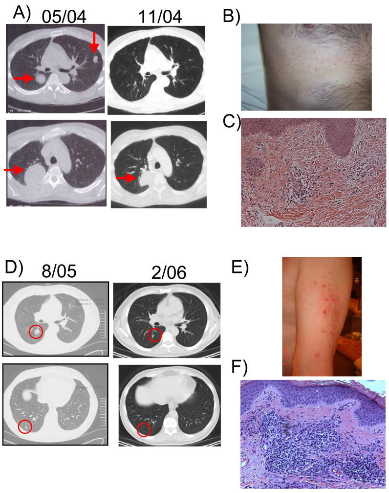
Four patients demonstrated an objective response (Table 1). One patient (NRA3) in the first cohort receiving tremelimumab at 3 mg/kg monthly had a partial response of bulky lung metastasis. This was the first therapy for metastatic melanoma for this patient. Four lung metastasis completely regressed within 4 months, and a large paraspinal mass initially measuring over 16 cm in maximum diameter regressed 55% (Figure 1a). His only toxicity during this period was grade 2 pruritic skin rash, which at biopsy revealed a multifocal acantholytic dyskeratosis consistent with Grover’s disease of the skin (Figure 1b and c), a condition we have previously seen with tremelimumab but not with DC vaccines alone (A. Ribas, unpublished). Nine months after starting the combined therapy, a CT scan suggested that the residual paraspinal mass had increased 1 cm in maximum diameter. A decision was made to surgically resect this lesion. The patient is currently disease-free 58+ months later.
Figure 1.
Examples of clinical events. A, B and C) Tumor regression and skin toxicity in patient NRA3. A) CT scan images of lung metastasis before (left column) and after (right column) starting treatment with MART-1/DC with concomitant tremelimumab at 3 mg/kg monthly. B) Skin eruption developed after 9 months of initiation of therapy. Discrete, erythematous, non-follicular, pruritic papules involving the upper part of the trunk. C) Histological features showing acantholytic dyskeratosis consistent with Grover’s disease (hematoxylin-eosin, original magnification 20x). D, E and F) Tumor regression and skin toxicity in patient NRA9. D) CT scan images of lung metastasis before (left column) and after (right column) starting treatment with MART-1/DC with concomitant tremelimumab at 10 mg/kg monthly. E) Skin eruption developed after 3 years of initiation of therapy. Erythematous maculopaular lesions coalescing to form thin plaques with some scaling. Most affected areas were the proximal extensor surfaces of the arms and legs followed by the trunk F) Histological features showing a mixed granulomatous inflammation (hematoxylin-eosin, original magnification 20x).
One patient in cohort C, who had lung metastasis progressing after multi-agent chemotherapy, had a complete response which is ongoing at 44+ months (NRA9, Figure 1d). This patient developed recurrent grade 2 diarrhea while on monthly doses of tremelimumab for one and a half years. The diarrhea improved when switched to maintenance dosing at 15 mg/kg every 3 months. The patient developed grade 2 skin rash clinically consistent with Grover’s disease during the first year of dosing. During the third year on maintenance dosing the patient developed a new pruritic skin rash (maculopapular lesions coalescing to form thin plaques with some scaling). Most affected areas were the proximal extensor surfaces of the arms and legs followed by the trunk. Skin biopsy showed a mixed granulomatous inflammation (Figure 1e and f).
Two patients in cohort E also had objective responses to therapy. One (NRA14) had a single in-transit subcutaneous lesion of 1 cm which appeared after 3 prior resections of other similar lesions in the same leg. This patient had previously received a vaccine consisting of gp100209-217, MART-126-35, and tyrosinase368-376 peptides together with interleukin 12 (IL-12) (29) 2 years before and then progressed. This in-transit lesion completely regressed within one month of initiating study participation and the patient continues tumor-free 28+ months later. His only toxicity was grade 1 pruritic skin rash, which at biopsy was consistent with lichenoid dermatitis. The other patient (NRA15) had CT and PET positive metastatic lymph nodes up to 2 cm in maximum diameter, and was treatment naïve at the time of starting participation in this protocol. The lymph node metastasis have nearly completely regressed and the patient continues to be tumor-free 28+ months later while on maintenance dosing with tremelimumab. He has not had any evidence of side effects to this combined therapy.
Pathological Analysis
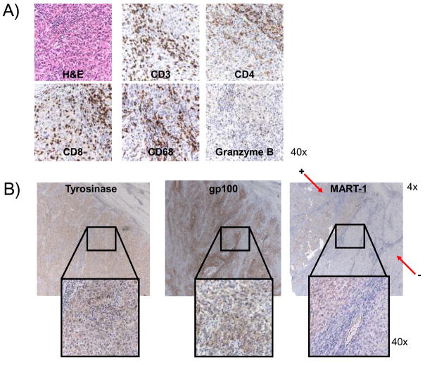
The partially regressed paraspinal mass from patient NRA3 was analyzed for the presence of immune infiltrating cells upon surgical resection. There were large areas of infiltration by CD3+ cells, with a higher ratio of CD8+ cells than CD4+ cells (Figure 2). These cells were consistent with cytotoxic T lymphocytes (CTLs) since there was a diffuse cytoplasmic staining with granzyme B, a granule that mediates cytotoxic signals delivered by CD8+ CTLs. There were also frequent macrophages, stained with the CD68 marker. When stained with the melanoma markers MART-1, HMB45 (gp100), tyrosinase and S-100, a large portion of the mass was negative for MART-1. However, the same areas negative for MART-1 were positive for the other melanosomal antigens. This finding suggests that an area of this resected mass included antigenic variant melanoma cells having lost MART-1 expression by IHC.
Figure 2.
Pathological analysis of a partially regressed mass from patient NRA3. A) High power (40x) images of hematoxylin-eosin (H&E) and immune subset immunohistochemistry (IHC) stains, including CD3 staining for T cells, CD4 staining for T helper subset, CD8 staining for cytotoxic T lymphocyte (CLT) subset, CD68 for macrophages and granzyme B for CTL-associated cytotoxic granules. B) Low (4x) and high power (40x) images of IHC staining of the resected mass for the melanosomal antigen tyrosinase (left column) and gp100 (stained with the HMB45 antibody, middle column) showing uniform staining, and the melanosomal antigen MART-1 (right column) showing large areas without brown IHC staining.
Immune Monitoring for Melanoma-specific T Cell Responses
T cell responses to the immunizing MART-126-35 peptide were assessed using the MHC tetramer and the ELISPOT assays. Changes from baseline were scored following our algorithm to detect statistically significant antigen-specific T cell expansions (6, 7). Six patients, including 2 out of the 4 patients with an objective response to therapy, had increases in MART-126-35-specific CD8 positive T cells by the MHC tetramer assay, and 5 patients, including 2 out of the 4 patients with an objective response, using the ELISPOT assay (Table 3). There was no trend of differential expansion of MART-126-35-specific T cells among study cohorts, suggesting that tremelimumab does not directly increase the number of CD8+ T cells specific for this melanosomal antigen in peripheral blood in a dose-response fashion when combined with DC-based vaccination. Also, there was no clear increasing trend in the number of peripheral blood cells specific for other melanosomal antigens (tyrosinase368-376 and gp100209-217), nor to control infectious disease antigens (CMV pp65495-503 and EBV BMLF1259-267).
Table 3.
Results of immune monitoring with the MHC tetramer and ELISPOT assays. Patient codes are listed based on at least one time point with MHC Tetramer or ELISPOT assay results beyond the 99% reference change value (RCV) for each peptide epitope and immune monitoring assay.
| Assay | Epitope | No Objective Tumor Response | Objective Tumor Response | ||
|---|---|---|---|---|---|
| < 99% RCV | > 99% RCV | < 99% RCV | > 99% RCV | ||
| MHC Tetramer | MART126-35 | NRA 4, 7, 8, 10, 12, 13, 16 | NRA 1, 2, 5, 11 | NRA 3, 15 | NRA 9, 14 |
| tyrosinase368-376 | NRA 4, 5, 8, 11, 13, 16 | NRA 1, 2, 7, 10, 12 | NRA 3, 9, 14, 15 | - | |
| gp100209-217 | NRA 2, 4, 5, 7, 8, 11, 12, 13, 16 | NRA 1, 10 | NRA 3, 9, 14, 15 | - | |
| EBV BMLF1259-267 | NRA 1, 2, 4, 7, 8, 10, 11, 12, 13, 16 | NRA 5 | NRA 3, 9, 15 | NRA 14 | |
| CMV pp65495-503 | NRA 1, 2, 4, 7, 8, 10, 11, 12, 13, 16 | NRA 5 | NRA 9, 14, 15 | NRA 3 | |
| ELISPOT | MART126-35 | NRA 1, 2, 5, 7, 8, 10, 12, 13 | NRA 4, 11, 16 | NRA 3, 9 | NRA 14, 15 |
| tyrosinase368-376 | NRA 4, 5, 7, 8, 10 | NRA 1, 2, 11, 12, 13, 16 | NRA 3, 9, 15 | NRA 14 | |
| gp100209-217 | NRA 4, 5, 7, 8, 10, 11, 13 | NRA 1, 2, 12, 16 | NRA 3, 9 | NRA 14, 15 | |
| EBV BMLF1259-267 | NRA 1, 2, 4, 5, 7, 8, 10, 12, 13, 16 | NRA 11 | NRA 3, 9 | NRA 14, 15 | |
| CMV pp65495-503 | NRA 1, 4, 5, 7, 8, 10, 12, 16 | NRA 2, 11, 13 | NRA 3, 9 | NRA 14, 15 | |
Gene Expression Profiling in Peripheral Blood
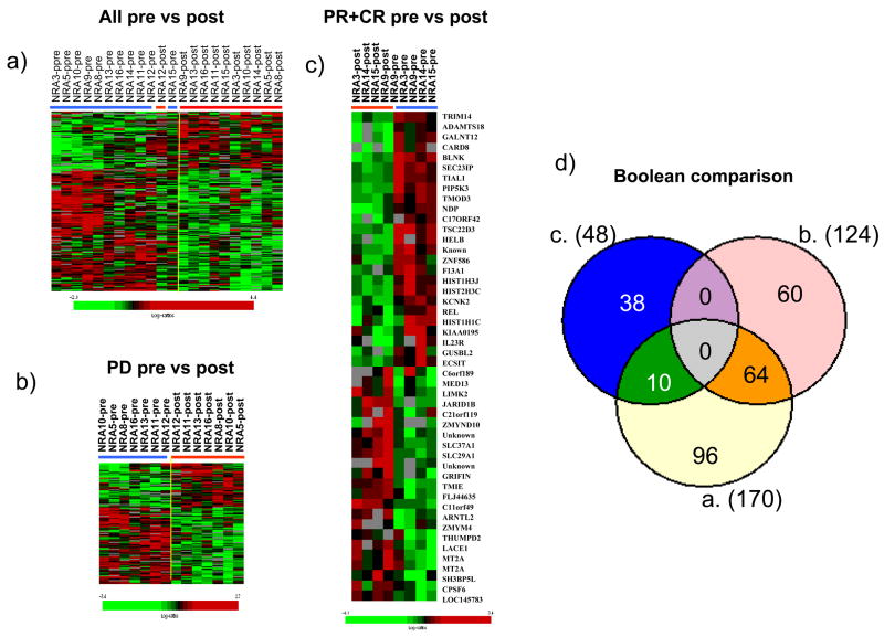
Since the antigen-specific immune monitoring assays did not allow a clear definition of effects in blood that would differentiate clinical responders from non-responders, we undertook an exploratory whole genome profiling in PBMC obtained at baseline and 2–3 months after the initial dose. Unsupervised cluster analysis did not identify clear segregation between pre- and post-treatment samples, suggesting that a relatively small proportion of genes in peripheral blood cells were affected at the transcriptional level by the treatment. We then compared pre- with post-treatment PBMC samples independently of the clinical outcome. This class comparison identified 170 differentially expressed genes (p<0.005, pp<0.022). Among them, 115 genes were down regulated and 55 up regulated (Figure 3a). To stratify genes potentially responsible for tumor rejection, we grouped samples according to clinical outcome and evaluated treatment-induced gene expression in samples from patients experiencing progressive disease (PD) or those who experienced PR or CR. Comparison of pre- with post-treatment samples in patients with an objective response identified 48 genes differentially expressed (p<0.005, permutation not significant), 10 of which were found to be in common with the 124 genes significantly altered (p<0.005, pp=0.062) in patients with PD (Figure 3c and Table S1). We finally compared pre-treatment samples from patients experiencing PR or CR versus PD to explore if we could define baseline gene expression profiles that could later identify patients with a response or resistance. Out of baseline up regulated genes, genes associated with immune function were highly enriched (14/55 in CR and 3/12 in PR, Table S2). Seven genes were B cell signal transduction and function-related genes, including CD24, helicase (DNA) B (HELB) and B-cell linker (BLNK, Table S2).
Figure 3.
Heat map describing tremelimumab-induced PBMC gene expression; blue and red bars represent pre-treatment post-treatment samples, respectively. A) All pre- and post-treatment samples are displayed showing 170 transcripts whose expression was significantly (p<0.005) altered by treatment (paired two sample t test, permutation p<0.022). B) Samples for patients with progressive disease (PD) were compared between pre- and post-treatment collections (p<0.005, 124 genes). C) Clinical responder’s (PR+CR) pre- versus post-treatment analysis (p<0.005, 48 genes). D) Venn diagram displaying overlap of genes within the three groups (all patients, PD and CR+PR).
Discussion
The combination of tumor antigen-pulsed DC and the CTLA4 blocking monoclonal antibody tremelimumab can be safely administered to humans and results in antitumor activity. The maximum tolerated dose (MTD) of this combination was 107 MART-1/DC with concomitant tremelimumab at 6 mg/kg when administered monthly, but in this clinical trial we could not determine an MTD when tremelimumab was administered every 3 months at doses up to 15 mg/kg. Neither one of the two patients that developed DLTs had an objective response to therapy, suggesting that worse autoimmune or inflammatory toxicity is not directly associated with response to this combined therapy. In addition, tumor responses in this study did not follow the proposed immune-related response criteria (30), although the late increase in size of a lung lesion in patient NRA3 without further progression may reflect a transient inflammatory response. This pilot clinical trial cannot discern between responses to the DC vaccines alone or to tremelimumab alone from responses to the combination. One of the four patients with an objective response received dosing of tremelimumab at 3 mg/kg, a dose that achieves plasma levels of 30 μg/ml for less than 2 weeks. This concentration is below the target plasma level set up prospectively in the first-in-human testing of this antibody (3, 4), suggesting that this response may not be explained solely by single agent tremelimumab. The other three responding patients received doses of tremelimumab at 10 or 15 mg/kg, which are known to induce tumor response in approximately 7–10% of patients in single agent studies (5, 31, 32). Therefore, these other responses may be explained by tremelimumab alone, although they seem to be at the higher end of what can be reasonably expected with this agent as single therapy.
We could not detect a dose-response effect of tremelimumab on the expansion of MART-126-35-specific T cells. It is possible that the immune stimulating effect of CTLA4 blocking monoclonal antibodies has a more marked effect on CD4+ T helper cells than on CD8+ CTLs, as described in preclinical models (33), which would explain the lack of expansion of MART-126-35-specific CD8+ T cells with increasing doses of tremelimumab. In addition, we could not confirm our prior observations that determinant spreading is an immunological feature of patients with a clinical response after single tumor antigen peptide-specific immunization (14, 15), although there were expansions of non-MART-1-specific T cells beyond the assay variability. In the absence of a detectable MART-1 driver clone, it is difficult to consider expansion of other tumor antigen-specific cells as subdominant clones triggered by determinant spreading. Overall, the level of MART-126-35-specific T cell expansion in peripheral blood with this combination was low, which is a common finding in other tumor antigen peptide-specific clinical trials in patients with metastatic disease despite objective clinical responses (34) in particular when given with high dose IL-2 (13, 35). One possibility is that the vaccine-primed effector cells accumulate in draining lymph nodes and tumor lesions at much higher frequencies than in peripheral blood as has been shown for MAGE-3 vaccination (34). Supporting this concept, we have previously described an increased frequency of melanoma-specific T cells in tumor samples of patients responding to single agent tremelimumab (8), even though we do not detect expansions in peripheral blood with this treatment (7). In fact, it has been reported that CTLA4 may have a key role in establishing durable interactions between antigen-speticic T cells and their targets (36), suggesting that vaccine-primed lymphocytes should accumulate in tumors as opposed to circulate in blood. An indirect evidence of this possibility in the current clinical trial is the development of large areas of MART-1 negative tumor escape variant cells in a responding patient with a slowly regressing large lung mass.
In this study we used DC generated by GM-CSF and IL-4 in vitro differentiation from monocytes without an additional maturation step. A large body of preclinical data suggests that DC that have been maturated with monocyte-conditioned media or a cytokine cocktail are superior to non-matured cells in their ability to stimulate T cell responses (37, 38). However, it is still unclear what the optimal DC maturation conditions are for clinical use. Initial reports suggested favorable outcomes with matured DC (39), but a larger clinical testing of matured DC resulted in very low melanoma tumor responses (40). Furthermore, a clinical trial directly comparing matured and non-matured DC vaccination failed to demonstrate improved immunological or clinical potency of the matured DC (41). For this study we chose to use the same conditions for DC generation that had resulted in long lived objective responses in occasional patients with metastatic melanoma in our prior experience (14, 15), with the goal of testing if the concomitant administration of a CTLA4 blocking monoclonal antibody would result in improvement of these DC-based vaccines.
Changes in gene expression profiling in blood samples obtained from patients receiving tumor immunotherapy strategies have been seldomly described, although we belive they are of key importance to understanding how the immunotherapeutic approach impacts on the recipient’s immune system (42, 43). In our limited set of patients we explored basic variations in transcription induced by the combined treatment and, at the same time, tested the possibility that pre-treatment signatures may provide insights into future biomarker discovery (44). Of interest to us were genes consistently up regulated in pre-treatment samples from patients who experienced an objective response, which if validated could serve as prognostic factors in future studies. The finding of immune-related genes, in particular related to B cell function, were up regulated at baseline in patients who went onto an objective response may correlate with the detection of increased humoral responses in patients responding to CTLA4-blocking antibodies described by others (45, 46). We fully acknowledge that the low number of samples from clinical responders in our series demands further validation studies in larger cohorts, where gene expression profiling in blood should be compared to gene expression profiling in tumors, where it should be most relevant.
In conclusion, the combination of MART-1/DC with concomitant tremelimumab is feasible in patients with metastatic melanoma, especially when tremelimumab is administered every 3 months, and results in durable objective clinical responses at the higher range of the expected objective tumor response rates with either therapy alone. Therefore, this combination warrants further study in patients with advanced malignant melanoma.
Supplementary Material
Acknowledgments
The clinical trial was conducted under a research grant from Pfizer Inc. This work was additionally supported by the Melanoma Research Foundation, the Harry J. Lloyd Charitable Trust, P50 CA086306, U54 CA119347 and RN2-00902-1 New Faculty Award 2 from the California Institute for Regenerative Medicine (all to A.R.). The UCLA General Clinical Research Center (G-CRC) is supported by USPHS Grant M01-RR-0865. The UCLA Flow Cytometry Core Facility is supported by the NIH awards CA-16042 and AI-28697.
Footnotes
Statement of Translational Relevance
Antitumor responses induced by the activation of the immune system to tumor antigens results in a low percentage of clinical responses, but most of these are durable. A major limitation of tumor immunotherapy is immune cell-instrinsic negative regulatory networks. In a phase 1 clinical trial we provided immune activation signals derived by tumor antigen presented by dendritic cells (DC) together with the blockade of a major negative regulator of the immune system, the cytotoxic T lymphocyte-associated antigen 4 (CTLA4). We describe the safety, immune effects and clinical antitumor activity of this combination in patients with metastatic melanoma. Our results provide evidence that the combination is safe and results in clinically-relevant and durable tumor regressions, which should warrant the continued clinical testing of this combination.
Conflict of interest disclosure. Dr. Antoni Ribas has received commercial research grants and honoraria from Pfizer Inc.; Dr. John A. Glaspy has received speaker’s bureau/honoraria from Pfizer Inc.; Dr. Jesus Gomez-Navarro is employed by Pfizer Inc. The remaining authors have no conflicts of interest to report.
References
- 1.Chambers CA, Kuhns MS, Egen JG, Allison JP. CTLA-4-mediated inhibition in regulation of T cell responses: mechanisms and manipulation in tumor immunotherapy. Annu Rev Immunol. 2001;19:565–94. doi: 10.1146/annurev.immunol.19.1.565. [DOI] [PubMed] [Google Scholar]
- 2.Leach DR, Krummel MF, Allison JP. Enhancement of antitumor immunity by CTLA-4 blockade. Science. 1996;271:1734–6. doi: 10.1126/science.271.5256.1734. [DOI] [PubMed] [Google Scholar]
- 3.Ribas A, Hanson DC, Noe DA, et al. Tremelimumab (CP-675,206), a cytotoxic T lymphocyte associated antigen 4 blocking monoclonal antibody in clinical development for patients with cancer. Oncologist. 2007;12:873–83. doi: 10.1634/theoncologist.12-7-873. [DOI] [PubMed] [Google Scholar]
- 4.Ribas A, Camacho LH, Lopez-Berestein G, et al. Antitumor activity in melanoma and anti-self responses in a phase I trial with the anti-cytotoxic T lymphocyte-associated antigen 4 monoclonal antibody CP-675,206. J Clin Oncol. 2005;23:8968–77. doi: 10.1200/JCO.2005.01.109. [DOI] [PubMed] [Google Scholar]
- 5.Camacho LH, Antonia S, Sosman J, et al. Phase I/II trial of Tremelimumab in patients with metastatic melanoma. J Clin Oncol. 2009;27:1075–81. doi: 10.1200/JCO.2008.19.2435. [DOI] [PubMed] [Google Scholar]
- 6.Comin-Anduix B, Gualberto A, Glaspy JA, et al. Definition of an immunologic response using the major histocompatibility complex tetramer and enzyme-linked immunospot assays. Clin Cancer Res. 2006;12:107–16. doi: 10.1158/1078-0432.CCR-05-0136. [DOI] [PubMed] [Google Scholar]
- 7.Comin-Anduix B, Lee Y, Jalil J, et al. Detailed analysis of immunologic effects of the cytotoxic T lymphocyte-associated antigen 4-blocking monoclonal antibody tremelimumab in peripheral blood of patients with melanoma. J Transl Med. 2008;6:22. doi: 10.1186/1479-5876-6-22. [DOI] [PMC free article] [PubMed] [Google Scholar]
- 8.Ribas A, Comin-Anduix B, Economou JS, et al. Intratumoral Immune Cell Infiltrates, FoxP3, and Indoleamine 2,3-Dioxygenase in Patients with Melanoma Undergoing CTLA4 Blockade. Clin Cancer Res. 2009;15:390–9. doi: 10.1158/1078-0432.CCR-08-0783. [DOI] [PubMed] [Google Scholar]
- 9.Kawakami Y, Eliyahu S, Sakaguchi K, et al. Identification of the immunodominant peptides of the MART-1 human melanoma antigen recognized by the majority of HLA-A2-restricted tumor infiltrating lymphocytes. J Exp Med. 1994;180:347–52. doi: 10.1084/jem.180.1.347. [DOI] [PMC free article] [PubMed] [Google Scholar]
- 10.Lee PP, Yee C, Savage PA, et al. Characterization of circulating T cells specific for tumor-associated antigens in melanoma patients. Nat Med. 1999;5:677–85. doi: 10.1038/9525. [DOI] [PubMed] [Google Scholar]
- 11.Valmori D, Dutoit V, Lienard D, et al. Tetramer-guided analysis of TCR beta-chain usage reveals a large repertoire of melan-A-specific CD8+ T cells in melanoma patients. J Immunol. 2000;165:533–8. doi: 10.4049/jimmunol.165.1.533. [DOI] [PubMed] [Google Scholar]
- 12.Nestle FO, Alijagic S, Gilliet M, et al. Vaccination of melanoma patients with peptide- or tumor lysate-pulsed dendritic cells [see comments] Nat Med. 1998;4:328–32. doi: 10.1038/nm0398-328. [DOI] [PubMed] [Google Scholar]
- 13.Rosenberg SA, Yang JC, Schwartzentruber DJ, et al. Immunologic and therapeutic evaluation of a synthetic peptide vaccine for the treatment of patients with metastatic melanoma [see comments] Nat Med. 1998;4:321–7. doi: 10.1038/nm0398-321. [DOI] [PMC free article] [PubMed] [Google Scholar]
- 14.Butterfield LH, Ribas A, Dissette VB, et al. Determinant Spreading Associated with Clinical Response in Dendritic Cell-based Immunotherapy for Malignant Melanoma. Clin Cancer Res. 2003;9:998–1008. [PubMed] [Google Scholar]
- 15.Ribas A, Glaspy JA, Lee Y, et al. Role of dendritic cell phenotype, determinant spreading, and negative costimulatory blockade in dendritic cell-based melanoma immunotherapy. J Immunother. 2004;27:354–67. doi: 10.1097/00002371-200409000-00004. [DOI] [PubMed] [Google Scholar]
- 16.Tchekmedyian S, Glasby J, Korman A, Keler T, Deo Y, Davis T. MDX-010 (human anti-CTLA4): a phase I trial in malignant melanoma. Proc Am Soc Clin Oncol (Abstract 56) 2002 [Google Scholar]
- 17.Ribas A, Timmerman JM, Butterfield LH, Economou JS. Determinant spreading and tumor responses after peptide-based cancer immunotherapy. Trends Immunol. 2003;24:58–61. doi: 10.1016/s1471-4906(02)00029-7. [DOI] [PubMed] [Google Scholar]
- 18.Lehmann PV, Forsthuber T, Miller A, Sercarz EE. Spreading of T-cell autoimmunity to cryptic determinants of an autoantigen. Nature. 1992;358:155–7. doi: 10.1038/358155a0. [DOI] [PubMed] [Google Scholar]
- 19.Gordon LK, Ribas A, Nusinowitz S, et al. Surveillance of the eye and vision in a clinical trial of MART1-transformed dendritic cells for metastatic melanoma. Control Clin Trials. 2004;25:400–7. doi: 10.1016/j.cct.2004.06.002. [DOI] [PubMed] [Google Scholar]
- 20.Straatsma BR, Nusinowitz S, Young TA, et al. Surveillance of the eye and vision in clinical trials of CP-675,206 for metastatic melanoma. Am J Ophthalmol. 2007;143:958–69. doi: 10.1016/j.ajo.2007.02.035. [DOI] [PubMed] [Google Scholar]
- 21.Therasse P, Arbuck SG, Eisenhauer EA, et al. New guidelines to evaluate the response to treatment in solid tumors [see comments] J Natl Cancer Inst. 2000;92:205–16. doi: 10.1093/jnci/92.3.205. [DOI] [PubMed] [Google Scholar]
- 22.Jaeger E, Bernhard H, Romero P, et al. Generation of cytotoxic T-cell responses with synthetic melanoma-associated peptides in vivo: implications for tumor vaccines with melanoma-associated antigens. Int J Cancer. 1996;66:162–9. doi: 10.1002/(SICI)1097-0215(19960410)66:2<162::AID-IJC4>3.0.CO;2-0. [DOI] [PubMed] [Google Scholar]
- 23.Schuler-Thurner B, Schultz ES, Berger TG, et al. Rapid Induction of Tumor-specific Type 1 T Helper Cells in Metastatic Melanoma Patients by Vaccination with Mature, Cryopreserved, Peptide-loaded Monocyte-derived Dendritic Cells. J Exp Med. 2002;195:1279–88. doi: 10.1084/jem.20012100. [DOI] [PMC free article] [PubMed] [Google Scholar]
- 24.Wang E, Miller LD, Ohnmacht GA, Liu ET, Marincola FM. High-fidelity mRNA amplification for gene profiling. Nat Biotechnol. 2000;18:457–9. doi: 10.1038/74546. [DOI] [PubMed] [Google Scholar]
- 25.Wang E. RNA amplification for successful gene profiling analysis. J Transl Med. 2005;3:28. doi: 10.1186/1479-5876-3-28. [DOI] [PMC free article] [PubMed] [Google Scholar]
- 26.Simon R, Lam A, LI MC, Ngan M, Menenzes A, Zhao Y. Analysis of Gene Expression Data Using BRB-Array Tools. Cancer Informatics. 2007;2:11–7. [PMC free article] [PubMed] [Google Scholar]
- 27.Jin P, Zhao Y, Ngalame Y, et al. Selection and validation of endogenous reference genes using a high throughput approach. BMC Genomics. 2004;5:55. doi: 10.1186/1471-2164-5-55. [DOI] [PMC free article] [PubMed] [Google Scholar]
- 28.Eisen MB, Spellman PT, Brown PO, Botstein D. Cluster analysis and display of genome-wide expression patterns. Proc Natl Acad Sci U S A. 1998;95:14863–8. doi: 10.1073/pnas.95.25.14863. [DOI] [PMC free article] [PubMed] [Google Scholar]
- 29.Hamid O, Solomon JC, Scotland R, et al. Alum with interleukin-12 augments immunity to a melanoma peptide vaccine: correlation with time to relapse in patients with resected high-risk disease. Clin Cancer Res. 2007;13:215–22. doi: 10.1158/1078-0432.CCR-06-1450. [DOI] [PubMed] [Google Scholar]
- 30.Hodi FS, Hoos A, Ibrahim R, et al. Novel efficacy criteria for antitumor activity to immunotherapy using the example of ipilimumab, an anti-CTLA-4 monoclonal antibody. J Clin Oncol. 2008;26 abstr 3008. [Google Scholar]
- 31.Ribas A, Antonia S, Sosman J, et al. Results of a phase II clinical trial of 2 doses and schedules of CP-675,206, an anti-CTLA4 monoclonal antibody, in patients (pts) with advanced melanoma. ASCO Annual Meeting Proceedings. 2007;25:8523. [Google Scholar]
- 32.Ribas A, Hauschild A, Kefford R, et al. Phase III, Open-Label, Randomized, Comparative Study of Tremelimumab (CP-675,206) and Chemotherapy (Temozolomide or Dacarbazine) in Patients with Advanced Melanoma. Journal of Clinical Oncology. 2008;26 abstr LBA9011. [Google Scholar]
- 33.Gattinoni L, Ranganathan A, Surman DR, et al. CTLA-4 dysregulation of self/tumor-reactive CD8+ T-cell function is CD4+ T-cell dependent. Blood. 2006;108:3818–23. doi: 10.1182/blood-2006-07-034066. [DOI] [PMC free article] [PubMed] [Google Scholar]
- 34.Lurquin C, Lethe B, De Plaen E, et al. Contrasting frequencies of antitumor and anti-vaccine T cells in metastases of a melanoma patient vaccinated with a MAGE tumor antigen. J Exp Med. 2005;201:249–57. doi: 10.1084/jem.20041378. [DOI] [PMC free article] [PubMed] [Google Scholar]
- 35.Sosman JA, Carrillo C, Urba WJ, et al. Three phase II cytokine working group trials of gp100 (210M) peptide plus high-dose interleukin-2 in patients with HLA-A2-positive advanced melanoma. J Clin Oncol. 2008;26:2292–8. doi: 10.1200/JCO.2007.13.3165. [DOI] [PMC free article] [PubMed] [Google Scholar]
- 36.Schneider H, Downey J, Smith A, et al. Reversal of the TCR stop signal by CTLA-4. Science. 2006;313:1972–5. doi: 10.1126/science.1131078. [DOI] [PubMed] [Google Scholar]
- 37.Romani N, Reider D, Heuer M, et al. Generation of mature dendritic cells from human blood. An improved method with special regard to clinical applicability. J Immunol Methods. 1996;196:137–51. doi: 10.1016/0022-1759(96)00078-6. [DOI] [PubMed] [Google Scholar]
- 38.Jonuleit H, Kuhn U, Muller G, et al. Pro-inflammatory cytokines and prostaglandins induce maturation of potent immunostimulatory dendritic cells under fetal calf serum-free conditions. Eur J Immunol. 1997;27:3135–42. doi: 10.1002/eji.1830271209. [DOI] [PubMed] [Google Scholar]
- 39.Schuler-Thurner B, Dieckmann D, Keikavoussi P, et al. Mage-3 and influenza-matrix peptide-specific cytotoxic T cells are inducible in terminal stage HLA-A2.1+ melanoma patients by mature monocyte-derived dendritic cells. J Immunol. 2000;165:3492–6. doi: 10.4049/jimmunol.165.6.3492. [DOI] [PubMed] [Google Scholar]
- 40.Schadendorf D, Ugurel S, Schuler-Thurner B, et al. Dacarbazine (DTIC) versus vaccination with autologous peptide-pulsed dendritic cells (DC) in first-line treatment of patients with metastatic melanoma: a randomized phase III trial of the DC study group of the DeCOG. Ann Oncol. 2006;17:563–70. doi: 10.1093/annonc/mdj138. [DOI] [PubMed] [Google Scholar]
- 41.Bercovici N, Haicheur N, Massicard S, et al. Analysis and characterization of antitumor T-cell response after administration of dendritic cells loaded with allogeneic tumor lysate to metastatic melanoma patients. J Immunother. 2008;31:101–12. doi: 10.1097/CJI.0b013e318159f5ba. [DOI] [PubMed] [Google Scholar]
- 42.Panelli MC, Wang E, Phan G, et al. Gene-expression profiling of the response of peripheral blood mononuclear cells and melanoma metastases to systemic IL-2 administration. Genome Biol. 2002;3 doi: 10.1186/gb-2002-3-7-research0035. RESEARCH0035. [DOI] [PMC free article] [PubMed] [Google Scholar]
- 43.Panelli MC, Stashower ME, Slade HB, et al. Sequential gene profiling of basal cell carcinomas treated with imiquimod in a placebo-controlled study defines the requirements for tissue rejection. Genome Biol. 2007;8:R8. doi: 10.1186/gb-2007-8-1-r8. [DOI] [PMC free article] [PubMed] [Google Scholar]
- 44.Butterfield LH, Disis ML, Fox BA, et al. A systematic approach to biomarker discovery; Preamble to “the iSBTc-FDA taskforce on Immunotherapy Biomarkers”. J Transl Med. 2008;6:81. doi: 10.1186/1479-5876-6-81. [DOI] [PMC free article] [PubMed] [Google Scholar]
- 45.Yuan J, Gnjatic S, Li H, et al. CTLA-4 blockade enhances polyfunctional NY-ESO-1 specific T cell responses in metastatic melanoma patients with clinical benefit. Proc Natl Acad Sci U S A. 2008;105:20410–5. doi: 10.1073/pnas.0810114105. [DOI] [PMC free article] [PubMed] [Google Scholar]
- 46.Fong L, Kwek SS, O’Brien S, et al. Potentiating endogenous antitumor immunity to prostate cancer through combination immunotherapy with CTLA4 blockade and GM-CSF. Cancer Res. 2009;69:609–15. doi: 10.1158/0008-5472.CAN-08-3529. [DOI] [PMC free article] [PubMed] [Google Scholar]
Associated Data
This section collects any data citations, data availability statements, or supplementary materials included in this article.


