ABSTRACT
Tumors at the petrous apex are associated with a variety of symptoms, which most often involve the trigeminal nerve. The authors present a rare case of a small epidermoid tumor in Meckel's cave that caused medically refractory trigeminal neuralgia. The surgical challenge associated with approaches to such lesions is discussed. The skull base tumor was excised completely through a small temporal craniotomy. The practicality of neuronavigation in reaching the petrous apex using a small extradural window is presented.
Keywords: Epidermoid tumor, Meckel's cave, petrous apex, trigeminal nerve, trigeminal neuralgia
Johann Friedrich Meckel, a German anatomist (1724 to 1774), first described the encasement of the trigeminal ganglion in the petrous apex region.1 Meckel's cave houses the gasserian ganglion along with the three divisions of the trigeminal nerve. This region is closely related to the cavernous sinus, with which it shares several dural layers and cranial nerves. The location is a seat to many benign and infectious lesions, which can present with bizarre irritative and compressive cranial nerve symptoms and signs. The authors discuss a rare case of Meckel's cave epidermoid causing trigeminal neuralgia (TN) and review literature on various approaches to the lesion.
CASE REPORT
A 25-year-old woman presented with a 1-year history of paroxysmal lancinating pain in the V2, V3 distribution of the left trigeminal nerve. She was treated with carbamazepine, amitriptyline, gabapentin, and baclofen, but had no relief from neuralgic symptoms.
Radiology
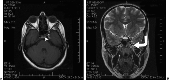
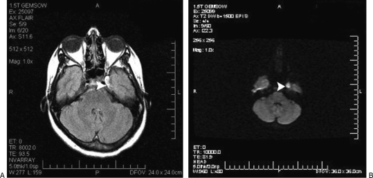
A magnetic resonance imaging (MRI) scan done at our institute showed a lesion in the region of the petrous apex, which was isointense to hyperintense on T1-weighted image (WI), hyperintense on T2-WI, and exhibited no contrast enhancement (Fig. 1). It was hyperintense on fluid attenuated inversion recovery (FLAIR) and diffusion-weighted sequences (Fig. 2).
Figure 1.
(A) Axial T1-weighted image (WI) showing as isointense to hyperintense in left Meckel's cave (white arrowhead). (B) Coronal T2-WI showing a hyperintense lesion. Curved arrow shows the extradural corridor to the lesion.
Figure 2.
The lesion (white arrowhead) is hyperintense on (A) axial fluid attenuated inversion recovery (FLAIR) sequence and (B) diffusion-weighted image.
Summary of Operative Management
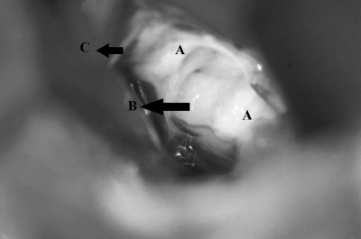
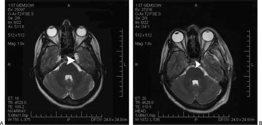
The tumor was approached extradurally at the middle cranial fossa base. A lumbar cerebrospinal fluid (CSF) drain was placed to relax the dura and to secure its easy dissection from the middle cranial fossa base. Stealth (Medtronic, Minneapolis, USA) neuronavigation was used to localize the lesion. The dura overlying the lesion at the petrous apex was opened longitudinally and parallel to the direction of the tentorium to avoid damage to the trigeminal nerve. The tumor was seen in Meckel's cave and was easily separable from the gasserian ganglion and V3 nerve (Fig. 3). It was excised completely (Fig. 4).
Figure 3.
Operative photograph showing epidermoid tumor (A) engulfing the V3 division (B); the margins of incised dura (C) are seen in the periphery.
Figure 4.
Axial T2-weighted image shows (A) tumor before surgery and (B) postoperatively the same area after complete tumor excision. White arrowhead points to the site of tumor.
The patient had minimal V3 hypoesthesia postoperatively but had complete relief from TN symptoms. At 1-year follow-up, she had no recurrent symptoms and was not disabled by facial numbness.
DISCUSSION
Tumors of Meckel's cave account for <0.5% of all intracranial lesions and are usually associated with fifth nerve dysfunction.2,3 Beck and Menezes, who described the pathology and presentation of lesions at Meckel's cave, had one patient with an epidermoid tumor in their series of 12 cases.2
Epidermoid tumors represent 0.2 to 1.3% of all intracranial tumors.3,4 These tumors arise from ectopic ectodermal cells, which are retained as inclusion cysts at the time of closure of the neural grooves or pharyngeal arches. There are 23 reported cases of Meckel's cave epidermoid tumors in indexed literature. Out of these, only three previous cases had presented with TN.4,5,6 This contrasts with cerebellopontine angle epidermoids, which are reported in 0.2 to 5.5% of patients with TN.7
The incidence of TN is reported to be 4.3 per 100,000 people.8 Trigeminal neuropathy may involve the entire course of the nerve, from brainstem nuclei to peripheral branches. Cirak et al divided the nerve into 4 divisions viz, extracranial, cavernous sinus, Meckel's cave and brainstem, to simplify the differential diagnosis.8 Trigeminal neuropathy arising within Meckel's cave and cavernous sinus is due to meningiomas, trigeminal schwannomas, epidermoid cysts, metastases, amyloid deposits, pituitary adenomas, and vascular pathologies such as dural arteriovenous fistula, cavernomas, and aneurysms.8,9,10 Our case was refractory to treatment with a combination of membrane-stabilizing drugs.
Epidermoid tumors are known to creep around or engulf intracranial structures. Cranial nerve deficits are thought to arise from resultant ischemia.11 In addition, leakage of tumor content is known to induce inflammation. Surprisingly, very few patients present with irritative symptoms, but more often report cranial nerve palsies or compressive symptoms.3 On computed tomography (CT) scans, they are characteristically hypodense, nonenhancing lesions that resemble arachnoid cysts.12 There is a possibility that small epidermoid tumors may be overlooked on CT studies.
Surgery
The petrous apex forms the extradural part of the petroclinoid region and provides a unique window to access lesions in this area.13 Meckel's cave lies in the posteromedial region of the middle cranial fossa and contains the gasserian ganglion and the subarachnoid trigeminal cistern.2,3,13 We used neuronavigation to access the small lesion, using an infratemporal extradural approach at the skull base to minimize damage to surrounding meningovenous and neural structures. Magnetic resonance images were used for neuronavigation, with seven fiducials placed around the area of interest centered at the petrous apex. Although the drainage of CSF would change MRI coordinates of the lesion, bony landmarks at the petrous apex provide a reasonable guide to the lesion.
Proper orientation to the dural lining of cavernous sinus and venous structures at the petrous apex is required to protect the carotid artery, cranial nerves, and petrosal sinus.
Approaches to Meckel's Cave
The various approaches to Meckel's cave epidermoid tumors are summarized in Table 1. Nadkarni, who used an infratemporal interdural approach, mentions that tumor dissection from neural structures was not possible due to adhesions.4 Miyazawa, who has reviewed literature on Meckel's cave epidermoids, advocated using trigeminal sensory-evoked response to limit neural injury.3 Large lesions can also be approached using a subtemporal intradural approach.2 Ohta used an orbitozygomatic approach for a large tumor causing oculomotor palsy and facial hypesthesia. However, the capsule was not excised to protect neurovascular structures in the cavernous sinus.14
Table 1.
Summary of Various Approaches Described for Meckel's Cave Epidermoid Tumor
| Author | Site of Lesion | Symptoms | Approach | Outcome |
|---|---|---|---|---|
| TSER, trigeminal sensory-evoked response; TN, trigeminal neuralgia. | ||||
| Beck and Menezes2 | Left Meckel's cave | V, VIII involvement and partial VII nerve palsy | Temporal craniotomy & subtemporal intradural approach | Residual V, VIII nerve symptoms |
| Miyazawa et al3 | Left Meckel's cave | V and VI nerve palsy | Subtemporal intradural approach & resection using TSER | Reduced facial numbness and diplopia |
| Ohta H et al14 | Right Meckel's cave | III and V nerve palsy | Orbitozygomatic approach | Improvement in III and V nerve symptoms |
| Nadkarni et al4 | Right Meckel's cave | Facial numbness, tingling | Subtemporal interdural | Relief from trigeminal sensory symptoms |
| Present case | Left Meckel's cave | TN | Subtemporal extradural approach to petrous apex using neuronavigation | Relief from TN, but minimal V3 numbness |
CONCLUSIONS
Magnetic resonance imaging should be routinely performed in case of refractory TN, to identify a compressive pathology. This can be beneficial, especially when microvascular decompression through the posterior fossa is contemplated for such cases. Small tumors at the middle fossa base can be excised with image guidance with reasonable accuracy using a small extradural surgical window.
REFERENCES
- Tubbs R S, Salter E G, Oakes W J. Bony anomaly of Meckel's cave. Clin Anat. 2006;19:75–77. doi: 10.1002/ca.20163. [DOI] [PubMed] [Google Scholar]
- Beck D W, Menezes A H. Lesions in Meckel's cave: variable presentation and pathology. J Neurosurg. 1987;67:684–689. doi: 10.3171/jns.1987.67.5.0684. [DOI] [PubMed] [Google Scholar]
- Miyazawa N, Yamazaki H, Wakao T, Nukui H. Epidermoid tumors of Meckel's cave: case report and review of the literature. Neurosurgery. 1989;25:951–955. [PubMed] [Google Scholar]
- Nadkarni T, Dindorkar K, Muzumdar D, Goel A. Epidermoid tumor within Meckel's cave–case report. Neurol Med Chir (Tokyo) 2000;40:74–76. doi: 10.2176/nmc.40.74. [DOI] [PubMed] [Google Scholar]
- Kapila A, Steinbaum S, Chakeres D W. Meckel's cave epidermoid with trigeminal neuralgia: CT findings. J Comput Assist Tomogr. 1984;8:1172–1174. doi: 10.1097/00004728-198412000-00027. [DOI] [PubMed] [Google Scholar]
- Mehta D S, Malik G B, Dar J. Trigeminal neuralgia due to cholesteatoma of Meckel's cave. Case report. J Neurosurg. 1971;34:572–574. doi: 10.3171/jns.1971.34.4.0572. [DOI] [PubMed] [Google Scholar]
- Meng L, Yuguang L, Feng L, Wandong S, Shugan Z, Chengyuan W. Cerebellopontine angle epidermoids presenting with trigeminal neuralgia. J Clin Neurosci. 2005;12:784–786. doi: 10.1016/j.jocn.2004.09.023. [DOI] [PubMed] [Google Scholar]
- Cirak B, Kiymaz N, Arslanoglu A. Trigeminal neuralgia caused by intracranial epidermoid tumor: report of a case and review of the different therapeutic modalities. Pain Physician. 2004;7:129–132. [PubMed] [Google Scholar]
- Du R, Binder D K, Halbach V, Fischbein N, Barbaro N M. Trigeminal neuralgia in a patient with a dural arteriovenous fistula in Meckel's cave: case report. Neurosurgery. 2003;53:216–221. doi: 10.1227/01.neu.0000069535.42897.1f. [DOI] [PubMed] [Google Scholar]
- Yu E, de Tilly L N. Amyloidoma of Meckel's cave: a rare cause of trigeminal neuralgia. AJR Am J Roentgenol. 2004;182:1605–1606. doi: 10.2214/ajr.182.6.1821605. [DOI] [PubMed] [Google Scholar]
- Isaacson B, Kutz J W, Roland P S. Lesions of the petrous apex: diagnosis and management. Otolaryngol Clin North Am. 2007;40:479–519. doi: 10.1016/j.otc.2007.03.003. [DOI] [PubMed] [Google Scholar]
- Batra A, Tripathi R P, Singh A K, Tatke M. Petrous apex arachnoid cyst extending into Meckel's cave. Australas Radiol. 2002;46:295–298. doi: 10.1046/j.1440-1673.2002.01063.x. [DOI] [PubMed] [Google Scholar]
- Ozveren M F, Uchida K, Aiso S, Kawase T. Meningovenous structures of the petroclival region: clinical importance for surgery and intravascular surgery. Neurosurgery. 2002;50:829–836. doi: 10.1097/00006123-200204000-00027. [DOI] [PubMed] [Google Scholar]
- Ohta H, Ottomo M, Nakamura T, Yokota A. A case of epidermoid tumor inside the Meckel's cave. No Shinkei Geka. 1997;25:943–947. [Article in Japanese] [PubMed] [Google Scholar]


