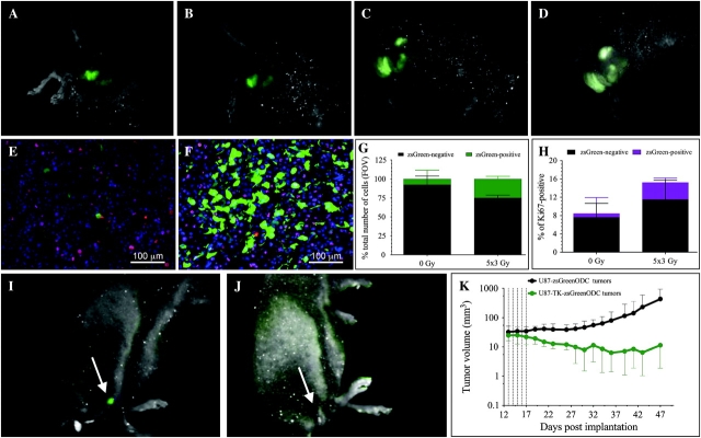
Erratum: “In Vivo Imaging, Tracking, and Targeting of Cancer Stem Cells” by Vlashi et al. [J Natl Cancer Inst 2009;101(5):350–359]. In Figure 4, G, H, and K, the uncertainty represented by the error bars corresponded to the SEM when it was referred to in the legend as the 95% confidence interval. The corrected figure showing 95% confidence intervals is shown below. We regret the error.
Figure 4.
The effect of fractionated radiation on cancer-initiating cells in vivo. A–D) U87MG-ZsGreen-cODC–expressing tumors subjected to fractionated radiation and imaged before treatment (A), after a 3-Gy radiation exposure (B), after 5- × 3-Gy radiation exposures (C), or 72 hours after the last fraction (D). E, F) High-magnification views of untreated tumors (E), in which the proliferating, Ki67-positive population of cells displays high proteasome activity with only a few low proteasome activity (ZsGreen-positive) cells, and tumors treated with daily fractions of 3 Gy (F), in which the number of ZsGreen-positive cells increased substantially (G) as did the percentage of cells that were positive for Ki67 (H). Counterstaining with 4′,6-diamidino-2-phenylindole (blue). Mean values and 95% confidence intervals (CIs) are shown for two independent experiments. I–K) Mice with tumors derived from U87MG cells expressing a fusion protein of thymidine kinase, ZsGreen and the carboxyl terminus of the murine ornithine decarboxylase degron, treated with ganciclovir (5 intraperitoneal injections of 50 mg/kg starting on day 12 after implantation [I] and 18 days after initiation of treatment [J]). K) Growth of the tumors in the mice treated with ganciclovir. Tumor volume (mm3) was assessed with calipers and are shown as means ± 95% CIs (n = 5 mice per group).


