Abstract
Background
Our objective is to evaluate the Medtronic CGMS® continuous glucose monitoring system and plasma glucose (PG) measurement performed in a monitoring schedule as tools to identify individuals with type 1 diabetes at risk when diving.
Methods
We studied 24 adults, 12 type 1 diabetes subjects and 12 controls, during 5 recreational scuba dives performed on 3 consecutive days. The CGMS was used by all participants on all the days and all the dives. Comparisons were made between PG performed in a monitoring schedule during the days of diving, self-monitored blood glucose (SMBG) performed 2 weeks prior to diving, and the CGMS during the study.
Results
One hundred seventeen dives were performed. Hypoglycemia (<70 mg/dl) was found in six individuals and on nine occasions. However, no symptoms of hypoglycemia were present during or immediately postdiving. In one case, repetitive hypoglycemia prediving gave rise to a decision not to dive. None of the dives were aborted. The number of hypoglycemic episodes, 10 min prediving or immediately postdiving, were related to the duration of diabetes, r = 0.83 and p =0.01, and the percentage of SMBG values below target (<72 mg/dl), r = 0.65 and p =0.02. Moreover, the number of hypoglycemic episodes was also related to the total duration below low limit (<70 mg/dl), measured by the CGMS, r =0.74 and p =0.006.
Conclusion
Safe dives are possible to achieve by well-informed, well-controlled individuals with type 1 diabetes. Using downloaded SMBG, CGMS, and repetitive PG in a monitoring schedule, it is possible to identify those subjects who are suitable for diving.
Keywords: blood glucose, CGMS, type 1 diabetes mellitus, diving
Background
The risk of hypoglycemia is a major concern for divers with diabetes. A severe hypoglycemic event could lead to reduced consciousness and the subsequent risk of drowning, which could be fatal and cause severe problems for the diving partner. In many countries, recreational diving is an absolute contraindication for type 1 and type 2 diabetes requiring insulin treatment. This position has softened since the early 1990s. Divers with type 2 diabetes on diets or oral agents have been allowed to participate in the British SubAqua Club, the Sub-Aqua Association, and the Scottish Sub-Aqua Club since 1991. A further softening took place 3 years later when the same organizations stated that divers with diabetes were allowed to dive under certain carefully controlled circumstances.1 On the other hand, in Australia and New Zealand, the South Pacific Underwater Medicine Society published a statement in 19922 opposing diving for all individuals with diabetes, apart from diet-controlled individuals with diabetes—this is still current opinion.
Observational data relating to logged dives of subjects with diabetes without ill effects or problems have been published. A voluntary survey conducted by the Divers Alert Network reported 48,663 dives by 110 divers with diabetes.3 The Diving Diseases Research Centre in the United Kingdom performed a study collecting data from 230 divers with diabetes having 5348 logged dives.4 There were no deaths and no episodes of decompression illness, and hypoglycemic events were present in very small numbers. No case ended adversely.
The observational data that have been published speak in favor of the safety of divers with type 1 diabetes. A repetitive monitoring schedule predive, assessing glucose levels via finger pricking at planned times of 60, 30, and 10 min predive, and immediately postdive was developed by George Burghen [Camp DAVI (Diabetes Association of the Virgin Islands)], and these guidelines were tested by Lerch et al.5
In the Lerch et al. study, 7 divers with type 1 diabetes were compared with 7 healthy controls under closely controlled conditions while performing 11 dives during a period of 6 days. Before diving (10 min predive), all divers with diabetes were told to aim for plasma glucose (PG) between 162 and 221 mg/dl. No cases of hypoglycemia were reported in this study.
Due to the possible consequences, hypoglycemia is the major risk factor for those diving with diabetes. In addition to the avoidance of hypoglycemia while diving, repetitive episodes of hypoglycemia should be avoided on the days before diving, since this could blunt the hormonal response during subsequent exercise or hypoglycemia.6
The CGMS® continuous glucose monitoring device (Medtronic, Minneapolis, MN) offers an opportunity to assess the glucose levels in different contexts. Chico et al. showed that the CGMS was a useful tool to avoid episodes of hypoglycemia and to detect episodes of hypoglycemic unawareness.7
To the best of our knowledge, this article is the first of its kind using the CGMS during diving conditions.
We believe that no prior study has explored the change in glucose levels during sports diving in individuals with type 1 diabetes or healthy volunteers.
Today, there are approximately 10 million individuals worldwide who are active divers. A fair estimation is that at least 100,000 of these are on insulin treatment. Since there are many divers with diabetes who are active divers and also many that would like to become recreational divers, it is very important to evaluate the potential risk of glucose variations in connection with and during recreational diving.
This knowledge is needed in order to identify glycemic levels suitable for recreational diving and to formulate recommendations for safe diving for those with diabetes who are permitted to dive.
A self-monitored blood glucose (SMBG) monitoring schedule in combination with continuous glucose monitoring could be a useful tool for divers with type 1 diabetes. This article addresses the general values of these combined methods.
Research Design and Methods
Participants
The participants were recruited from both Sweden and Norway via advertisements in national journals. Twenty-four adult divers volunteered to participate in the study. Twelve individuals with type 1 diabetes and twelve healthy controls were included. For characteristics, see Table 1.
Table 1.
Characteristics of Study Population
| Characteristics | Type 1 diabetes | Healthy controls | P |
|---|---|---|---|
| Number (n) | 12 | 12 | – |
| Gender (female/male) | 0/12 | 1/11 | – |
| Age (years) and mean (range) | 31(18–49) | 33 (21–52) | NS |
| Duration of diabetes (years) and mean (range) | 12.2 (0.3–30) | – | – |
| Weight (kg) and mean (range) | 84 (73–100) | 85 (75–105) | NS |
| BMI (kg/m2) and mean (range) | 24.4 (21.2–27.2) | 26.0 (21.0–29.1) | NS |
| A1C (Mono–S) and mean (range)a | 6.3 (4.9–8.6) | – | – |
| A1C (NGSPb) and mean (range)a | 7.1 (5.8–9.2) | – | – |
| Treatment (CSII/MDI) | 3/9 | – | – |
| Insulin per day (U/kg) | 0.64 (0.33–0.89) | – | – |
Reference range for A1C in Sweden (Mono-S) 3.6–5.0%.
Mono-S values converted to DCCT/NGSP units.
All subjects gave their written, informed consent prior to participation in the study. The ethics committee at Uppsala University approved the study protocol.
Study eligibility required scuba diving certification. The exclusion criteria were secondary complications (macroangiopathy and active proliferative retinopathy) as well as known hypoglycemic unawareness episodes.
Design
The 24 participants were divided into two groups—diabetes subjects and control subjects. In all, five recreational scuba dives were performed on three consecutive days in 8 to 11 °C seawater. All dives were performed by each subject. Dive 1 was a shallow surface practice. The remaining dives were made at a depth of 18 to 22 m with a duration of 42 to 52 min. All divers wore dry suits on every dive.
Plasma glucose and CGMS were used in parallel by all participants. All PG was performed by medical staff during the project. Comparisons were made between these two glucose measurement methods. Comparisons were also made between the number of hypoglycemic episodes and the results from downloaded SMBG values, performed 2 weeks prior to diving, as well as the results from the glucose readings with the CGMS.
Throughout the study, the individuals with diabetes adjusted their insulin dosages according to their own judgement. Continuous subcutaneous insulin infusion (CSII) was disconnected during dives. Immediately prior to the dives, 15–30 g of carbohydrates, in the form of fruit, was given in amounts depending on glucose levels: 15 g when at 140–230 mg/dl and 30 g when at 70–140 mg/dl.
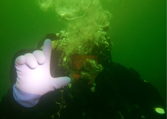
Some of the present guidelines suggest that the diver should surface to ingest glucose if signs of hypoglycemia are present.5 Since this could be difficult to accomplish in a rapid, safe manner due to depth and current, all divers were trained to signal “L” (low) for hypoglycemia (Figure 1). All participants were, after training, also provided a fructose/glucose formulation (Enervitene®, Enervit, Italy) to use orally below surface if signs of hypoglycemia were present during the dive. No such episode with signs of hypoglycemia was present during the following dives.
Figure 1.
Photo of a diver giving the “L” (low) signal for hypoglycemia.
Individuals with repeated low glucose levels 60 and 10 min predive (<70 mg/dl) were not permitted to dive.
Glycosylated Hemoglobin
Diabetes control was assessed at the beginning of the trial by measuring glycosylated hemoglobin [(A1C, Mono-S, ref. value 3.6–5.0%) Bio Rad Variant II, Bio Rad Laboratories AB, Sweden]. For individuals with diabetes, A1C measured with the reference system Mono-S is approximately 1% lower than the National Glycohemoglobin Standardization Program (NGSP).8,9
Glucose Measurements
Self-Monitored Blood Glucose Measurements and Plasma Glucose Measurements
Capillary blood was the source for all reference measurements.
SMBG is the measurement performed with individual home glucose meters during the 14 days immediately prior to diving. The SMBG were downloaded using Diasend (Aidera, Göteborg, Sweden) in order to show statistical parameters in each case. All PG, defined as the capillary measurements during the 3 days, were analyzed with a HemoCue® monitor used together with HemoCue monitor microcuvettes (HemoCue, Ängelholm, Sweden). During the study, glucose was measured 90, 60, and 10 min predive and immediately postdive—in all, 6–8 measurements per day. Each dive started 90 min postmeal. All PG values were used to calibrate the CGMS.
Continuous Glucose Monitoring System
The CGMS consists of a small monitor connected with a cable to a sensor that is placed in subcutaneous tissue. The CGMS measures subcutaneous interstitial glucose (IG). A glucose sensor signal is acquired every 10 s, and an average of 30 values during 5 min are stored in the monitor. The sensor was inserted in the subcutaneous tissue, approximately 10 cm lateral of the umbilicus, by specially trained personnel. The CGMS was used on 3 consecutive days during all dives on all divers.
The range of IG detection is 40–400 mg/dl. The calculated variables were the mean absolute difference (MAD), frequency of hypo- (<70 mg/dl) and hyperglycemia (>180 mg/dl), and duration above (>180 mg/dl) and below (<70 mg/dl) low and within limits, expressed in percentages, day by day.
Data Analysis
Correlation analysis [t-test (two-tailed) and Pearson's correlation (two-tailed)] was used to compare the number of hypoglycemic episodes during the monitoring schedule (PG) related to diving with the results from SMBG and CGMS. The same correlation analysis [t-test (two-tailed) and Pearson's correlation (two-tailed)] was used to compare the results from monitored PG and SMBG values. The t-test (two-tailed) was used to evaluate the glucose difference pre- and postdive within and between the diabetes group and the control group. The Wilcoxon signed-ranks test and the Mann-Whitney test were used in cases where the variables were not distributed normally. All PG values were compared with corresponding CGMS values and the difference was expressed as the MAD.
Results
Of the 120 planned dives, 117 were performed (diabetes: 58 and control: 59). None of the dives were aborted. Hypoglycemia (PG <70 mg/dl) was found 10 min predive or immediately postdive in six individuals and on nine occasions, all mild hypoglycemia, mean 56 (40–68) mg/dl. Three of these hypoglycemic episodes (58–68 mg/dl) were found predive in three individuals and six (40–67 mg/dl) were found postdive in six individuals. In relation to all dives, three individuals were found to have hypoglycemic levels on two occasions, either 10 min predive or immediately postdive. However, no symptoms of hypoglycemia were present during diving or immediately postdive. In the group with diabetes, one individual was not allowed to dive due to repetitive hypoglycemia and one due to hyperglycemia together with a headache. In the control group, one individual arrived too late to participate in the first dive.
SMBG two weeks prior to the dive study was performed using daily finger pricking tests, mean number 4.2 ± 2.8 with a mean glucose value of 150 ± 43 mg/dl. The number of hypoglycemic episodes 10 min predive or immediately postdive was related to the percentage of SMBG values below target (<72 mg/dl) (r = 0.65 and p = 0.02) and also to the duration of diabetes (r = 0.83 and p = 0.01).
The mean PG was 158 ± 34 mg/dl, a value not statistically different from the mean SMBG value measured 2 weeks prior to diving. The group means of PG in the diabetes subjects, during the 3 days comprising 5 scuba dives, were as follows: Day 1, 182 ± 54 mg/dl; Day 2, 146 ± 32 mg/dl; and Day 3, 151 ± 38 mg/dl.
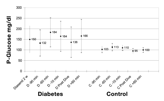
Figure 2 shows the distribution of glucose levels, SMBG values, and the PG values measured in the monitoring schedule. There was a significant difference at each test time: p < .005.
Figure 2.
Distribution of glucose levels (SMBG measured 14 days prior to dive and PG measured in a monitoring schedule). Values expressed as mean and standard deviation for all five dives aggregated and shown in the study groups: type 1 diabetes and control. Time (min) in relation to dive. There was a significant difference between the groups each test time: p < .005.
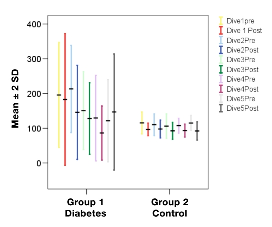
The mean differences between PG 10 min predive and immediately postdive for the diabetes and control groups are presented in Figure 3. There was a significant difference between the groups related to the difference predive-postdive: Dives 1–3 and 5, p < .05, and Dive 4, p < .001.
Figure 3.
Distribution of glucose levels (PG), mean and standard deviation, in the study groups: type 1 diabetes and control. Values represented are as follows: 10 min predive and immediately postdive with all 5 dives in consecutive order. There was a significant difference between the groups: Dives 1–3 and 5, p < .05, and Dive 4, p < .001.
The mean drop in PG in the two groups is presented in Table 2. There was a significant difference between the groups. The mean absolute individual prechange-postchange in PG was found to be 31 ± 68 mg/dl in the group with diabetes and 14 ± 16 mg/dl in the control group, p < .05. The change in the group with diabetes corresponds to a 12 ± 42% decrease of the predive value and a 12 ± 14% decrease in the controls.
Table 2.
Distribution of Differences in PG Predive-Postdive in the Study Groups: Diabetes and Control.a
| Dive | Diabetes (n = 12) | Control (n = 12) | P | ||||||||
|---|---|---|---|---|---|---|---|---|---|---|---|
| Rangeb (mg/dl) | Mean (mg/dl) | SEM | Rangeb (mg/dl) | Mean (mg/dl) | SEM | ||||||
| total | min | max | total | min | max | ||||||
| 1 | 281 | −166 | 115 | −25 | 68 | 50 | −50 | 0 | −18 | 14 | <.05 |
| 2 | 238 | −135 | 103 | −60 | 61 | 58 | −38 | 20 | −9 | 20 | <.05 |
| 3 | 264 | −196 | 68 | −29 | 67 | 48 | −41 | 7 | −14 | 14 | <.05 |
| 4 | 127 | −119 | 9 | −47 | 47 | 45 | −34 | 11 | −14 | 13 | <.05 |
| 5 | 294 | −128 | 166 | 11 | 83 | 65 | −58 | 7 | −20 | 20 | <.05 |
| All | 108 | −74 | 34 | −31 | 32 | 39 | −25 | 14 | −14 | 11 | <.05 |
Mean and standard error of mean (SEM) shown.
Range illustrates the difference between the highest reduction and highest increase.
During the 3 days, a total of 28 sensors were used by the 24 participants, which means that only 4 sensors had to be replaced. All changes of sensors were made due to alarms and/or calibration errors.
According to the values registered with the CGMS, the divers with diabetes spent 26.9 ± 17.0% of their study time with glucose values above the high limit (>180 mg/dl) and 15.3 ± 16.7% below the low limit (<70 mg/dl).
A correlation was seen between the number of hypoglycemic readings (PG) and the total duration below low limit (<70 mg/dl), measured by the CGMS (r = 0.74 and p = 0.006).
The overall mean correlation between the CGMS and PG was r = 0.93 ± 0.04 in the group with diabetes, and the mean correlation coefficients for each day were as follows: Day 1, r = 0.77 ± 0.27; Day 2, r = 0.94 ± 0.07; and Day 3, r = 0.96 ± 0.05. The overall MAD was 14.4 ± 6%, and the corresponding figures for each day were as follows: Day 1, 23.2 ± 19.3%; Day 2, 11.6 ± 4.5%, and Day 3, 11.2 ± 5.7%. The overall MAD within the control group was 8.6 ± 1.7%, and the corresponding figures for each day were as follows: Day 1, 8.2 ± 1.9%; Day 2, 8.8 ± 2.1%, and Day 3, 8.7 ± 3.2%.
Conclusions
The risk of hypoglycemia is a major concern for divers with diabetes. An event occurring under water is a greater threat than on land. Technological and pharmacological improvements have reduced the risk of hypoglycemia in insulin-treated subjects with type 1 and type 2 diabetes. However, the current recommendation by a large number of diving medicine physicians is to disqualify an individual with type 1 diabetes from participating in sports diving.
In this study, PG was measured 90, 60, and 10 min predive and immediately postdive. For a diver with diabetes, it is important to understand the glucose trend, to take appropriate action, and perhaps to also refrain from an individual dive. In our study, we showed that this monitoring schedule identified episodes of hypoglycemia. The monitoring schedule is of great importance since episodes of hypoglycemic unawareness could be detected as well as treated. A gradually reduced glucose level prior to dive could be avoided. It was also possible to stop an individual from diving due to repetitive hypoglycemia.
We found that hypoglycemia before or after diving was related to the duration of diabetes. Moreover, the number of hypoglycemic episodes was related to the duration of diabetes, the percentage of SMBG below 72 mg/dl 2 weeks prior to diving, and the total duration below low limit (<70 mg/dl), registered by the CGMS. Thus individuals with a longer duration of diabetes run a higher risk of hypoglycemia in connection with diving. The number of hypoglycemic episodes related to diving relates to the percentage of SMBG values below target performed 14 days prior to diving. This suggests that there is a relation of glucose profile variability independent from exercise per se. These findings are supported by Cox et al.10 who showed that severe hypoglycemia is often identifiable from SMBG, where a specific blood glucose fluctuation pattern precedes severe hypoglycemia. We therefore propose that SMBG be downloaded prior to diving in order to identify individuals with diabetes who run a high risk of having hypoglycemia related to diving.
The CGMS is a useful tool for assessing daily glucose fluctuations, but it has certain limitations. It measures glucose concentrations in the extracellular fluid rather than in the intravascular space. The relationship between glucose concentrations in these two compartments is not straightforward and may change according to physiological variations in insulin concentration and glucose uptake, utilization, and elimination.11,12 These limitations are balanced by the ability to record continuous glucose data and detect unrecognized hypoglycemic episodes. To the best of our knowledge, no published studies have yet assessed glucose levels during recreational diving. We used MAD instead of correlation in order to evaluate the quality of the CGMS readings. The correlation is sensitive to the range of glucose levels and is therefore improved by greater glucose variability and calibrations at both high and low glucose measurements. MAD, on the other hand, measures the percentage difference irrespective of the level of measurements. MAD could be used for the evaluation of quality when retrospective calibrations are performed, as in this study. It would not be applicable for real-time data calibration and display. In this study, the quality of the CGMS readings are good.
In the control group, MAD was lower compared with the group with diabetes. Moreover, in the group with diabetes, MAD was also higher on Day 1 compared with subsequent days, while this difference was not seen in the control group. A possible reason for this could be a more unstable sensor function at the start and greater differences between sensor signal and blood glucose measurements when calibration is performed the first time, but this has to be further investigated.
In addition to the benefits of using the CGMS during repetitive dives, we also propose that the CGMS be used prior to diving as a means of detecting frequent hypoglycemia as well as hypoglycemic unawareness. Hypoglycemic unawareness could, in fact, blunt a normal hormonal response to a hypoglycemic event during exercise.6 In our study, we also show that a number of hypoglycemic episodes were present but without any symptoms.
We show that it is possible to identify individuals with type 1 diabetes who are suitable for diving. Modern technology using downloaded home glucose meter values and the CGMS are important tools for reducing the risk of hypoglycemia in connection with diving.
Abbreviations
- CSII
continuous subcutaneous insulin infusion
- IG
interstitial glucose
- MAD
mean absolute difference
- MDI
multiple daily injections
- NGSP
National Glycohemoglobin Standardization Program
- PG
plasma glucose
- SEM
standard error of mean
- SMBG
self-monitored blood glucose
Funding
This work was supported by Aidera, Medtronic, and Novo Nordisk.
References
- 1.Bryson P, Edge C, Lindsay D, Willshurst P. The case for diving diabetics. SPUMS J. 1994;24:11–13. [Google Scholar]
- 2.Davies D. SPUMS statement on diabetes. SPUMS J. 1992;22(1):31–32. [Google Scholar]
- 3.GdeL Dear, Pollock NW, Uguccioni DM, Dovenbarger J, Feinglos MN, Moon RE. Plasma glucose responses in recreational divers with insulin-requiring diabetes. Undersea Hyperb Med. 31(3):2004, 291–301. [PubMed] [Google Scholar]
- 4.Edge CJ, St Ledger -Dowse M, Bryson P. Scuba diving with diabetes mellitus—the UK experience 1991-2001. Undersea Hyperb Med. 2005;32(1):27–37. [PubMed] [Google Scholar]
- 5.Lerch M, Lutrop C, Thurm U. Diabetes and diving: can the risk of hypoglycemia be banned? SPUMS J. 1996;26(2):62–66. [Google Scholar]
- 6.Galassetti P, Tate D, Neill RA, Morrey S, Wasserman DH, Davis SN. Effect of antecedent hypoglycaemia on counterregulatory responses to subsequent euglycaemic exercise in type 1 diabetes. Diabetes. 2003;52(7):1761–1769. doi: 10.2337/diabetes.52.7.1761. [DOI] [PubMed] [Google Scholar]
- 7.Chico A, Vidal-Rios P, Subira M, Novialis A. The continuous glucose monitoring system is useful for detecting unrecognized hypoglycaemias in patients with type 1 and type 2 diabetes but is not better than frequent capillary glucose measurements for improving metabolic control. Diabetes Care. 2003;26(4):1153–1157. doi: 10.2337/diacare.26.4.1153. [DOI] [PubMed] [Google Scholar]
- 8.Arnqvist H, Wallensteen M, Jeppsson JO. Standards for long-term measures of blood sugar are established. Lakartidningen. 1997;94(50):4789–4790. [PubMed] [Google Scholar]
- 9.Hoelzel W, Weykamp C, Jeppsson JO, Miedema K, Barr JR, Goodall I, Hoshino T, John WG, Kobold U, Little R, Mosca A, Mauri P, Paroni R, Susanto F, Takei I, Thienpont L, Umemoto M, Wiedmeyer HM. IFCC Working Group on HbA1c Standardization IFCC reference system for measurement of hemoglobin A1c in human blood and the national standardization schemes in the United States, Japan, and Sweden: a method-comparison study. Clin Chem. 2004;50(1):166–174. doi: 10.1373/clinchem.2003.024802. [DOI] [PubMed] [Google Scholar]
- 10.Cox DJ, Gonder-Frederick L, Ritterband L, Clarke W, Kovatchev BP. Prediction of severe hypoglycaemia. Diabetes Care. 2007;30(6):1370–1373. doi: 10.2337/dc06-1386. [DOI] [PubMed] [Google Scholar]
- 11.Aussedat B, Dupire-Angel M, Gifford R, Klein JC, Wilson GS, Reach G. Interstitial glucose concentration and glycemia: implications for continuous subcutaneous glucose monitoring. Am J Physiol Endocrinol Metab. 2000;278(4):E716–E728. doi: 10.1152/ajpendo.2000.278.4.E716. [DOI] [PubMed] [Google Scholar]
- 12.Kulcu E, Tamada JA, Reach G, Potts RO, Lescho MJ. Physiological differences between interstitial glucose and blood glucose measured in human subjects. Diabetes Care. 2003;26:2405–2409. doi: 10.2337/diacare.26.8.2405. [DOI] [PubMed] [Google Scholar]


