Abstract
Colonization of the intestinal epithelium by enterohaemorrhagic Escherichia coli (EHEC) is characterized by an attaching and effacing (A/E) histopathology. The locus of enterocyte effacement (LEE) pathogenicity island encodes many genes required for the A/E phenotype including the global regulator of EHEC virulence gene expression, Ler. The LEE is subject to a complex regulatory network primarily targeting ler transcription. The RNA chaperone Hfq, implicated in post-transcriptional regulation, is an important virulence factor in many bacterial pathogens. Although post-transcriptional regulation of EHEC virulence genes is known to occur, a regulatory role of Hfq in EHEC virulence gene expression has yet to be defined. Here, we show that a hfq mutant expresses increased levels of LEE-encoded proteins prematurely, leading to earlier A/E lesion formation relative to wild type. Hfq indirectly affects LEE expression in exponential phase independent of Ler by negatively controlling levels of the regulators GrlA and GrlR through post-transcriptional regulation of the grlRA messenger. Moreover, Hfq negatively affects LEE expression in stationary phase independent of GrlA and GrlR. Altogether, Hfq plays an important role in coordinating the temporal expression of the LEE by controlling grlRA expression at the post-transcriptional level.
Keywords: enterohaemorrhagic Escherichia coli, Hfq, LEE, grlRA
Introduction
Enterohaemorrhagic Escherichia coli (EHEC) O157:H7 is a human pathogen that causes diarrhea, haemorrhagic colitis and haemolytic uremic syndrome (Nataro and Kaper, 1998). EHEC colonization of the intestinal epithelial surface is accompanied by a characteristic attaching and effacing (A/E) histopathology marked by localized destruction of brush border microvilli, intimate attachment to enterocytes and rearrangement of cytoskeletal components beneath adherent bacteria (Frankel et al., 1998). The genes required for the A/E phenotype are primarily contained in the 35–45 kb locus of enterocyte effacement (LEE) pathogenicity island, which is conserved among EHEC strains and other A/E pathogens including enteropathogenic E. coli (EPEC), atypical EPEC, rabbit EPEC, Escherichia albertii and Citrobacter rodentium (McDaniel et al., 1995; Nataro and Kaper, 1998; Huys et al., 2003; Rasko et al., 2008).
The LEE contains five major operons (LEE1 to LEE5) that encode a type III secretion system (TTSS), the adhesion factor intimin, a translocated intimin receptor (Tir), translocator proteins, secreted effectors and regulators (McDaniel et al., 1995; Elliott et al., 1998; Perna et al., 1998; Garmendia et al., 2005). Proper coordination of virulence gene expression is crucial for the bacterial pathogen to successfully colonize the host. Under conditions suboptimal for infection, the global modulator H-NS silences LEE expression (Bustamante et al., 2001; Umanski et al., 2002; Haack et al., 2003; Barba et al., 2005). The LEE1-encoded regulator Ler is required for transcriptional activation of LEE genes and virulence genes located outside the LEE (Mellies et al., 1999; Friedberg et al., 1999; Elliott et al., 2000; Sperandio et al., 2000; Sanchez-SanMartin et al., 2001; Haack et al., 2003; Abe et al., 2008) by relieving H-NS-mediated silencing (Sperandio et al., 2000; Bustamante et al., 2001; Umanski et al., 2002; Haack et al., 2003; Torres et al., 2007; Mellies et al., 2008). A complex regulatory network controlling LEE1 expression reflects the importance of Ler in virulence gene expression. Factors including H-NS, IHF, Fis, Hha, GrlA, GrlR, BipA, Pch, EtrA, EivF, GrvA, QseA, and the alarmone ppGpp integrate environmental signals to regulate EHEC LEE1 transcription in response to changing environmental and host conditions (for reviews see Kaper et al., 2004; Mellies et al., 2007). The LEE-encoded regulators GrlA and GrlR activate and repress LEE transcription, respectively (Barba et al., 2005; Iyoda et al., 2006). Specifically, GrlA binds to the EHEC LEE1 promoter to activate ler transcription (Laaberki et al., 2006; Russell et al., 2007; Huang and Syu, 2008). Ler and GrlA then form a positive transcriptional regulatory loop in part by counteracting H-NS repression (Barba et al., 2005). GrlR interacts with GrlA to prevent the formation of higher order complexes between GrlA and LEE1 promoter DNA, thereby fine-tuning GrlA-mediated activation of ler transcription (Creasey et al., 2003; Jobichen et al., 2007; Huang and Syu, 2008). Consequently, GrlA and GrlR control activation of LEE transcription by optimizing Ler expression. Since GrlA is required for transcriptional activation of LEE1 (Barba et al., 2005), factors controlling GrlA expression are important for ler expression, and thereby for initiating expression of the Ler regulon.
Many transcriptional regulators are subject to post-transcriptional regulation mediated by small RNAs (sRNAs) in conjunction with Hfq, enabling the bacteria to rapidly respond to changes in the environment. Hfq is a conserved global post-transcriptional regulator, originally identified as an essential factor for phage Qβ replication (Franze de Fernandez et al., 1968), which functions in a hexameric ring-shaped structure as an RNA chaperone (Møller et al., 2002; Schumacher et al., 2002; Moll et al., 2003; Geissmann and Touati, 2004; Brennan and Link, 2007) by mediating the binding of sRNAs preferentially to single-stranded AU-rich regions of messenger RNA (mRNA) targets (Zhang et al., 2002; Brescia et al., 2003; Kawamoto et al., 2006). Small RNAs often bind to the 5’-untranslated leader region of cognate messengers to modulate their stability and/or translation (for reviews see Valentin-Hansen et al., 2004; Majdalani et al., 2005; Repoila and Darfeuille, 2009; Waters and Storz, 2009). A classic example is Hfq-mediated regulation of the global regulators RpoS and H-NS by sRNAs including DsrA. Upon binding to rpoS and hns mRNAs, DsrA stimulates translation and decreases messenger stability, respectively (for reviews see Lease and Belfort, 2000; Repoila and Gottesman, 2003; Majdalani et al., 2005). Since the RpoS and H-NS regulons are important for the general stress response, a hfq mutant shows pleitropic phenotypes (Tsui et al., 1994). In recent years, Hfq has been established as an important virulence factor in bacterial pathogens including Salmonella enterica serovar Typhimurium (Sittka et al., 2007; Wilson et al., 2007), Shigella sonnei (Mitobe et al., 2008), Vibrio cholerae (Ding et al., 2004), Pseudonomas aeruginosa (Sonnleitner et al., 2003), Brucella abortus (Robertson and Roop, Jr., 1999), Yersinia enterocolitica (Nakao et al., 1995), Legionella pneumophila (McNealy et al., 2005), Listeria monocytogenes (Christiansen et al., 2006), Neisseria meningitidis (Fantappiè et al., 2009), Francisella tularensis (Meibom et al., 2009) and uropathogenic E. coli (Kulesus et al., 2008). Although post-transcriptional regulation for EHEC virulence gene expression occurs (Roe et al., 2003; 2004; 2007; Walters and Sperandio, 2006; Campellone et al., 2007; Lodato and Kaper, 2008) a regulatory role of Hfq in EHEC virulence gene expression has yet to be defined. Here, we demonstrate that Hfq negatively affects LEE expression in exponential phase by regulating the expression of the LEE-encoded regulators GrlA and GrlR at the post-transcriptional level by destabilizing grlRA mRNA. In addition, we show that Hfq negatively affects LEE expression in stationary phase independent of grlRA. Our data suggest that Hfq plays an important role in controlling GrlA and GrlR levels to prevent induction of LEE expression during exponential growth, and thereby ensure proper temporal expression of the LEE.
Results
Hfq affects EHEC acid resistance, motility and protein secretion
To evaluate the effect of hfq on EHEC virulence, we constructed an in-frame deletion of hfq in the EHEC O157:H7 EDL933 stx mutant background, TUV93-0. Acid resistance is an essential virulence trait of EHEC O157:H7 as it enables survival of bacteria passing the acidic gastric barrier of the host (Price et al., 2000). Since Hfq affects the expression of RpoS and H-NS regulons, Hfq is important for bacteria to respond to various environmental stresses such as low pH conditions they encounter during infection (Lin et al., 1995; Atlung and Ingmer, 1997; Valentin-Hansen et al., 2004). To determine the acid tolerance of an EHEC hfq mutant, we compared survival rates of stationary-phase cells of wild type and hfq mutant strains after one-hour of exposure to pH 2.5. The hfq mutant was significantly (P < 0.004) more sensitive to acid shock compared to the wild type and the complemented hfq mutant (Table 1), indicating an important role of hfq in acid tolerance of EHEC. The acid sensitive phenotype of the EHEC hfq mutant is likely due to strongly reduced RpoS levels (data not shown), which is consistent with previous findings for hfq mutants of E. coli K-12 and Salmonella enterica Typhimurium (Muffler et al., 1996; Brown and Elliott, 1996).
TABLE 1.
Acid resistance and motility of a hfq mutant.
| Acid resistancea,c |
Motilityb,c |
|
|---|---|---|
| TUV93-0, pBR322 | 5 ± 0.2 | 25 ± 5 |
| TUV93-0 hfq, pBR322 | < 0.001 | 10 ± 0 |
| TUV93-0 hfq, pAMH100 | 9 ± 1 | 19 ± 3 |
Percent survival of cells after one hour of incubation in LB at pH 2.5.
Cell motility in 0.3% agar expressed as the diameter of the bacterial swarming ring in mm.
Values represent the means ± SD of three independent experiments.
The deletion of hfq impairs motility of the pathogens Pseudomonas aeruginosa, Salmonella enterica Typhimurium and uropathogenic E. coli (Sonnleitner et al., 2003; Sittka et al., 2007; Kulesus et al., 2008). To test whether Hfq affects motility of EHEC, wild type and hfq mutant strains were grown on semi-solid agar plates. The hfq mutant showed significantly (P < 0.02) decreased motility compared to the wild type, as determined by the diameter of the swarming ring, whereas motility was restored in a complemented hfq mutant (Table 1).
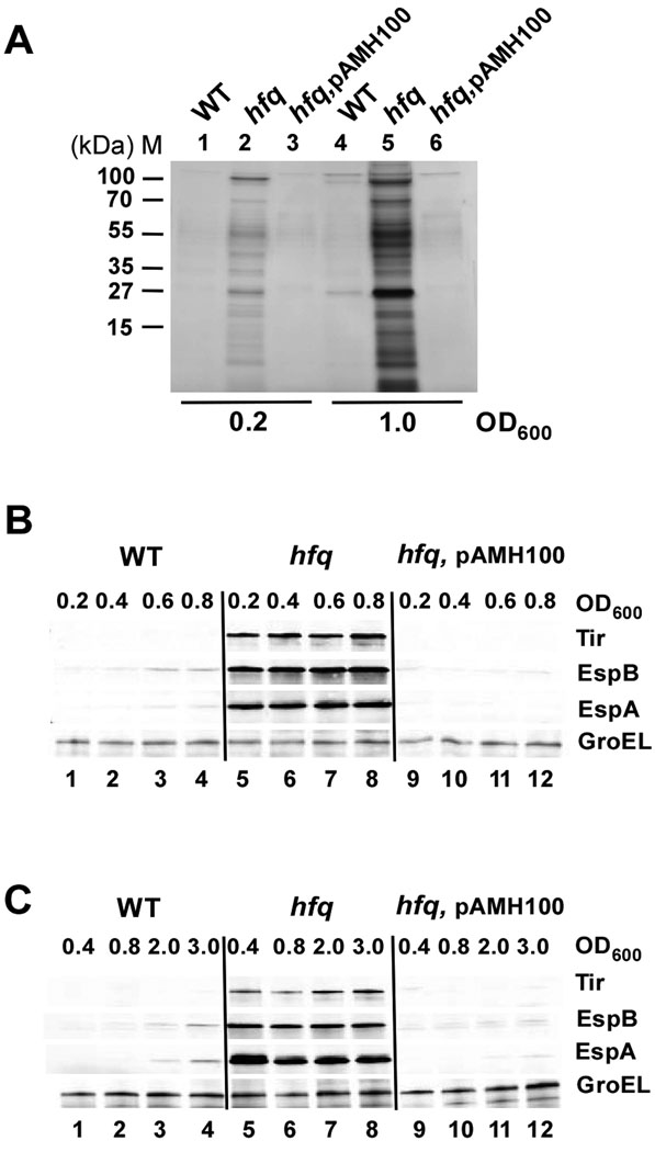
Protein secretion and subsequent translocation of effectors into the host cell by the TTSS apparatus is crucial for A/E lesion formation to occur, and thereby for virulence of EHEC (Garmendia et al., 2005). Indeed, deletion of hfq in Salmonella enterica Typhimurium abolishes secretion of effectors encoded by the pathogenicity island-1 (SPI-1), thereby reducing virulence (Sittka et al., 2007). To assess the role of hfq in EHEC protein secretion, we compared the abundance of proteins in culture supernatants from TUV93-0 wild type and hfq mutant strains grown aerobically in DMEM. Proteins from equal volumes of culture supernatant were resolved by SDS-PAGE and visualized by silver staining. The hfq mutant exhibited increased levels of protein secretion in both exponential and transition phases compared to wild type (Fig. 1A, compare lanes 1 and 4 with 2 and 5), whereas the complemented hfq mutant had protein secretion restored to wild type levels (Fig. 1A, lanes 3 and 6). Comparison of the protein profiles revealed that the hfq mutant prematurely releases proteins in levels that exceeded those for wild type in the transition to stationary phase (Fig. 1A, compare lanes 2 and 5 with lane 4). The most abundant protein released to culture supernatants by the hfq mutant was identified as EspB by Western blot analyses (data not shown), suggesting a central role of Hfq in controlling LEE protein levels. Since Western blot analyses further revealed significantly increased levels of the cytoplasmic chaperonin GroEL in culture supernatants of the hfq mutant (data not shown), the increased abundance of proteins in these supernatants is most likely due to increased membrane perturbation, which is consistent with the involvement of Hfq in the regulation of the σE-mediated envelope stress response (Guisbert et al., 2007; Valentin-Hansen et al., 2007).
Figure 1.
Hfq affects the abundance of proteins in culture supernatants and LEE expression in EHEC. (A) Proteins present in culture supernatants of EHEC TUV93-0 wild type, hfq mutant and the mutant strain complemented with hfq from pAMH100 grown in DMEM at 37°C to an optical density at 600 nm (OD600) ~ 0.2 (lanes 1–3) and 1.0 (lanes 4–6). Proteins corresponding to 300 µl of culture supernatant were prepared as described in Experimental procedures, resolved in a 12% SDS-PAGE and visualized by silver staining. (B and C) The temporal expression profiles of LEE-encoded proteins in wild type TUV93-0, a hfq mutant and a complemented hfq mutant were determined by Western blot analyses as described in Experimental procedures. Equal amounts of total protein including both whole cell and secreted proteins, was prepared from cultures grown at 37°C in DMEM (B) and LB (C) to the cell densities indicated, resolved by 4–20% SDS-PAGE and blotted onto nylon membranes. Levels of Tir, EspA, EspB and GroEL were detected by Western blot analyses using polyclonal antisera against the respective proteins. GroEL served as an internal control for the total protein amount.
LEE expression occurs prematurely at increased levels in a hfq mutant
To test whether Hfq affects LEE expression, we examined the temporal expression pattern of LEE-encoded proteins in TUV93-0 wild type, a hfq mutant and a complemented hfq mutant grown in both DMEM and LB using Western blot analyses (Fig. 1B and C, respectively). The LEE-encoded proteins EspA, EspB and Tir were expressed in a growth phase-dependent manner in the wild type with highest abundance in late exponential and stationary phases (Fig. 1B and C, lanes 1–4). Tir levels in the wild type were only detected by overexposing the immunoblot (data not shown). The hfq mutant expressed EspA, EspB and Tir prematurely in exponential phase and throughout growth at increased levels compared to wild type (Fig. 1B and C, lanes 5–8). Quantification of the immunoblots revealed that protein levels of EspA, EspB and Tir were increased by respectively 30, 14 and 16-fold in the hfq mutant grown in DMEM, and 19, 5 and 5-fold in the mutant grown in LB, compared to maximum levels detected for the wild type (Fig. 1B and C, lane 5 relative to 4). Consistent with our findings, while this paper was under review, an EHEC hfq mutant was reported to express elevated levels of EspA and EspB (Hirakawa et al., 2009). The increased abundance of LEE-encoded proteins in the hfq mutant was abolished upon complementation with pAMH100 but not the empty vector pBR322 (Fig. 1B and C, lanes 9–12; data not shown). Moreover, the hfq mutant expressed the LEE5-encoded chaperone CesT and the LEE-encoded effector EspG prematurely at increased levels (data not shown). These results suggest that Hfq plays an important role in coordinating the temporal expression of both LEE-and non-LEE-encoded proteins. The increased expression of proteins in the hfq mutant was accompanied by a longer generation time (g) compared to wild type. Because the hfq mutant grew slower in DMEM (g,WT ~ 25 min and g,hfq ~ 60 min) than in LB (g,WT ~ 25 min and g,hfq ~ 40 min), LB medium was used for cell growth in the experiments below unless stated otherwise. The decreased growth phenotype of the hfq mutant, shared by other bacterial pathogens (Ding et al., 2004; Sittka et al., 2007; Attia et al., 2008), might be due to drastically increased energy consumption caused by elevated LEE expression and subsequent protein secretion since a hfq ler double mutant, devoid of LEE expression, exhibited similar growth as an E. coli K-12 hfq mutant strain (data not shown).
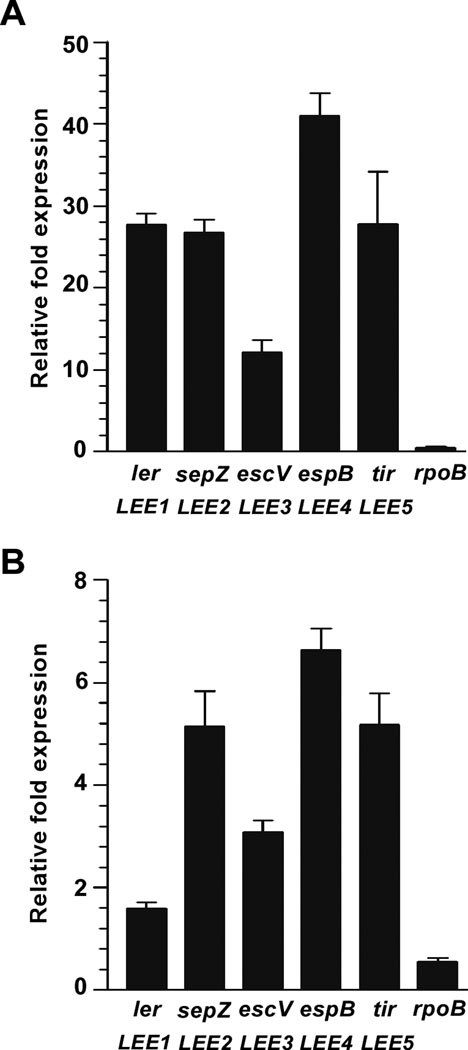
To determine whether Hfq affects LEE expression on the transcriptional or post-transcriptional level, we examined LEE transcription in wild type and hfq mutant strains grown to exponential and stationary phases in LB. The relative expression of ler (LEE1), sepZ (LEE2), escV (LEE3), espB (LEE4), tir (LEE5) and the control rpoB was measured by quantitative real-time PCR (qRT-PCR) and normalized to 16S rRNA levels. Transcription of LEE in the hfq mutant grown to the exponential phase was significantly increased by 12 to 41-fold relative to wild type TUV93-0 (P < 0.002), whereas levels of the control transcript rpoB did not change significantly (Fig. 2A). Moreover, levels of LEE-encoded transcripts of the hfq mutant in exponential phase significantly exceeded those of wild type in stationary phase, which represent the maximum levels for the wild type, by 3 to 7-fold (P < 0.002). LEE transcription in stationary phase in the hfq mutant relative to wild type was also significantly increased but to a lesser degree than in exponential phase by 3 to 6-fold for LEE2-5 (P < 0.002), while LEE1 transcript levels did not change significantly (Fig. 2B). The finding that transcript levels of LEE2-5 in the hfq mutant were the same in exponential and stationary phase (data not shown) correlates with the continuously increased levels of LEE proteins in the hfq mutant throughout growth (fig. 1B and C, lanes 5–8). Thus, Hfq negatively affects LEE transcription especially during exponential growth. As Ler is an activator of LEE expression (Elliott et al., 2000), increased abundance of the ler transcript in exponential phase, and consequently increased Ler levels, most likely accounts for the increased LEE transcription in the hfq mutant.
Figure 2.
LEE transcript levels are increased in a hfq mutant. Quantitative real-time PCR measurements of LEE transcripts in total RNA isolated from TUV93-0 wild type and hfq mutant strains grown at 37°C in LB to OD600 ~ 0.6 and 3.0 were performed as described in Experimental procedures. The relative fold expression representing the change in transcript levels of ler, sepZ, escV, espB and tir in a hfq mutant relative to wild type grown to OD600 ~ 0.6 (A) and 3.0 (B) was determined. Detection of the rpoB transcript served as a control for a gene which expression is not significantly stimulated in a hfq mutant. Results represent means and standard deviations from triplicate experiments. Error bars show the standard deviations of ΔΔCT values. Levels of 16S rRNA were used to normalize the CT values of target genes to variations in bacterial numbers.
Hfq negatively affects Ler levels
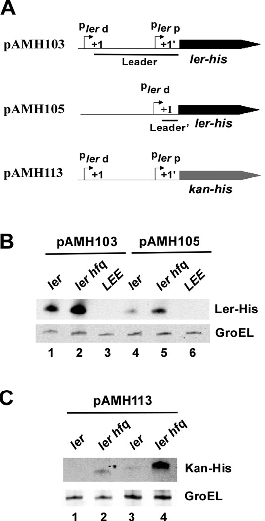
Since Hfq often affects gene expression by controlling the levels of global regulators, Hfq could potentially affect LEE transcription through post-transcriptional regulation of ler. The LEE1 operon in EHEC is expressed from two promoters, the proximal and distal promoters, with transcriptional start sites located 32 nt and 163 nt upstream of the ler translation initiation codon, respectively (Sperandio et al., 2002; Yerushalmi et al., 2008). The ler messenger is primarily expressed from the distal promoter (Sperandio et al., 2002), resulting in a transcript encoding a 163 nt-long untranslated 5’-untranslated leader region, which provides a potential target for Hfq-mediated regulation by small RNAs. The length of a leader region was shown to determine the efficiency of interaction between Hfq, a small RNA and target mRNA (Soper and Woodson, 2008; Updegrove et al., 2008). To test whether the ler mRNA leader region is required for the Hfq-mediated regulation of LEE1, we deleted the region between the transcription initiation sites of the two promoters including the proximal promoter and ~130 nt of the leader region. The resulting construct, pAMH105, expressed a ler transcript encoding a truncated 5’ leader from the distal promoter (Fig. 3A). Western blot analyses of His-tagged Ler, expressed from the two native ler promoters (pAMH103) and from the distal ler promoter in the presence of a truncated leader region (pAMH105), revealed a two to three-fold increase in Ler expression from both constructs in ler mutant backgrounds in the absence of hfq (Fig. 3B, compare lanes 1 and 4 with 2 and 5), whereas no Ler was detected in a LEE-deleted strain as expected (Fig. 3B, lanes 3 and 6). This result indicates that the ler messenger leader region is not required for Hfq-mediated regulation of Ler levels. The five-fold decrease in Ler expression observed from the construct encoding a truncated leader sequence (pAMH105) relative to the wild type construct (pAMH103) (Fig. 3B compare lanes 1 and 4) could be due to the absence of the proximal ler promoter in pAMH105 or alternatively due to a regulatory effect of the ler leader region on Ler expression.
Figure 3.
Hfq indirectly affects Ler levels. (A) Schematic outline of constructs used to determine ler-his expression from ler distal and proximal promoters (pAMH103), a ler promoter with 120 nt of the 160 nt leader sequence including the proximal promoter region deleted (pAMH105) and kan-his expressed from a native ler promoter (pAMH113). Pler d and Pler p designate the distal and proximal ler promoter, respectively. (B) The ler leader region is not required for Hfq-mediated regulation of Ler levels. Western blot analysis of Ler-His expressed from pAMH103 (lanes 1–3) and pAMH105 (lanes 4–6) in TUV93-0 ler, TUV93-0 ler hfq and EDL933 ΔLEE strains grown in LB at 37°C to OD600 ~ 0.4. (C) Expression of Kan-His from the ler promoter is negatively affected by hfq. Western blot analysis of Kan-His expressed from pAMH113 in TUV93-0 ler and ler hfq strains grown in LB at 37°C to OD600 ~ 0.4 (lanes 1–2) and 3.0 (lanes 3–4). Ler-His, Kan-His and GroEL were detected using antibodies against the His-tag and GroEL, respectively. GroEL served as an internal loading control.
To test whether Ler is required for a Hfq-mediated increase in expression from the ler promoter, we examined levels of Kan-His expressed from an in-frame kanamycin cassette under the control of a native ler promoter (pAMH113, fig. 3A) in ler and ler hfq mutant backgrounds. Western blot analyses revealed increased levels of Kan-His in the ler hfq double mutant in both exponential and stationary phase cells by four and twelve-fold, respectively, compared to the ler mutant (Fig. 3C, compare lanes 1–2 and 3–4). Furthermore, we measured transcript levels by qRT-PCR analyses using primers specific to the ler leader sequence of a corresponding chromosome-encoded in-frame kanamycin cassette, expressed from the native ler promoter during exponential growth of TUV93-0 Δler::kan and its hfq mutant derivate. Results revealed a four to five-fold (4.6 ± 0.2, P < 0.01) increase in transcription from the ler promoter in the ler hfq mutant compared to the ler mutant. These findings indicate that Hfq negatively affects LEE1 expression independent of Ler, possibly by controlling levels of a transcriptional regulator of LEE1. It should be noted that Hfq controls the transcriptional repressor of LEE1, H-NS (Porter et al., 2005). However, as Hfq and DsrA act in concert to negatively regulate H-NS, levels of H-NS are increased in a hfq mutant (Lease et al., 1998; Sledjeski et al., 2001), and LEE expression would therefore be expected to decrease. Thus, the effect of Hfq on LEE expression is unlikely to be mediated by H-NS.
Hfq affects LEE expression in EPEC
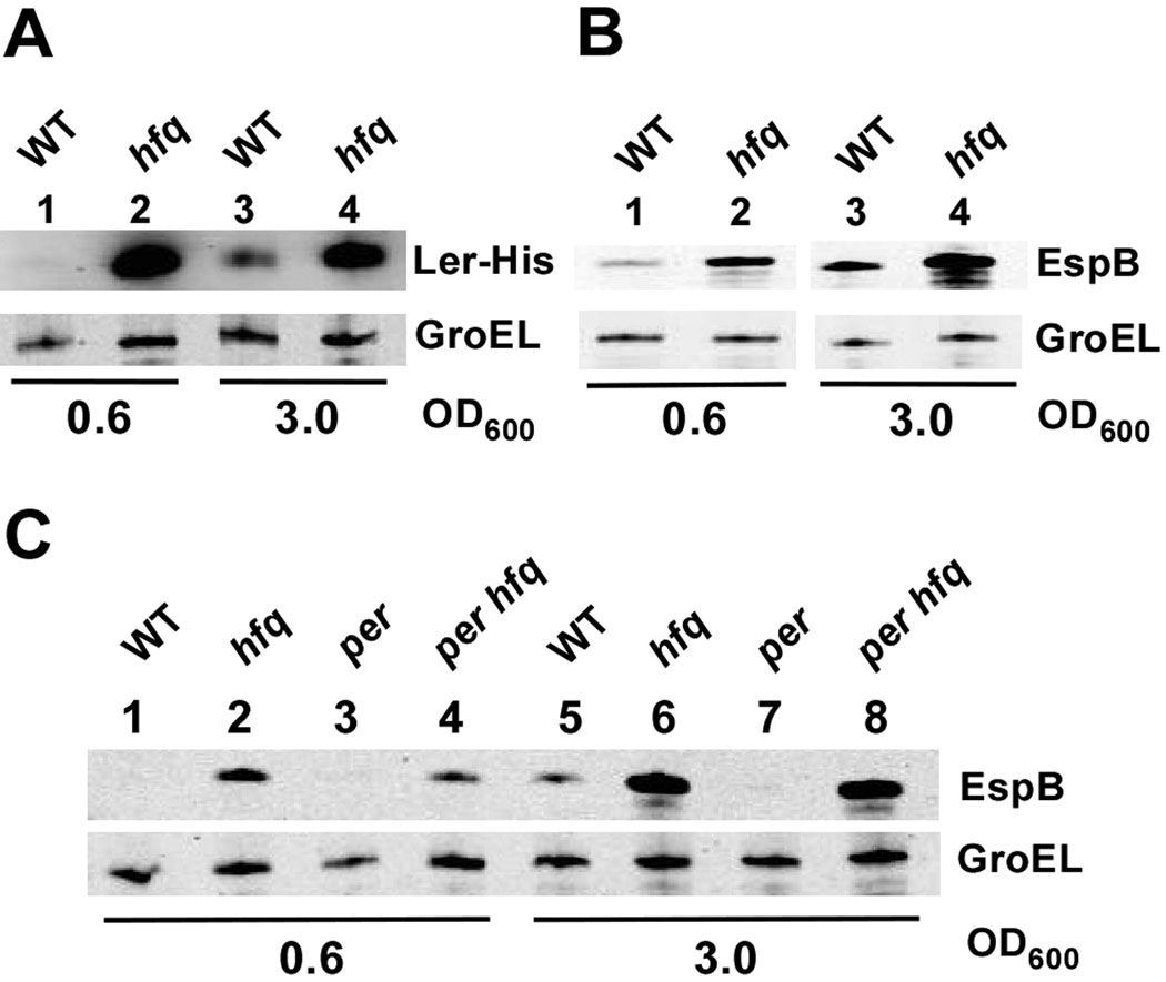
Because transcriptional regulation of LEE1 in EHEC and EPEC is similar (Mellies et al., 2007), Hfq is likely to affect LEE expression in EPEC as well. To investigate whether Hfq also negatively modulates LEE expression in EPEC, we deleted hfq in EPEC strain E2348/69 using lambda-red-mediated recombination as described in Experimental procedures. Western blot analyses revealed increased expression of EPEC Ler-His from pAMH104 in exponential and stationary phase cells of the hfq mutant compared to wild type (Fig. 4A, compare lanes 1 and 3 with 2 and 4). Hence, levels of EspB in the hfq mutant were also increased (Fig. 4B, lanes 2 and 4). Among the multiple transcriptional activators described for EPEC LEE1 is PerC, which is encoded by the perABC operon located on the EPEC adherence factor plasmid pEAF (Mellies et al., 1999; Porter et al., 2004). To test whether Hfq affects LEE expression in EPEC through regulation of per, we deleted hfq in the E2348/69 per mutant derivative, JPN15, and examined the expression of EspB by Western blot analyses. Results revealed a similar increase in EspB levels in hfq per double and hfq single mutants (Fig. 4C, compare lanes 2 and 6 with 4 and 8), indicating that the abundant LEE expression in a hfq mutant mostly occurs independent of per. However, EspB levels in exponential phase cells of the hfq per double mutant were three-fold lower than in the hfq mutant (Fig. 4C, lanes 2 and 4), yet significantly increased relative to the per single mutant (Fig. 4C, lane 3). Thus, we cannot rule out the possibility that Hfq in part affects EPEC LEE expression by indirectly affecting per expression. Previous work from our lab demonstrated that transcription of perC is indirectly negatively regulated by the acid resistance regulator GadX (Shin et al., 2001), which inhibits the expression of the per activator PerA and thereby PerC under low pH conditions (Shin et al., 2001; Porter et al., 2004). Since the Hfq-associated small RNA GadY positively regulates gadX mRNA stability (Opdyke et al., 2004), Hfq most likely affects EPEC LEE expression at low pH in conjunction with GadY by indirectly affecting perA, and thereby perC expression, through post-transcriptional regulation of gadX. However, we did not address the effect of Hfq during growth at low pH as it was beyond the scope of this study.
Figure 4.
Hfq affects LEE expression in EPEC E2348/69. Wild type and hfq mutant strains grown in LB at 37°C to OD600 ~ 0.6 (lanes 1–2) and 3.0 (lanes 3–4) were assayed for Ler-His expressed from pAMH104 (A) and EspB (B) by Western blot analyses using His and EspB specific antibodies, respectively. (C) Hfq affects LEE expression independent of per. Western blot analyses to measure EspB levels in E2348/69, E2348/69 hfq, JPN15 (per) and JPN15 hfq strains, grown in LB at 37°C to OD600 ~ 0.6 (lanes 1–4) and OD600 ~ 3.0 (lanes 5–8), were performed using antibodies specific to EspB and GroEL.
Hfq negatively controls LEE expression through grlRA
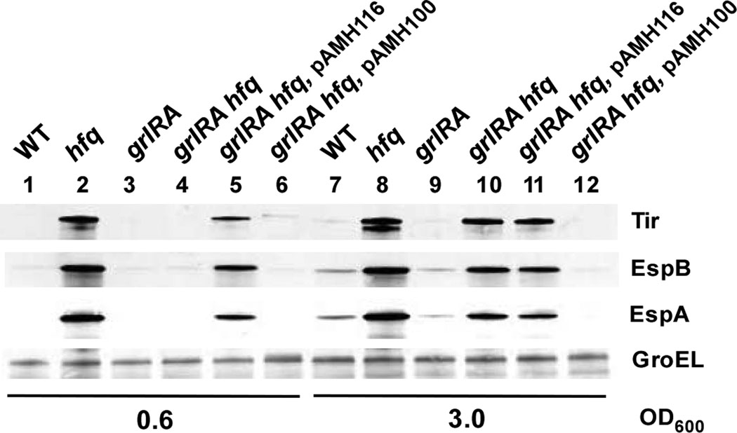
Besides Ler, the LEE encodes at least three regulators (GrlA, GrlR and Mcp) that affect LEE expression (Deng et al., 2004; Tsai et al., 2006). Whereas Mcp affects Ler function, GrlA and GrlR regulate LEE1 transcription. Since GrlA controls the spatiotemporal transcription of LEE-encoded genes by optimizing LEE1 transcription, and thus Ler expression (Barba et al., 2005), we raised the question of whether Hfq mediates the regulation of LEE expression through GrlA and GrlR. To address this issue, we deleted the grlRA operon in EHEC TUV93-0, and subsequently introduced an in-frame deletion of hfq creating a grlRA hfq double mutant. To determine the role of grlRA in the Hfq-mediated regulation of LEE, we examined protein levels of Tir, EspA and EspB in exponential (Fig. 5, lanes 1–6) and stationary phase cells (Fig. 5, lanes 7–12). Interestingly, the prematurely increased expression of LEE-encoded proteins in the hfq mutant (Fig. 5, lane 2) was absent in the grlRA hfq double mutant in exponential phase (Fig. 5, lane 4), indicating that grlRA is epistatic to hfq in regulating LEE expression. As expected, complementing the grlRA hfq mutant with grlRA from pAMH116 restored LEE protein levels nearly to that of the hfq mutant (Fig. 5, lane 5), whereas complementation with either hfq from pAMH100 or the empty vector did not affect protein levels (Fig. 5, lane 6; data not shown). Moreover, complementing the grlRA hfq mutant with grlA expressed in trans from an arabinose inducible promoter was sufficient to restore LEE expression to levels of a hfq mutant (data not shown), suggesting that the effect of Hfq on LEE expression in exponential phase is mediated by grlA. Interestingly, the effect of grlA on the Hfq-mediated regulation of LEE expression was abolished in stationary phase cells, as LEE protein levels in the grlRA hfq mutant were increased to the levels of a hfq mutant (Fig. 5, compare lanes 8 and 10). Complementation of stationary phase grlRA hfq cells with grlRA from pAMH116 did not affect levels of LEE-encoded proteins (Fig. 5, compare lanes 10 and 11), whereas complementation with hfq from pAMH100 decreased protein levels to that of the grlRA mutant as anticipated (Fig. 5, compare lanes 9 and 12). Furthermore, qRT-PCR analyses revealed a 44-fold (44 ± 0.7, P < 0.0007) increase in ler transcript levels in a hfq mutant relative to a hfq grlRA mutant in exponential phase, whereas ler transcription was unaffected in stationary phase (0.75 ± 0.17, P = 0.08), indicating that grlRA affects ler transcript levels primarily in exponential phase. These results indicate that the Hfq-mediated regulation of LEE expression through grlRA occurs specifically in exponential phase, and that Hfq affects LEE expression in stationary phase independent of grlRA.
Figure 5.
Hfq negatively regulates levels of LEE-encoded proteins through grlRA. The effect of grlRA on LEE expression was measured by Western blot analyses of TUV93-0 wild type, a hfq mutant, a grlRA mutant, a hfq grlRA double mutant and a hfq grlRA double mutant strain complemented with grlRA (pAMH116) and hfq (pAMH100) grown in LB to exponential (OD600 ~ 0.6, lanes 1–6) and stationary phase (OD600 ~ 3.0, lanes 7–12). Levels of Tir, EspA, EspB and GroEL were detected using polyclonal antibodies against the respective proteins. GroEL served as an internal control for total protein levels.
The increased levels of EspA, EspB and Tir in the grlRA hfq mutant in stationary phase could be due to either increased transcript abundance or protein stability. To determine whether the grlRA-independent effect of Hfq on LEE expression in stationary phase was due to increased transcription, we used qRT-PCR to compare espB transcript levels in exponential and stationary phase cells of the hfq grlRA mutant. Results revealed a seven-fold (7 ± 1.5, P < 0.01) increase in espB transcript levels in stationary compared to exponential phase, indicating that increased EspB protein levels in stationary phase cells of the hfq grlA mutant are due to increased espB transcript abundance. Furthermore, we measured levels of ler transcript in exponential and stationary phase cells of the hfq grlA mutant using qRT-PCR, and found that ler transcript levels were five-fold (5 ± 0.7, P < 0.0001) increased in the hfq grlRA mutant in stationary relative to exponential phase. Thus, increased levels of ler transcripts in stationary phase, followed by an increase in LEE transcription, could account for the observed increase in LEE-encoded proteins in stationary phase cells of the hfq grlRA mutant (Fig. 5, lane 10). To test whether RpoS is involved in the Hfq-mediated regulation of LEE expression in stationary phase, we expressed RpoS in trans from an arabinose inducible promoter in the hfq mutant to levels comparable with that of the wild type strain, and determined levels of EspB and Tir by Western blot analyses. Results revealed increased levels of EspB and Tir in the hfq mutant regardless of the presence of RpoS (data not shown), suggesting that RpoS is not implicated in the Hfq-mediated regulation of LEE expression in stationary phase.
Hfq regulates grlRA expression independent of Ler
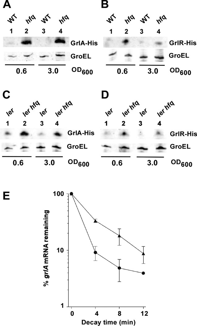
The increased expression of LEE in exponential phase hfq mutant cells was absent in the hfq grlRA double mutant, raising the question of whether Hfq affects expression of grlRA. To investigate this, we used Western analyses to examine protein levels of His-tagged GrlA (pAMH117) and GrlR (pAMH118) in TUV93-0 wild type and hfq mutant backgrounds. Both exponential and stationary phase cells of the hfq mutant expressed significantly increased levels of GrlA-His (Fig. 6A, lanes 2 and 4) and GrlR-His (Fig. 6B, lanes 2 and 4) compared to wild type, indicating that Hfq negatively affects GrlA and GrlR protein levels. Likewise, Western analyses revealed increased levels of GrlA-His in a hfq mutant of EPEC strain E2348/69 grown to exponential and stationary phases (data not shown), suggesting a similar regulatory mechanism by Hfq in these EHEC and EPEC backgrounds.
Figure 6.
Hfq affects grlRA expression independent of Ler. The expression of GrlA (A and C) and GrlR (B and D) was determined in TUV93-0 wild type and hfq backgrounds in the presence (A and B) and in the absence (C and D) of ler by Western analyses using a His specific antibody. TUV93-0 wild type, hfq and ler mutant strains, harboring plasmids encoding grlRA-his (pAMH117) and grlR-his (pAMH118), were grown in LB at 37°C to exponential (A–D lanes 1–2) and stationary phases (A–D lanes 3–4). His-tagged GrlR and GrlA proteins were detected by Western blot analyses using a His specific antibody. Detection of GroEL served as an internal control. (E) Hfq affects grlA mRNA stability. Wild type TUV93-0 and hfq mutant strains were grown in LB at 37°C to exponential phase (OD600 ~ 0.6), transcription was blocked by rifampicin (0 min) and aliquots were collected at the time indicated, and total RNA was extracted as described in Experimental procedures. Analysis of grlA mRNA stability was carried out using qRT-PCR to measure the amount of grlA transcript normalized to levels of 16S rRNA. Data represent the mean ± SD percentage of grlA transcript remaining in three independent RNA samples isolated from wild type (circles) and the hfq mutant (triangles) at the time indicated after rifampicin addition relative to time 0. The percentage of transcript remaining for each time point was significantly different in RNA isolated from wild type and hfq mutant strains as determined by the t-test (P < 0.01). Half-lives of grlA mRNA in wild type and hfq mutant strains were calculated from the lin-log graph to 1.5 min and 3 min, respectively.
Ler-independent expression of grlRA from its native promoter as reflected by transcriptional activation of EHEC LEE was observed in E. coli K-12 (Russell et al., 2007). This along with our finding that Hfq negatively regulated expression of LEE1 in the absence of ler (Fig. 3C) raised the question of whether Hfq regulates grlRA independent of Ler. To address this, we examined the expression of His-tagged GrlA and GrlR in ler and ler hfq mutant strains by Western blot analyses. Results revealed an increased expression of GrlA-His (Fig. 6C, lanes 2 and 4) and GrlR-His (Fig. 6D, lanes 2 and 4) in the hfq mutant background in the absence of ler. Thus, Hfq affects LEE expression in exponential phase through negative regulation of GrlA and GrlR levels independent of Ler.
Hfq controls grlRA transcript abundance post-transcriptionally by affecting messenger stability
To examine if increased protein levels of GrlA and GrlR in the hfq mutant was due to increased transcript levels, we used qRT-PCR to quantify the amount of grlA and grlR transcripts in total RNA isolated from wild type and hfq mutant strains grown to exponential phase in LB. Indeed, levels of grlA and grlR transcripts were increased in the hfq mutant compared to wild type by sixteen-fold (16 ± 1, P < 0.003) and eleven-fold (11 ± 1, P < 0.001), respectively. The increase in grlA transcript levels was less severe with a five-fold (5 ± 1, P < 0.001) increase in the hfq ler double mutant compared to the ler mutant. This might be due to the absence of a Ler-GrlA positive regulatory loop in the hfq ler background. The abundance of grlRA transcript in the hfq mutant could be due to either increased transcription or increased messenger stability. First, we determined whether the increased grlRA transcript level in a hfq mutant was due to increased transcription from the grlRA operon promoter. To this end, we measured the level of transcription from the grlRA promoter in exponentially grown grlRA and grlRA hfq mutant strains, which have the coding region of grlRA replaced with a scar sequence that was introduced upon deletion, by qRT-PCR analyses using oligos specific to the grlRA leader region and the scar sequence, respectively, as described in Experimental procedures. Transcriptional activity from the grlRA promoter was similar in the grlRA and grlRA hfq mutant backgrounds, as reflected by a relative fold difference in transcript levels of about one (1.2 ± 0.1, P = 0.2), indicating that Hfq more likely affects grlRA messenger stability. Second, to determine grlRA mRNA stability in wild type and hfq mutant strains, we grew the strains in LB to exponential phase, then treated cells with rifampicin to stop additional transcription, and extracted RNA at various times thereafter. The amount of grlA transcript remaining at 0, 4, 8 and 12 minutes upon transcription inhibition was measured by qRT-PCR using oligos specific to grlA. The grlA messenger was more stable in the hfq mutant, with 33% of transcripts remaining compared to 9% in the wild type, after four minutes of rifampicin exposure (Fig. 6E). The 16S rRNA levels remained unchanged in both strains at all time points tested (data not shown). The increased stability of the grlA messenger in the hfq mutant was reflected by a two-fold longer RNA half-life compared to the wild type. Since Northern blot analyses of total RNA using probes specific to grlR, grlA and grlRA revealed similar RNA banding patterns for RNA isolated from wild type and hfq mutant strains (data not shown), the stability of the grlA transcript in a hfq mutant likely also reflects that of the grlR transcript, which is apparent as grlR and grlA are in an operon (Barba et al., 2005). Thus, Hfq controls grlRA expression post-transcriptionally by negatively modulating messenger stability.
Hfq-mediated regulation of grlRA requires a factor located outside of the LEE
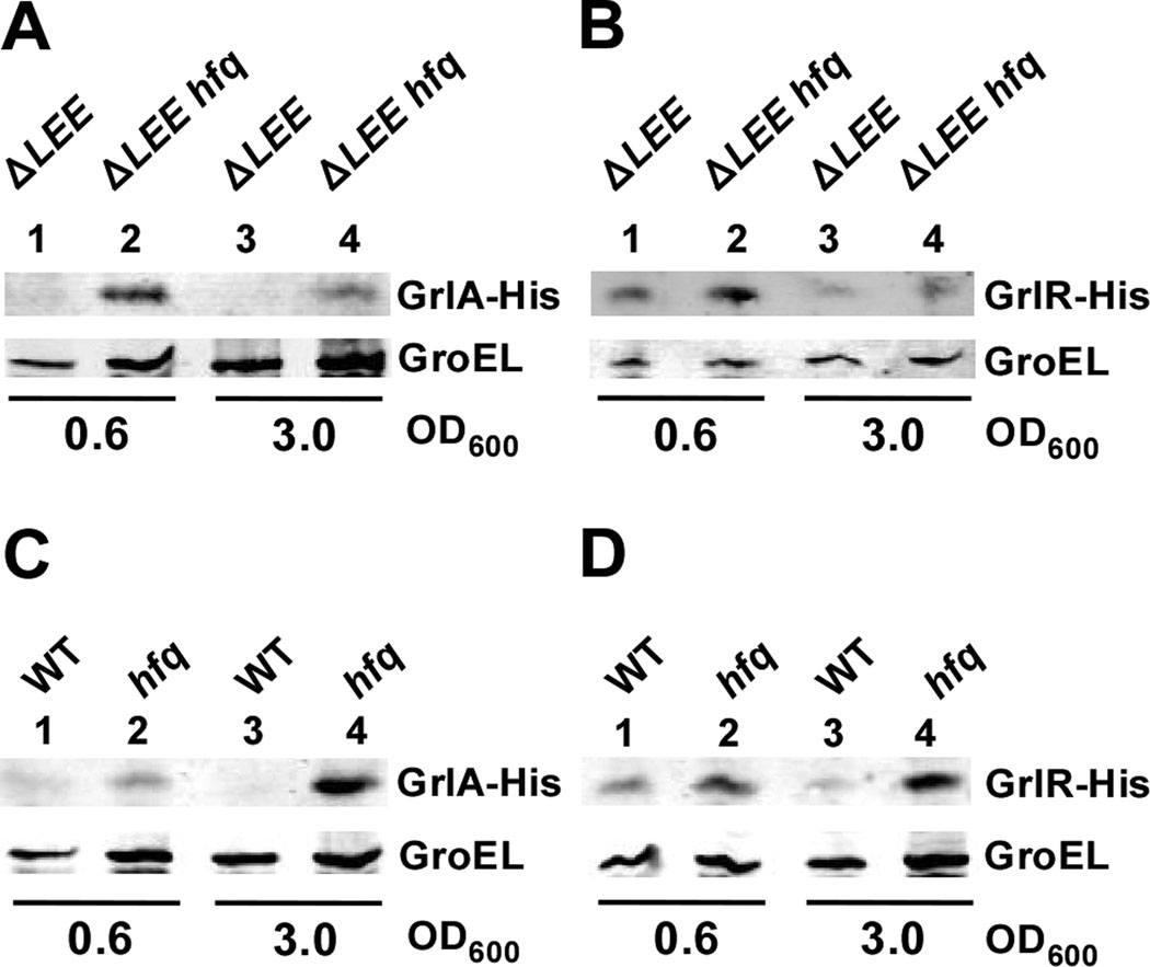
Hfq often affects transcript stability in concerted action with sRNAs. A potential factor, such as a sRNA, mediating the Hfq-dependent regulation of grlRA is either located on a pathogenicity island or on the K-12 backbone of the EHEC chromosome. To test whether a potential sRNA is located outside of the LEE pathogenicity island, we constructed a hfq mutant derivate of a LEE-deleted EHEC EDL933 strain and measured the expression of GrlA and GrlR from pAMH125 and pAMH126, respectively, in ΔLEE and ΔLEE hfq backgrounds. Western analysis revealed increased levels of GrlA and GrlR in the ΔLEE hfq mutant relative to the ΔLEE strain especially in exponential phase (Fig. 7A and B, lanes 2 and 4), suggesting that a potential sRNA is located outside of the LEE either on another pathogenicity island or on the K-12 backbone of the chromosome. The LEE operons are expressed in E. coli K-12 (Mellies et al., 1999), raising the question of whether hfq also influences grlRA expression in this background. To address this, we examined the expression of GrlA and GrlR from pAMH117 and pAMH118, respectively, in wild type and hfq mutant derivatives of the E. coli K-12 strain MG1655. Western analyses revealed increased expression of both GrlA and GrlR in the MG1655 hfq mutant strain compared to wild type (Fig. 7C and D, lanes 2 and 4), indicating that a factor(s) such as a sRNA that potentially is required for the Hfq-mediated regulation of grlRA is located on the K-12 backbone of the chromosome rather than on an EHEC specific genetic island.
Figure 7.
The Hfq-mediated regulation of grlRA requires a factor that is located outside the LEE. Western blot analyses were used to detect levels of GrlA (A and C) and GrlR (B and D) in ΔLEE and ΔLEE hfq mutant derivates of EHEC EDL933 (A and B) and wild type and hfq mutant backgrounds of E. coli K-12 MG1655 (C and D) grown in LB at 37°C to OD600 ~ 0.6 (lanes 1–2) and 3.0 (lanes 3–4). His-tagged GrlR and GrlA were expressed from pAMH117/pAMH118 and pAMH125/pAMH126 in K-12 and EHEC backgrounds, respectively. His-tagged proteins were detected using a His specific antibody as described in Experimental procedures. GroEL served as an internal control for total protein levels.
Hfq controls the timing of A/E lesion formation
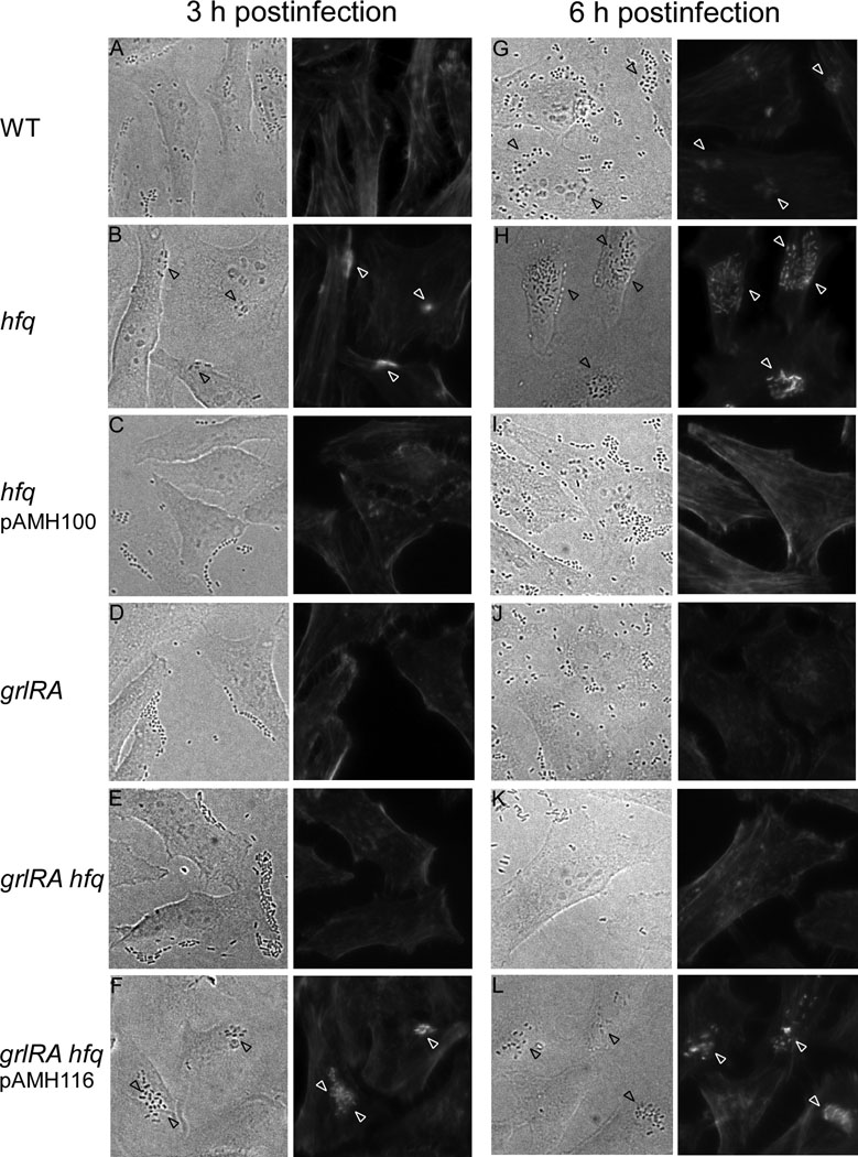
The expression of the LEE-encoded factors is required for EHEC to trigger actin polymerization, and thus form A/E lesions on the eukaroytic epithelial cell. Since the hfq mutant strain prematurely expresses LEE-encoded proteins at increased levels in a grlRA-dependent manner, we predicted that Hfq would affect the kinetics of A/E lesion formation. To characterize the effect of Hfq on the ability of EHEC to form A/E lesions in vitro, we infected HeLa cells with wild type, hfq, grlRA and hfq grlRA mutant derivatives of strain TUV93-0 for 2, 3, 4 and 6 h. We then determined the presence of A/E lesions using the fluorescent actin staining (FAS) test, where the A/E lesions are visualized as condensed actin beneath the adherent bacteria on infected cells which actin filaments were FITC-phalloidin stained. Figure 8 shows phase-contrast and fluorescence actin images representing FAS assays at 3h and 6 h postinfection. The A/E lesion phenotype of wild type TUV 93-0 was diminished in exponential (3 h postinfection) compared to stationary phase (6 h postinfection) (Fig. 8, panels A and G), which is consistent with a previous study (Campellone et al., 2007). The hfq mutant formed A/E lesions prematurely at 3 h postinfection compared to wild type and the complemented mutant (Fig. 8, panels A–C). The premature A/E lesion phenotype was diminished in a hfq grlRA double mutant and restored when complementing with grlRA from pAMH116 (Fig. 8, panels E–F), which is consistent with Hfq affecting LEE expression through grlRA (Fig. 5). Similar results were obtained for FAS assays carried out 2 h postinfection (data not shown). At 6 h postinfection both wild type and hfq mutant strains formed A/E lesions with the hfq mutant forming a greater number of A/E lesions (Fig. 8, panels G–H). The absence of A/E lesions upon infection with the complemented hfq mutant could be due to a two-fold increase in Hfq levels expressed from the mid-copy number plasmid pAMH100 (Fig. 8, panel I). Consistent with data from 3 h postinfection, the enhanced A/E lesion phenotype of the hfq mutant at 6 h postinfection was diminished in a hfq grlRA double mutant and partially restored in a grlRA-complemented hfq grlRA mutant (Fig. 8, panels, K–L). Interestingly, regardless of the increased LEE expression in stationary phase (Fig. 5, lane 10), the hfq grlRA mutant was only capable of forming A/E lesions at 6 h postinfection, when complemented with grlRA. It was previous reported that GrlA-dependent repression of the flagellar operon through flhD is crucial for bacterial adhesion to HeLa cells (Iyoda et al., 2006). Thus, the inability of the hfq grlRA mutant to form A/E lesions at 6 h postinfection could be due to elevated flhD expression, leading to decreased cell adhesion of this mutant. Altogether, the regulatory effect of Hfq on LEE expression correlates with the A/E phenotype of a hfq mutant strain.
Figure 8.
Hfq affects the A/E phenotype through grlRA. FAS test using HeLa cells infected for 3 h (A–F) and 6 h (G–L) with TUV93-0, TUV93-0 hfq, TUV93-0 hfq pAMH100, TUV93-0 grlRA, TUV93-0 grlRA hfq and TUV93-0 grlRA hfq pAMH116. The actin cytoskeleton of infected HeLa cells was stained with FITC-phalloidin. Representative phase-contrast (left panels) and fluorescence actin (right panels) images are shown. Black and white arrowheads indicate bacteria and A/E lesions, respectively.
Discussion
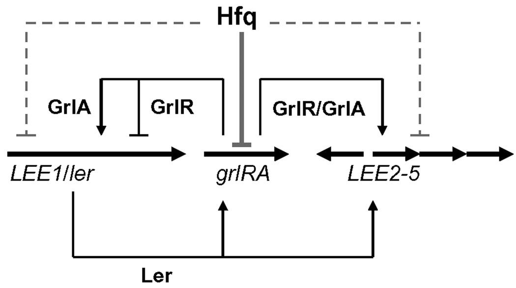
Coordinating gene expression in response to environmental stimuli is important for the bacterial pathogen to survive in hostile environments in the host and to evade the host immune response, thereby successfully colonizing the host intestine. Consequently, virulence specific regulators often are subject to both transcriptional and post-transcriptional control through complex regulatory networks that integrate environmental signals. The global regulator Ler coordinates virulence gene expression to ensure optimal fitness of bacteria during infection by differentially regulating expression of the LEE-encoded TTSS and a group four capsule to ensure transient shielding of intimin and the TTSS early in infection (Shifrin et al., 2008). Furthermore, the GrlR-GrlA regulatory system inversely controls the expression of the TTSS, enterohaemolysin and flagella (Iyoda et al., 2006; Saitoh et al., 2008). Whereas the complex regulatory network controlling the induction of LEE1/ler expression in response to environmental signals has been extensively studied, the regulatory basis for grlRA expression is less defined. Our study on the effect of the RNA chaperone Hfq on EHEC virulence gene expression revealed an important role of Hfq in controlling LEE expression. We demonstrated that Hfq negatively affects LEE expression in exponential phase through post-transcriptional regulation of grlRA, and in stationary phase independent of the GrlR-GrlA regulatory pathway as depicted in our current model of Hfq-mediated regulation of the LEE (Fig. 9). We further showed that the regulatory effect of Hfq on LEE is reflected by a premature and enhanced A/E histopathological phenotype of a hfq mutant strain.
Figure 9.
Model depicting the regulation of LEE by Hfq, GrlA, GrlR and Ler. Black arrows and T-lines represent positive and negative regulation, respectively. Gray solid and broken T-lines indicate Hfq-mediated regulation in exponential and stationary phase, respectively. Ler is a positive regulator of LEE2-5 and grlRA expression, whereas GrlA and GrlR activate and repress LEE1 transcription, respectively, as described in the introduction. Moreover, GrlA and GrlR positively affect transcription of LEE2 and LEE4 independent of Ler (Russell et al., 2007). Hfq negatively affects grlRA expression by modulating transcript stability to prevent expression of GrlA and GrlR, and thereby the initiation of the Ler-GrlA positive regulatory loop and subsequent LEE expression during exponential growth. In addition, Hfq negatively affects LEE4 and LEE5 expression independent of grlRA in stationary phase. To emphasis the impact of Hfq on LEE expression, the regulation of LEE by additional regulators is excluded from this model (for a detailed model of LEE regulation see Mellies et al., 2007).
Many virulence genes acquired by horizontal transfer, including LEE island-encoded genes (Perna et al., 1998), are silenced by H-NS (Deng et al., 2001; Navarre et al., 2006). H-NS levels change during growth with high levels present in exponential phase followed by decreased levels in stationary phase, consequently, the expression of the H-NS regulon including the LEE is growth phase-dependent (Ali et al., 1999; Roe et al., 2003; Hansen et al., 2005). Counter silencing by DNA-binding H-NS paralogues such as Ler, is among the mechanisms by which H-NS-mediated repression is overcome (reviewed by Stoebel et al., 2008). Thus, increased Ler levels are most likely required to antagonize H-NS-mediated repression for LEE expression to occur in exponential phase. We demonstrated that the requirement of grlRA for LEE expression to occur in a hfq mutant during exponential growth is diminished in stationary phase, suggesting that GrlA is primarily required for the induction of ler expression during conditions when H-NS levels are increased. Indeed, a recent study showed that GrlA positively controls ler expression in a H-NS-dependent pathway (Laaberki et al., 2006). The Hfq-mediated control of grlRA expression occurs independent of ler (Fig. 6C and D), indicating that induction of grlRA expression occurs independent from the Ler-GrlA positive regulatory pathway. Thus, expression of GrlA is most likely crucial for initiating the Ler-GrlA positive regulatory loop.
In line with the finding that Hfq prevents the induction of LEE expression in exponential phase through grlRA, we showed that Hfq negatively regulates grlRA transcript abundance and consequently GrlR and GrlA protein levels (Fig. 6A and B). We demonstrated that the grlA transcript during exponential phase is more stable in a hfq mutant compared to wild type (Fig. 6E), indicating that Hfq affects grlRA expression on the post-transcriptional level by destabilizing grlRA mRNA. Hfq-mediated destabilization of mRNAs often involves the concerted action with sRNAs most of which promote target mRNA decay (Aiba, 2007; Repoila and Darfeuille, 2009). However, whether Hfq regulation of grlRA transcript stability involves a sRNA has yet to be determined. While Hfq acts as an adaptor between the sRNAs and RNaseE, the sRNAs recruit the degradosome to target mRNAs, thereby promoting rapid degradation of mRNA (Morita et al., 2005). We showed that Hfq also negatively affects GrlA and GrlR levels in an E. coli K-12 background (Fig. 7), suggesting that a sRNA, which along with Hfq potentially mediates the decay of grlRA, is encoded by the K-12 backbone of the EHEC and EPEC chromosomes. Interstingly, a previous study identified a negative regulatory cis-acting element, located between positions +143 to +397 relative to the transcription initiation site of grlR, which in addition to H-NS negatively affects grlRA expression (Barba et al., 2005). This element could provide a potential target for post-transcriptional regulation by a sRNA. Alternatively, Hfq also affects messenger stability by regulating polyadenylation-dependent messenger decay (Mohanty et al., 2004). We are currently investigating whether the Hfq-mediated destabilization of grlRA mRNA involves a sRNA.
Regulation by Hfq in conjunction with sRNAs is an important means to control temporal expression of virulence-specific regulators by bacterial pathogens including Vibrio cholerae and Salmonella enterica Typhimurium, where Hfq and sRNAs affect levels of the virulence gene master regulator HapR (Lenz et al., 2004) and the major activator of the SPI-1-encoded TTSS and effector genes HilA (Sittka et al., 2007), respectively. Here we show, to our knowledge for the first time, that Hfq controls the temporal expression of virulence genes in EHEC and EPEC through post-transcriptional control of the LEE-encoded regulators GrlA and GrlR. We showed that LEE expression in exponential phase is negatively regulated by the Hfq-mediated regulation of GrlA and GrlR (Fig. 5 and Fig. 6). This suggests that a signal inducing GrlA expression during exponential growth is required to relieve the Hfq-mediated repression of grlRA, for LEE expression to occur in a growth phase-independent manner. Interestingly, qRT-PCR analyses revealed similar levels of grlRA mRNA in exponential and stationary phases (1.5 ± 0.1, P = 0.04), suggesting that a pool of grlRA mRNA, available throughout growth, enables cells to rapidly synthesize Grl proteins in response to environmental signals whenever needed. It has yet to be determined whether certain environmental conditions or the host induces such a signal, which is expected to negatively affect the abundance of a putative sRNA that could cause the destabilization of the grlRA messenger. LEE expression is activated by quorum sensing via the signaling molecules AI-3, epinephrine and norepinephrine, which signal through the QseEF two-component system to direct transcriptional activation of LEE1 by QseA (Sperandio et al., 1999; 2003; Walters and Sperandio, 2006; Sharp and Sperandio, 2007). Although Western blot analyses of EspB expression in a hfq grlRA mutant revealed that qseA is not epistatic to hfq in regulating LEE expression (data not shown), we cannot exclude the possibility that quorum sensing provides a signal to relieve the Hfq-mediated decay of grlRA mRNA. In addition to controlling grlRA mRNA stability in exponential phase (Fig. 6E), we showed that Hfq negatively regulates LEE expression in stationary phase by affecting ler transcript abundance and thereby Ler levels independent of grlRA (Fig. 5, data not shown). However, the mechanism behind Hfq-mediated regulation of ler in stationary phase has yet to be resolved. Moreover, Hfq might be involved in the post-transcriptional regulation of Tir and EspA, which is mediated by non-LEE-encoded factors and results in heterogeneous expression within an EHEC cell population (Roe et al., 2003; 2004).
We showed that Hfq negatively controls levels of GrlA in exponential phase to ensure proper temporal expression of the LEE. By regulating GrlA, Hfq in turn affects expression of the two global regulators, Ler and FlhD, important in coordinating virulence and flagellar gene expression, respectively (Iyoda et al., 2006; Abe et al., 2008). The important impact of Hfq on the virulence potential of EHEC is reflected by the following phenotypes of the hfq mutant: (i) The hfq mutant of EHEC has decreased motility (Table 1), which most likely is caused by increased GrlA levels (Iyoda et al., 2006) and might reduce the ability of the mutant to penetrate the intestinal mucus layer to colonize the host. (ii) The hfq mutant expresses strongly reduced RpoS levels leading to vulnerability to a variety of stress conditions including acid stress (Table 1), which diminishes its ability to survive passage through the acidic gastric barrier (Lin et al., 1995; Price et al., 2000). (iii) The hfq mutant expresses the virulence-associated regulators Ler, GrlR and GrlA prematurely at increased levels (Fig. 3 and Fig. 6), thereby not only disrupting proper temporal expression of virulence genes but also expending energy unnecessarily on protein synthesis (Fig. 1). Untimely expression of the TTSS during infection may expose the bacteria to the host immune system (Shifrin et al., 2008). Thus, despite an increased ability to form A/E lesions on HeLa cells, the hfq mutant is expected to exhibit strongly reduced virulence due to the inability to appropriately coordinate the expression of stress response and virulence genes. In summary, our data establish a role of Hfq in virulence gene regulation of EHEC, where through negative post-transcriptional regulation of grlRA expression, Hfq ensures repression of LEE expression during exponential growth, and yet provides a means to induce grlRA and subsequently LEE expression if necessary. We are currently investigating the mechanism by which Hfq destabilizes the grlRA messenger with emphasis on identifying a potential Hfq-associated sRNA involved in the decay of grlRA mRNA.
Experimental procedures
Standard procedures
Standard DNA techniques, liquid media and agar plates were used as described (Sambrook and Russell, 2001). Restriction endonucleases, T4 DNA kinase and ligase were used as recommended by the manufacturer (New England Biolabs). DNA used for cloning and recombination purposes was PCR amplified using the high-fidelity DNA polymerases PfuUltra™ II Fusion HS or Easy-A™ (Stratagene). The University of Maryland Biopolymer Core Facility performed primer synthesis and DNA sequencing. Bacterial techniques were carried out as described (Miller, 1972). Bacteria were grown at 37°C in LB or DMEM (Invitrogen #11965) media supplemented with ampicillin (100 µg/ml), chloramphenicol (cm) (30 µg/ml), kanamycin (kan) (50 µg/ml) or tetracycline (20 µg/ml) as needed. HeLa cells (ATTC #CCL-2) were cultured in DMEM/F12 (Invitrogen #11330) supplemented with 10% fetal bovine serum (FBS), 100 U/ml penicillin and 100 µg/ml streptomycin at 37°C in 5% CO2.
Bacterial strain constructions
Bacterial strains used in this study are listed in Table 1. Gene deletions in E. coli K-12 MG1655, EHEC TUV93-0, EDL933 ΔLEE, EPEC E2348/69 and JPN15 were constructed using Lambda Red-mediated recombination using linear DNA fragments as described (Datsenko and Wanner, 2000; Murphy and Campellone, 2003). To generate deletions in hfq and grlRA, DNA fragments encoding a kanamycin resistance cassette flanked by Flipase Recognition Target (FRT) site-inverted repeats and sequences homologous to regions flanking the gene of interest, were PCR amplified from pKD13 using primer sets K4928/K5045 and K5663/K5664, respectively (oligonucleotides are listed in Table 3). The DNA fragments were electroporated into appropriate target strains, where the Red recombinase system was expressed from arabinose and IPTG inducible promoters on pKD46 and pKM201, respectively, to allow recombination into the chromosome. Recombinants were selected and purified on LB agar plates containing kanamycin at 37°C. The kanamycin resistance marker was eliminated from recombinant strains using a pCP20-encoded FLP flipase, which acts specifically on FRT sites when expressed at 42°C. The plasmids pKD46, pKM201 and pCP20 encode temperature-sensitive replicons and were cured by growing cells at 37°C or above. Strain EDL933 ΔLEE was deleted for hfq by recombining a DNA fragment, which was PCR amplified from TUV93-0 Δhfq::kan genomic DNA (gDNA) using primers K5445/K5446, into EDL933 ΔLEE expressing the Red recombinase system from pTP223. The resulting strains deleted for hfq (AMH100, AMH109-112, and AMH115) or grlRA (AMH108) carried deletions of 303 and 828 nt starting from the translation initiation codon, respectively, which in place of the disrupted gene was proceeded by an 81 nt-long in-frame scar sequence that encodes a 27 amino-acid residue peptide. The kanamycin cassette was retained in hfq derivatives of EPEC and EDL933 ΔLEE (AMH110-111 and AMH115). Deletion mutants with ler replaced by an in-frame kanamycin cassette were generated by recombining a DNA fragment, amplified from SE1101 gDNA with primers K1372/1420, into TUV93-0 and AMH100, creating strains AMH101 and AMH102, respectively. To generate the double mutant strains AMH102 and AMH109, an hfq deletion was introduced into the strains as described above. Gene deletions of hfq, ler and grlRA were verified by PCR amplification analysis using the respective primer sets K4785/4786, K1372/K1420 and K5665/K5666. In addition, hfq deletions were confirmed by Western blot analysis using an Hfq-specific polyclonal antibody (Kindly provided by Ding J. Jin).
TABLE 3.
Oligonucleotides used in this study.
| Name | Oligo sequence (5’ to 3’) |
|---|---|
| K1372 | CCGGAATTCCTGTAACTCGAATTAAGTAGAGT |
| K1420 | CGCGGATCCAGCTCACGTTATCGTTATCATT |
| K4785 | GAGACGTATCGTGCGCAAT |
| K4786 | GAACGCAGGATCGCTGGCT |
| K4797 | GCACCGGGATCCTCAGTGATGGTGATGGTGATGAATATTTTTCAGCGGTA TTATTTCTTCTT |
| K4928 | ACGCAGGATCGCTGGCTCCCCGTGTAAAAAAAACAGCCCGAAACCATTG TGTAGGCTGGAGCTGCTTCG |
| K4980 | GTCAGGATCCAGTAAGGCGAGCCTGCCTAAGG |
| K4981 | GTCAGAAGCTTGAACGCAGGATCGCTGGCT |
| K5045 | ATCGAAAGGTTCAAAGTACAAATAAGCATATAAGGA AAAGAGAGAATGTCCGGGGATCCGTCGACCT |
| K5377 | TGTGTAGGCTGGAGCTGCT |
| K5378 | CATATGAATATCCTCCTTAGTTCC |
| K5373 | CTTTTTTCAAATGTGTAAAATACATTATCA |
| K5374 | CTTAAAATATTAAAGCATGCGGAGAT |
| K5359 | CACATACAACAAGTCCATACATTC |
| K5360 | CGAGTCCATCATCAGGCACATTA |
| K5361 | GCAACCGCTGCTTTGGTTGG |
| K5362 | CGACATCAGCAACACTTTCCG |
| K5365 | TGCAAGTCGAACGGTAACAG |
| K5366 | AGTTATCCCCCTCCATCAGG |
| K5425 | GCACCGGGATCCTCAGTGATGGTGATGGTGATGAAACAATTCATCCAGT AAAATATAATATTTTATTTTC |
| K5445 | GATCTCGAGCGAAACAGTAGGAACGTCGGCTTTG |
| K5446 | CTAGAGCTCGGCAGAGCAAGGTTGGGAGTCATT |
| K5653 | GCGACTATGTAATTCCTGACTCAGT |
| K5656 | CCATAACTAACATATCTGACACGAAATG |
| K5663 | GAAAGGAGTGAGGTTAGTATGAAACTGAGTGAGTT ATGATTATGTCCGGGGATCCGTCGACCTGC |
| K5664 | GCTTTATTTTTATTCTTCTATAAAATATACTCAAAAAATTACGTCTAGTGT AGGCTGGAGCTGCTTCGA |
| K5665 | CACCGAAGCTTGCAACAGATATGTTTATACAGGCATCAT |
| K5666 | GCACCGGGATCCACTGGTAGATTTATCTACAGGTG |
| K5667 | GCACCGGGATCCTAGTGATGGTGATGGTGATG ACTCTCCTTTTTCCGCCTCATGATC |
| K5822 | GCACCGGGATCCTTAGTGATGGTGATGGTGATGTTTTAAATAAACTTGTG GCATTCCTGTGTTTT |
| K5831 | AGTGCTCGTTTTTCCCTTGA |
| K5832 | AGCGAAGAACTTTTGCCTCA |
| K5833 | CTCAGTGGATGAGAAGACAGGAG |
| K5834 | CCGCAGTAAGAGTACCTAGTACG |
| K5930 | GATGTTCCTGGACTTCCTGTAA |
| K5931 | CAACAGAGGTCTCAACACCATT |
| K6021 | GGTGGTTGTTTGATGAAATAGATGTGTC |
| K6022 | GCTATTAGCGACCTTATCAGGAAG |
| K6038 | GGTCTCCATTATTCTTGATATTGCTTATG |
| K6039 | GGAACTTCGAACTGCAGGTCG |
| K6046 | GCAATGAAGACTCCTGTGGGGA |
| K6047 | CATGAAGTATGATGTCCTCATCTTCA |
| K6088 | GCATCATCCCTTACCGTGGTTC |
| K6089 | GGATCTGCTCTGTGGTGTAGTTCA |
Plasmid constructions
Oligonucleotides used for cloning purposes are listed in Table 3.
pAMH100
A 1378 bp DNA fragment encoding hfq was PCR amplified from TUV93-0 gDNA using primers K4980/K4981, digested with BamHI and HindIII and cloned into the corresponding sites of pBR322. pAMH100 encodes hfq expressed from its three native promoters P1–3 and was used to complement a hfq mutant.
pAMH103 and pAMH104
The 796 bp DNA fragments encoding His-tagged EHEC and EPEC Ler expressed from native ler promoters were PCR amplified from TUV93-0 and E2348/69 gDNA, respectively, using primers K1372/K4797 and cloned into the EcoRI/BamHI sites of pBR322.
pAMH105
A plasmid encoding ler-his expressed from a modified ler promoter with ~120 nt of the ler leader sequence including the proximal ler promoter deleted, was obtained by combining two DNA fragments. The two fragments encode a 237 bp region upstream of the transcription initiation site of the distal ler promoter and a 425 bp region encoding the region downstream of the transcription initiation site of the proximal ler promoter comprising ler-his, respectively. The DNA fragments were PCR amplified from TUV93-0 gDNA using primer sets K1372/K5373 and K1420/K5374, phosphorylated using T4 polynucleotide kinase, respectively digested with EcoRI and BamHI, and cloned into the corresponding sites of pBR322.
pAMH113
A 856 bp fragment encoding kan-his under the control of the ler promoter was PCR amplified from SE1101 gDNA using primers K1372/K5425 and cloned into the EcoRI/BamHI sites of pBR322.
pAMH116
A 1601 bp DNA fragment encoding grlRA and the native promoter was PCR amplified from TUV93-0 gDNA using primers K5665/K5666 and cloned into the BamHI/HindIII sites of pBR322.
pAMH117
A 1337 bp DNA fragment encoding grlRA-his and the grlRA promoter region was amplified from TUV93-0 gDNA with primers K5665/K5667 and cloned into the BamHI/HindIII sites of pBR322.
pAMH118
A 869 bp DNA fragment encoding grlR-his proceeded by the grlRA promoter region was amplified from TUV93-0 gDNA using primers K5665/K5822 and cloned into the BamHI/HindIII sites of pBR322.
pAMH125
A 1032 bp DNA fragment encoding a cm resistance gene was amplified from pKD3 using primers K5377/K5378 and cloned into the MscI site of pAMH117.
pAMH126
A 1032 bp DNA fragment encoding a cm resistance gene was amplified from pKD3 using primers K5377/K5378 and cloned into the MscI site of pAMH118.
Assays for strain phenotype determinations
Motility assay
The motility of TUV93-0 pBR322, AMH100 pBR322 and AMH100 pAMH100 was determined as described (Bertin et al., 1994). Semi-solid tryptone plates (1% bactotryptone, 0.5% NaCl, 0.3% bactoagar, pH 7.4) containing ampicillin were inoculated with a drop of 1 X 10⁹ cells of each strain grown in LB medium at 37°C for 14 h. The motility of the strains was measured after incubation at 37°C for 10 h by measuring the diameter of the swarming ring.
Acid resistance assay
Stationary phase-induced acid resistance was determined for the TUV93-0 and hfq mutant strains carrying pBR322 or pAMH100 as described (Masuda and Church, 2002). Stationary phase cultures (18 h) grown at 37°C in LB medium containing ampicillin were diluted 1:1000 in warm LB, adjusted to pH 2.5 with HCl and incubated for 1 h at 37°C. Serial dilutions of cells in 1X phosphate-buffered saline (PBS, pH 7.4) before and after acid exposure were plated onto LB agar plates and incubated overnight at 37°C. Cell survival was measured as the percentage of colony forming units remaining after exposure to low pH compared to before exposure. Each assay was carried out independently at least three times.
Protein secretion
Overnight cultures of TUV93-0, AMH100 and AMH100 pAMH100 were diluted 1:1000 in DMEM and grown aerobically at 37°C. Culture supernatants were collected at the optical cell densities at 600 nm (OD600) values 0.2 and 1.0 by centrifugation followed by passage though a 0.22-µm pore size Millex GP filter (Millipore). Secreted proteins were precipitated with 20% (vol/vol) trichloric acid and 5% (vol/vol) deoxycholic acid for 15 min on ice, collected by centrifugation (16,100×g for 10 min at 4°C), and resuspended in 2X SDS Blue loading buffer (New England Biolabs) with 0.5 M Tris base added. Protein samples corresponding to 300 µl of culture supernatant were boiled for 5 min and analyzed by SDS-12% polyacrylamide gel electrophoresis (SDS-PAGE) along with a Pageruler™ Prestained protein Ladder Plus molecular weight marker (Fermentas). Proteins were visualized using the SilverQuest™ silver staining kit (Invitrogen) as recommended by the manufacturer.
Western analysis
Total protein was prepared from cultures grown in LB or DMEM to the optical cell densities indicated in the figure legends. Overnight cultures were diluted 1:1000 in medium with appropriate antibiotics added to strains carrying plasmids and grown aerobically at 37°C. Total protein was precipitated from cell culture samples with 5% (vol/vol) trichloric acid for 15 min on ice, collected by centrifugation at 16,100×g for 10 min at 4°C, and washed with 80% acetone. Precipitated protein was resuspended in 1X SDS Blue loading buffer (New England Biolabs) in volumes normalized to the optical densities of cultures and boiled for 5 min. Protein sample amounts equivalent to 0.03 OD600 units of culture were resolved by SDS-PAGE along with a MagicMark™ XP western protein standard (Invitrogen) using 10% NextGel (Amresco) and Criterion precast 4–20% Tris-HCl protein gels (BioRad) according to the manufacturers instructions. Proteins were transferred onto Immobilon-FL polyvinylidene difluoride membranes (Millipore) using a Trans-Blot SD Semi-Dry Transfer Cell (BioRad). Membranes were blocked overnight at 4°C with 5% non-fat drymilk in 1X PBS containing 0.1% Tween-20 (PBST). Polyclonal antibodies produced in rabbits against EspA, EspB, Tir, GroEL (Sigma) and Hfq (Sledjeski et al., 2001) were used diluted 1:6000 in 1X PBST. A monoclonal Tetra-His™ anti-mouse antibody (QIAGEN) was used in a 1:2000 dilution to detect His-tagged proteins. Alexa Fluor 680-conjugated goat anti-rabbit and Alexa Fluor 680-conjugated goat anti-mouse diluted 1:10000 in 1X PBST were used as secondary antibodies. Proteins were visualized and quantified using an Odyssey Infrared Imaging System with application software version 2.1 (Li-Cor) as recommended.
RNA isolation and qRT-PCR analysis
RNA isolation
Cultures of TUV93-0 and mutant derivatives were grown aerobically in LB at 37°C to OD600 values of 0.6 and 3.0. RNA used to determine grlA mRNA stability was isolated from cultures grown in LB at 37°C to an OD600 value of 0.6 (0 min) and 4, 8 and 12 min after rifampicin (250 µg/ml) addition. Total RNA was isolated from culture samples corresponding to ~ 2.3×10¹⁰ cells using hot phenol extraction as described (Dam Mikkelsen and Gerdes, 1997). Contaminating DNA in the RNA preparations was removed by treating three times with TURBO™ DNase (Ambion). RNA samples were quantified based on absorption at 260 nm. The integrity of RNA samples was verified by determining the ratio of absorption at 260 and 280 nm and by agarose gel electrophoresis. Removal of contaminating DNA in the RNA samples was confirmed by PCR analysis using oligos specific to rrsB (K5365/K5366, Table 3).
Quantitative Real-Time PCR
Quantitative Real-Time reverse transcription-PCR (qRT-PCR) was performed in a one-step reaction using the Superscript III™ Platinium SYBR Green One-Step qRT-PCR Kit (Invitrogen) and a Chromo 4™ real-time detector attached to a PTC-200 DNA engine base (MJ Research/BioRad). The qRT-PCR reactions contained 100 ng of total RNA in 25 µl reactions, prepared as described by the manufacturer, and was carried out using the following conditions for cDNA synthesis and amplification: 1 cycle at 50°C for 3 min, 1 cycle at 95°C for 5 min, and 40 cycles at 95°C for 15 s and 60°C for 30 sec. Transcripts encoding ler, sepZ, escV, espB, tir, grlA, grlR, rrsB, rpoB, the ler leader and the grlRA leader were detected using the primer pairs K5359/K5360, K5833/K5834, K5831/K5832, K5361/K5362, K5930/K5931, K5653/K5656, K6046/K6047, K5365/K5366, K6088/K6089, K6021/K6022 and K6038/K6039, respectively (Table 2). Template specificity for each primer pair was verified by melting curve analysis, where products were heated from 50°C to 95°C during monitoring of fluorescence. The qRT-PCR reaction was carried out in duplicates on RNA isolated from at least three independent cultures.
TABLE 2.
Strains and plasmids used in this study
| Strain or plasmid | Relevant characteristic(s) | Reference or source |
|---|---|---|
| Strains | ||
| TUV93-0 | EHEC O157:H7 strain EDL933Δstx1 Δstx2 | Donohue-Rolfe et al., 2000 |
| E2348/69 | Wild-type EPEC O127:H6 | Levine et al., 1978 |
| JPN15 | E2348/69 spontaneously cured of pMAR2 | Jerse et al., 1990 |
| MG1655 | Wild-type E. coli K-12 | Blattner et al., 1997 |
| EDL933 ΔLEE | EHEC O157:H7 EDL933 with LEE deleted | Posfai et al., 1997 |
| SE1101 | EHEC O157:H7 84–86 Δler::kan | Elliott et al., 2000 |
| AMH100 | TUV93-0 Δhfq | This study |
| AMH101 | TUV93-0 Δler::kan | This study |
| AMH102 | TUV93-0 Δhfq Δler::kan | This study |
| AMH108 | TUV93-0 ΔgrlRA | This study |
| AMH109 | TUV93-0 ΔgrlRA Δhfq | This study |
| AMH110 | E2348/69 Δhfq:kan | This study |
| AMH111 | JPN15 Δper Δhfq:kan | This study |
| AMH112 | MG1655 Δhfq | This study |
| AMH115 | EDL933 ΔLEE Δhfq:kan | This study |
| Plasmids | ||
| pBR322 | Medium-copy number cloning vector | Bolivar et al., 1977 |
| pCP20 | FLP-flipase expressing plasmid; repA101 (ts) PλR -FLP λ cI857 |
Cherepanov and Wackernagel, 1995 |
| pKD3 | FRT-flanked cm resistance cassette | Datsenko and Wanner, 2000 |
| pKD13 | FRT-flanked kan resistance cassette | Datsenko and Wanner, 2000 |
| pKD46 | λ Red recombinase expressing plasmid; repA101 (ts) ParaB-gam-bet-exo |
Datsenko and Wanner, 2000 |
| pKM201 | λ Red recombinase expressing plasmid; repA101 (ts) Ptac- gam-bet-exo |
Murphy and Campellone, 2003 |
| pTP223 | λ Red recombinase expressing plasmid; Plac- gam-bet-exo |
Poteete and Fenton, 1984 |
| pAMH100 | pBR322::hfqEHEC | This study |
| pAMH103 | pBR322::ler-hisEHEC | This study |
| pAMH104 | pBR322::ler-hisEPEC | This study |
| pAMH105 | pBR322::PlerΔPprox-ler-hisEHEC | This study |
| pAMH113 | pBR322::Pler(EHEC)-kan-his | This study |
| pAMH116 | pBR322::grlRAEHEC | This study |
| pAMH117 | pBR322::grlRA-hisEHEC | This study |
| pAMH118 | pBR322::grlR-hisEHEC | This study |
| pAMH125 | pAMH117::cm | This study |
| pAMH126 | pAMH118::cm | This study |
qRT-PCR data quantification and statistical analysis
Data were collected using the MJ Opticon Monitor analysis software version 3.1 (BioRad), normalized to transcript levels of rrsB (16S rRNA), and analyzed using the comparative critical cycle threshold (CT) method (Livak and Schmittgen, 2001). The expression levels of target genes in wild type and mutant derivatives were compared using the relative quantification method. Statistical significance between means was calculated using the unpaired t-test with a threshold P value of < 0.01. Relative expression levels are expressed as the means ± the standard deviation of the ΔΔCT value.
Fluorescent actin staining test
The ability of EHEC TUV93-0 and mutant derivatives to form A/E lesions on HeLa cell monolayers at 2, 3, 4 and 6 h post infection was evaluated using the fluorescent actin staining (FAS) test as described (Knutton et al., 1989). Bacterial strains were inoculated from single colonies on LB agar plates into tryptic soy broth (TSB), containing antibiotics as needed, and grown without aeration for 16–18 h at 37°C. Prior to infection, strains were diluted 1:5 into the infection medium (DMEM supplemented with 2% FBS and 0.5% mannose) and primed for infection by incubation at 37°C in 5% CO2 for 2 h. Semiconfuent HeLa cell monolayers were grown on glass coverslips, followed by addition of 2×10⁷ bacteria (4 h and 6 h infections) or 4×10⁷ bacteria (2 h and 3 h infections). Monolayers were fixed after 2, 3, 4 and 6 h of incubation with 4% formamide in PBS, washed three times with 1X PBS, permeabilized with 0.1% Triton X-100 in PBS, and stained with Alexa Fluor 488 phalloidin (Invitrogen) diluted 1:50. Coverslips were mounted on slides using Prolong Gold antifade reagent (Invitrogen) and the edges of the coverslip were sealed with cytoseal-60 (Richard-Allan Scientific). The FAS assay was carried out independently at least three times for each strain. The samples were visualized using an inverted Nikon TE-2000 microscope equipped with a 40X objective.
Acknowledgements
We thank John Leong for providing strain TUV93-0 and Ding J. Jin for the antiserum against Hfq. We thank Tamara Hilton for excellent technical assistance with tissue cell cultures and Wilfried Goetz at the Radiation Oncology Research Laboratory UMB SOM for allowing us the use of the microscope. We also thank Nathalie Basu, Jane Michalski and Susan Steyert for comments on the manuscript. This work was supported by NIH grants DK58957 and AI21657 to J.B.K.
References
- Abe H, Miyahara A, Oshima T, Tashiro K, Ogura Y, Kuhara S, et al. Global regulation by horizontally transferred regulators establishes the pathogenicity of Escherichia coli. DNA Res. 2008;15:25–38. doi: 10.1093/dnares/dsm033. [DOI] [PMC free article] [PubMed] [Google Scholar]
- Aiba H. Mechanism of RNA silencing by Hfq-binding small RNAs. Curr Opin Microbiol. 2007;10:134–139. doi: 10.1016/j.mib.2007.03.010. [DOI] [PubMed] [Google Scholar]
- Ali AT, Iwata A, Nishimura A, Ueda S, Ishihama A. Growth phase-dependent variation in protein composition of the Escherichia coli nucleoid. J Bacteriol. 1999;181:6361–6370. doi: 10.1128/jb.181.20.6361-6370.1999. [DOI] [PMC free article] [PubMed] [Google Scholar]
- Atlung T, Ingmer H. H-NS: a modulator of environmentally regulated gene expression. Mol Microbiol. 1997;24:7–17. doi: 10.1046/j.1365-2958.1997.3151679.x. [DOI] [PubMed] [Google Scholar]
- Attia AS, Sedillo JL, Wang W, Liu W, Brautigam CA, Winkler W, Hansen EJ. Moraxella catarrhalis expresses an unusual Hfq protein. Infect Immun. 2008;76:2520–2530. doi: 10.1128/IAI.01652-07. [DOI] [PMC free article] [PubMed] [Google Scholar]
- Barba J, Bustamante VH, Flores-Valdez MA, Deng W, Finlay BB, Puente JL. A positive regulatory loop controls expression of the locus of enterocyte effacement-encoded regulators Ler and GrlA. J Bacteriol. 2005;187:7918–7930. doi: 10.1128/JB.187.23.7918-7930.2005. [DOI] [PMC free article] [PubMed] [Google Scholar]
- Bertin P, Terao E, Lee EH, Lejeune P, Colson C, Danchin A, Collatz E. The H-NS protein is involved in the biogenesis of flagella in Escherichia coli. J Bacteriol. 1994;176:5537–5540. doi: 10.1128/jb.176.17.5537-5540.1994. [DOI] [PMC free article] [PubMed] [Google Scholar]
- Blattner FR, Plunkett G, III, Bloch CA, Perna NT, Burland V, Riley M, et al. The complete genome sequence of Escherichia coli K-12. Science. 1997;277:1453–1474. doi: 10.1126/science.277.5331.1453. [DOI] [PubMed] [Google Scholar]
- Bolivar F, Rodriguez RL, Greene PJ, Betlach MC, Heyneker HL, Boyer HW. Construction and characterization of new cloning vehicles. II. A multipurpose cloning system. Gene. 1977;2:95–113. [PubMed] [Google Scholar]
- Brennan RG, Link TM. Hfq structure, function and ligand binding. Curr Opin Microbiol. 2007;10:125–133. doi: 10.1016/j.mib.2007.03.015. [DOI] [PubMed] [Google Scholar]
- Brescia CC, Mikulecky PJ, Feig AL, Sledjeski DD. Identification of the Hfq-binding site on DsrA RNA: Hfq binds without altering DsrA secondary structure. RNA. 2003;9:33–43. doi: 10.1261/rna.2570803. [DOI] [PMC free article] [PubMed] [Google Scholar]
- Brown L, Elliott T. Efficient translation of the RpoS sigma factor in Salmonella typhimurium requires host factor I, an RNA-binding protein encoded by the hfq gene. J Bacteriol. 1996;178:3763–3770. doi: 10.1128/jb.178.13.3763-3770.1996. [DOI] [PMC free article] [PubMed] [Google Scholar]
- Bustamante VH, Santana FJ, Calva E, Puente JL. Transcriptional regulation of type III secretion genes in enteropathogenic Escherichia coli: Ler antagonizes H-NS-dependent repression. Mol Microbiol. 2001;39:664–678. doi: 10.1046/j.1365-2958.2001.02209.x. [DOI] [PubMed] [Google Scholar]
- Campellone KG, Roe AJ, Løbner-Olesen A, Murphy KC, Magoun L, Brady MJ, et al. Increased adherence and actin pedestal formation by dam-deficient enterohaemorrhagic Escherichia coli O157:H7. Mol Microbiol. 2007;63:1468–1481. doi: 10.1111/j.1365-2958.2007.05602.x. [DOI] [PubMed] [Google Scholar]
- Cherepanov PP, Wackernagel W. Gene disruption in Escherichia coli: TcR and KmR cassettes with the option of Flp-catalyzed excision of the antibiotic-resistance determinant. Gene. 1995;158:9–14. doi: 10.1016/0378-1119(95)00193-a. [DOI] [PubMed] [Google Scholar]
- Christiansen JK, Nielsen JS, Ebersbach T, Valentin-Hansen P, Søgaard-Andersen L, Kallipolitis BH. Identification of small Hfq-binding RNAs in Listeria monocytogenes. RNA. 2006;12:1383–1396. doi: 10.1261/rna.49706. [DOI] [PMC free article] [PubMed] [Google Scholar]
- Creasey EA, Delahay RM, Daniell SJ, Frankel G. Yeast two-hybrid system survey of interactions between LEE-encoded proteins of enteropathogenic Escherichia coli. Microbiology. 2003;149:2093–2106. doi: 10.1099/mic.0.26355-0. [DOI] [PubMed] [Google Scholar]
- Dam Mikkelsen N, Gerdes K. Sok antisense RNA from plasmid R1 is functionally inactivated by RNaseE and polyadenylated by poly(A) polymerase I. Mol Microbiol. 1997;26:311–320. doi: 10.1046/j.1365-2958.1997.5751936.x. [DOI] [PubMed] [Google Scholar]
- Datsenko KA, Wanner BL. One-step inactivation of chromosomal genes in Escherichia coli K-12 using PCR products. Proc Natl Acad Sci U S A. 2000;97:6640–6645. doi: 10.1073/pnas.120163297. [DOI] [PMC free article] [PubMed] [Google Scholar]
- Deng W, Li Y, Vallance BA, Finlay BB. Locus of enterocyte effacement from Citrobacter rodentium: sequence analysis and evidence for horizontal transfer among attaching and effacing pathogens. Infect Immun. 2001;69:6323–6335. doi: 10.1128/IAI.69.10.6323-6335.2001. [DOI] [PMC free article] [PubMed] [Google Scholar]
- Deng W, Puente JL, Gruenheid S, Li Y, Vallance BA, Vazquez A, et al. Dissecting virulence: systematic and functional analyses of a pathogenicity island. Proc Natl Acad Sci U S A. 2004;101:3597–3602. doi: 10.1073/pnas.0400326101. [DOI] [PMC free article] [PubMed] [Google Scholar]
- Ding Y, Davis BM, Waldor MK. Hfq is essential for Vibrio cholerae virulence and downregulates sigma expression. Mol Microbiol. 2004;53:345–354. doi: 10.1111/j.1365-2958.2004.04142.x. [DOI] [PubMed] [Google Scholar]
- Donohue-Rolfe A, Kondova I, Oswald S, Hutto D, Tzipori S. Escherichia coli O157:H7 strains that express Shiga toxin (Stx) 2 alone are more neurotropic for gnotobiotic piglets than are isotypes producing only Stx1 or both Stx1 and Stx2. J Infect Dis. 2000;181:1825–1829. doi: 10.1086/315421. [DOI] [PubMed] [Google Scholar]
- Elliott SJ, Sperandio V, Giron JA, Shin S, Mellies JL, Wainwright L, et al. The locus of enterocyte effacement (LEE)-encoded regulator controls expression of both LEE- and non-LEE-encoded virulence factors in enteropathogenic and enterohemorrhagic Escherichia coli. Infect Immun. 2000;68:6115–6126. doi: 10.1128/iai.68.11.6115-6126.2000. [DOI] [PMC free article] [PubMed] [Google Scholar]
- Elliott SJ, Wainwright LA, McDaniel TK, Jarvis KG, Deng YK, Lai LC, et al. The complete sequence of the locus of enterocyte effacement (LEE) from enteropathogenic Escherichia coli E2348/69. Mol Microbiol. 1998;28:1–4. doi: 10.1046/j.1365-2958.1998.00783.x. [DOI] [PubMed] [Google Scholar]
- Fantappiè L, Metruccio MM, Seib KL, Oriente F, Cartocci E, Ferlicca F, et al. The RNA chaperone Hfq is involved in stress response and virulence in Neisseria meningitidis and is a pleiotropic regulator of protein expression. Infect Immun. 2009;77:1842–1853. doi: 10.1128/IAI.01216-08. [DOI] [PMC free article] [PubMed] [Google Scholar]
- Frankel G, Phillips AD, Rosenshine I, Dougan G, Kaper JB, Knutton S. Enteropathogenic and enterohaemorrhagic Escherichia coli: more subversive elements. Mol Microbiol. 1998;30:911–921. doi: 10.1046/j.1365-2958.1998.01144.x. [DOI] [PubMed] [Google Scholar]
- Franze de Fernandez MT, Eoyang L, August JT. Factor fraction required for the synthesis of bacteriophage Qbeta-RNA. Nature. 1968;219:588–590. doi: 10.1038/219588a0. [DOI] [PubMed] [Google Scholar]
- Friedberg D, Umanski T, Fang Y, Rosenshine I. Hierarchy in the expression of the locus of enterocyte effacement genes of enteropathogenic Escherichia coli. Mol Microbiol. 1999;34:941–952. doi: 10.1046/j.1365-2958.1999.01655.x. [DOI] [PubMed] [Google Scholar]
- Garmendia J, Frankel G, Crepin VF. Enteropathogenic and enterohemorrhagic Escherichia coli infections: translocation, translocation, translocation. Infect Immun. 2005;73:2573–2585. doi: 10.1128/IAI.73.5.2573-2585.2005. [DOI] [PMC free article] [PubMed] [Google Scholar]
- Geissmann TA, Touati D. Hfq, a new chaperoning role: binding to messenger RNA determines access for small RNA regulator. EMBO J. 2004;23:396–405. doi: 10.1038/sj.emboj.7600058. [DOI] [PMC free article] [PubMed] [Google Scholar]
- Guisbert E, Rhodius VA, Ahuja N, Witkin E, Gross CA. Hfq modulates the σE-mediated envelope stress response and the σ32 -mediated cytoplasmic stress response in Escherichia coli. J Bacteriol. 2007;189:1963–1973. doi: 10.1128/JB.01243-06. [DOI] [PMC free article] [PubMed] [Google Scholar]
- Haack KR, Robinson CL, Miller KJ, Fowlkes JW, Mellies JL. Interaction of Ler at the LEE5 (tir) operon of enteropathogenic Escherichia coli. Infect Immun. 2003;71:384–392. doi: 10.1128/IAI.71.1.384-392.2003. [DOI] [PMC free article] [PubMed] [Google Scholar]
- Hansen AM, Qiu Y, Yeh N, Blattner FR, Durfee T, Jin DJ. SspA is required for acid resistance in stationary phase by downregulation of H-NS in Escherichia coli. Mol Microbiol. 2005;56:719–734. doi: 10.1111/j.1365-2958.2005.04567.x. [DOI] [PubMed] [Google Scholar]
- Hirakawa H, Kodama T, Takumi-Kobayashi A, Honda T, Yamaguchi A. Secreted indole serves as a signal for expression of type III secretion system translocators in enterohaemorrhagic Escherichia coli O157:H7. Microbiology. 2009;155:541–550. doi: 10.1099/mic.0.020420-0. [DOI] [PubMed] [Google Scholar]
- Huang LH, Syu WJ. GrlA of enterohemorrhagic Escherichia coli O157:H7 activates LEE1 by binding to the promoter region. J Microbiol Immunol Infect. 2008;41:9–16. [PubMed] [Google Scholar]
- Huys G, Cnockaert M, Janda JM, Swings J. Escherichia albertii sp. nov., a diarrhoeagenic species isolated from stool specimens of Bangladeshi children. Int J Syst Evol Microbiol. 2003;53:807–810. doi: 10.1099/ijs.0.02475-0. [DOI] [PubMed] [Google Scholar]
- Iyoda S, Koizumi N, Satou H, Lu Y, Saitoh T, Ohnishi M, Watanabe H. The GrlR-GrlA regulatory system coordinately controls the expression of flagellar and LEE-encoded type III protein secretion systems in enterohemorrhagic Escherichia coli. J Bacteriol. 2006;188:5682–5692. doi: 10.1128/JB.00352-06. [DOI] [PMC free article] [PubMed] [Google Scholar]
- Jerse AE, Yu J, Tall BD, Kaper JB. A genetic locus of enteropathogenic Escherichia coli necessary for the production of attaching and effacing lesions on tissue culture cells. Proc Natl Acad Sci U S A. 1990;87:7839–7843. doi: 10.1073/pnas.87.20.7839. [DOI] [PMC free article] [PubMed] [Google Scholar]
- Jobichen C, Li M, Yerushalmi G, Tan YW, Mok YK, Rosenshine I, et al. Structure of GrlR and the implication of its EDED motif in mediating the regulation of type III secretion system in EHEC. PLoS Pathog. 2007;3:e69. doi: 10.1371/journal.ppat.0030069. [DOI] [PMC free article] [PubMed] [Google Scholar]
- Kaper JB, Nataro JP, Mobley HL. Pathogenic Escherichia coli. Nat Rev Microbiol. 2004;2:123–140. doi: 10.1038/nrmicro818. [DOI] [PubMed] [Google Scholar]
- Kawamoto H, Koide Y, Morita T, Aiba H. Base-pairing requirement for RNA silencing by a bacterial small RNA and acceleration of duplex formation by Hfq. Mol Microbiol. 2006;61:1013–1022. doi: 10.1111/j.1365-2958.2006.05288.x. [DOI] [PubMed] [Google Scholar]
- Knutton S, Baldwin T, Williams PH, McNeish AS. Actin accumulation at sites of bacterial adhesion to tissue culture cells: basis of a new diagnostic test for enteropathogenic and enterohemorrhagic Escherichia coli. Infect Immun. 1989;57:1290–1298. doi: 10.1128/iai.57.4.1290-1298.1989. [DOI] [PMC free article] [PubMed] [Google Scholar]
- Kulesus RR, Diaz-Perez K, Slechta ES, Eto DS, Mulvey MA. Impact of the RNA chaperone Hfq on the fitness and virulence potential of uropathogenic Escherichia coli. Infect Immun. 2008;76:3019–3026. doi: 10.1128/IAI.00022-08. [DOI] [PMC free article] [PubMed] [Google Scholar]
- Laaberki MH, Janabi N, Oswald E, Repoila F. Concert of regulators to switch on LEE expression in enterohemorrhagic Escherichia coli O157:H7: interplay between Ler, GrlA, HNS and RpoS. Int J Med Microbiol. 2006;296:197–210. doi: 10.1016/j.ijmm.2006.02.017. [DOI] [PubMed] [Google Scholar]
- Lease RA, Belfort M. Riboregulation by DsrA RNA: trans-actions for global economy. Mol Microbiol. 2000;38:667–672. doi: 10.1046/j.1365-2958.2000.02162.x. [DOI] [PubMed] [Google Scholar]
- Lease RA, Cusick ME, Belfort M. Riboregulation in Escherichia coli: DsrA RNA acts by RNA:RNA interactions at multiple loci. Proc Natl Acad Sci U S A. 1998;95:12456–12461. doi: 10.1073/pnas.95.21.12456. [DOI] [PMC free article] [PubMed] [Google Scholar]
- Lenz DH, Mok KC, Lilley BN, Kulkarni RV, Wingreen NS, Bassler BL. The small RNA chaperone Hfq and multiple small RNAs control quorum sensing in Vibrio harveyi and Vibrio cholerae. Cell. 2004;118:69–82. doi: 10.1016/j.cell.2004.06.009. [DOI] [PubMed] [Google Scholar]
- Levine MM, Bergquist EJ, Nalin DR, Waterman DH, Hornick RB, Young CR, Sotman S. Escherichia coli strains that cause diarrhoea but do not produce heat-labile or heat-stable enterotoxins and are non-invasive. Lancet. 1978;1:1119–1122. doi: 10.1016/s0140-6736(78)90299-4. [DOI] [PubMed] [Google Scholar]
- Lin J, Lee IS, Frey J, Slonczewski JL, Foster JW. Comparative analysis of extreme acid survival in Salmonella typhimurium, Shigella flexneri, and Escherichia coli. J Bacteriol. 1995;177:4097–4104. doi: 10.1128/jb.177.14.4097-4104.1995. [DOI] [PMC free article] [PubMed] [Google Scholar]
- Lodato PB, Kaper JB. Post-transcriptional processing of the LEE4 operon in enterohaemorrhagic Escherichia coli. Mol Microbiol. 2008;71:273–290. doi: 10.1111/j.1365-2958.2008.06530.x. [DOI] [PMC free article] [PubMed] [Google Scholar]
- Livak KJ, Schmittgen TD. Analysis of relative gene expression data using real-time quantitative PCR and the 2−ΔΔCT method. Methods. 2001;25:402–408. doi: 10.1006/meth.2001.1262. [DOI] [PubMed] [Google Scholar]
- Majdalani N, Vanderpool CK, Gottesman S. Bacterial small RNA regulators. Crit Rev Biochem Mol Biol. 2005;40:93–113. doi: 10.1080/10409230590918702. [DOI] [PubMed] [Google Scholar]
- Masuda N, Church GM. Escherichia coli gene expression responsive to levels of the response regulator EvgA. J Bacteriol. 2002;184:6225–6234. doi: 10.1128/JB.184.22.6225-6234.2002. [DOI] [PMC free article] [PubMed] [Google Scholar]
- McDaniel TK, Jarvis KG, Donnenberg MS, Kaper JB. A genetic locus of enterocyte effacement conserved among diverse enterobacterial pathogens. Proc Natl Acad Sci U S A. 1995;92:1664–1668. doi: 10.1073/pnas.92.5.1664. [DOI] [PMC free article] [PubMed] [Google Scholar]
- McNealy TL, Forsbach-Birk V, Shi C, Marre R. The Hfq homolog in Legionella pneumophila demonstrates regulation by LetA and RpoS and interacts with the global regulator CsrA. J Bacteriol. 2005;187:1527–1532. doi: 10.1128/JB.187.4.1527-1532.2005. [DOI] [PMC free article] [PubMed] [Google Scholar]
- Meibom KL, Forslund AL, Kuoppa K, Alkhuder K, Dubail I, Dupuis M, et al. Hfq, a novel pleiotropic regulator of virulence-associated genes in Francisella tularensis. Infect Immun. 2009;77:1866–1880. doi: 10.1128/IAI.01496-08. [DOI] [PMC free article] [PubMed] [Google Scholar]
- Mellies JL, Barron AM, Carmona AM. Enteropathogenic and enterohemorrhagic Escherichia coli virulence gene regulation. Infect Immun. 2007;75:4199–4210. doi: 10.1128/IAI.01927-06. [DOI] [PMC free article] [PubMed] [Google Scholar]
- Mellies JL, Elliott SJ, Sperandio V, Donnenberg MS, Kaper JB. The Per regulon of enteropathogenic Escherichia coli: identification of a regulatory cascade and a novel transcriptional activator, the locus of enterocyte effacement (LEE)-encoded regulator (Ler) Mol Microbiol. 1999;33:296–306. doi: 10.1046/j.1365-2958.1999.01473.x. [DOI] [PubMed] [Google Scholar]
- Mellies JL, Larabee FJ, Zarr MA, Horback KL, Lorenzen E, Mavor D. Ler interdomain linker is essential for anti-silencing activity in enteropathogenic Escherichia coli. Microbiology. 2008;154:3624–3638. doi: 10.1099/mic.0.2008/023382-0. [DOI] [PMC free article] [PubMed] [Google Scholar]
- Miller JF. Experiments in Molecular Genetics. Cold Spring Harbor, NY: Cold Spring Harbor Laboratory Press; 1972. [Google Scholar]
- Mitobe J, Morita-Ishihara T, Ishihama A, Watanabe H. Involvement of RNA-binding protein Hfq in the post-transcriptional regulation of invE gene expression in Shigella sonnei. J Biol Chem. 2008;283:5738–5747. doi: 10.1074/jbc.M710108200. [DOI] [PubMed] [Google Scholar]
- Mohanty BK, Maples VF, Kushner SR. The Sm-like protein Hfq regulates polyadenylation dependent mRNA decay in Escherichia coli. Mol Microbiol. 2004;54:905–920. doi: 10.1111/j.1365-2958.2004.04337.x. [DOI] [PubMed] [Google Scholar]
- Moll I, Leitsch D, Steinhauser T, Bläsi U. RNA chaperone activity of the Sm-like Hfq protein. EMBO Rep. 2003;4:284–289. doi: 10.1038/sj.embor.embor772. [DOI] [PMC free article] [PubMed] [Google Scholar]
- Møller T, Franch T, Højrup P, Keene DR, Bächinger HP, Brennan RG, Valentin-Hansen P. Hfq: a bacterial Sm-like protein that mediates RNA-RNA interaction. Mol Cell. 2002;9:23–30. doi: 10.1016/s1097-2765(01)00436-1. [DOI] [PubMed] [Google Scholar]
- Morita T, Maki K, Aiba H. RNase E-based ribonucleoprotein complexes: mechanical basis of mRNA destabilization mediated by bacterial noncoding RNAs. Genes Dev. 2005;19:2176–2186. doi: 10.1101/gad.1330405. [DOI] [PMC free article] [PubMed] [Google Scholar]
- Muffler A, Fischer D, Hengge-Aronis R. The RNA-binding protein HF-I, known as a host factor for phage Qbeta RNA replication, is essential for rpoS translation in Escherichia coli. Genes Dev. 1996;10:1143–1151. doi: 10.1101/gad.10.9.1143. [DOI] [PubMed] [Google Scholar]
- Murphy KC, Campellone KG. Lambda Red-mediated recombinogenic engineering of enterohemorrhagic and enteropathogenic E. coli. BMC Mol Biol. 2003;4:11. doi: 10.1186/1471-2199-4-11. [DOI] [PMC free article] [PubMed] [Google Scholar]
- Nakao H, Watanabe H, Nakayama S, Takeda T. yst gene expression in Yersinia enterocolitica is positively regulated by a chromosomal region that is highly homologous to Escherichia coli host factor 1 gene (hfq) Mol Microbiol. 1995;18:859–865. doi: 10.1111/j.1365-2958.1995.18050859.x. [DOI] [PubMed] [Google Scholar]
- Nataro JP, Kaper JB. Diarrheagenic Escherichia coli. Clin Microbiol Rev. 1998;11:142–201. doi: 10.1128/cmr.11.1.142. [DOI] [PMC free article] [PubMed] [Google Scholar]
- Navarre WW, Porwollik S, Wang Y, McClelland M, Rosen H, Libby SJ, Fang FC. Selective silencing of foreign DNA with low GC content by the H-NS protein in Salmonella. Science. 2006;313:236–238. doi: 10.1126/science.1128794. [DOI] [PubMed] [Google Scholar]
- Opdyke JA, Kang JG, Storz G. GadY, a small-RNA regulator of acid response genes in Escherichia coli. J Bacteriol. 2004;186:6698–6705. doi: 10.1128/JB.186.20.6698-6705.2004. [DOI] [PMC free article] [PubMed] [Google Scholar]
- Perna NT, Mayhew GF, Posfai G, Elliott S, Donnenberg MS, Kaper JB, Blattner FR. Molecular evolution of a pathogenicity island from enterohemorrhagic Escherichia coli O157:H7. Infect Immun. 1998;66:3810–3817. doi: 10.1128/iai.66.8.3810-3817.1998. [DOI] [PMC free article] [PubMed] [Google Scholar]
- Poteete AR, Fenton AC. Lambda red-dependent growth and recombination of phage P22. Virology. 1984;15:161–167. doi: 10.1016/0042-6822(84)90281-2. [DOI] [PubMed] [Google Scholar]
- Porter ME, Mitchell P, Free A, Smith DG, Gally DL. The LEE1 promoters from both enteropathogenic and enterohemorrhagic Escherichia coli can be activated by PerC-like proteins from either organism. J Bacteriol. 2005;187:458–472. doi: 10.1128/JB.187.2.458-472.2005. [DOI] [PMC free article] [PubMed] [Google Scholar]
- Porter ME, Mitchell P, Roe AJ, Free A, Smith DG, Gally DL. Direct and indirect transcriptional activation of virulence genes by an AraC-like protein, PerA from enteropathogenic Escherichia coli. Mol Microbiol. 2004;54:1117–1133. doi: 10.1111/j.1365-2958.2004.04333.x. [DOI] [PubMed] [Google Scholar]
- Posfai G, Koob MD, Kirkpatrick HA, Blattner FR. Versatile insertion plasmids for targeted genome manipulations in bacteria: isolation, deletion, and rescue of the pathogenicity island LEE of the Escherichia coli O157:H7 genome. J Bacteriol. 1997;179:4426–4428. doi: 10.1128/jb.179.13.4426-4428.1997. [DOI] [PMC free article] [PubMed] [Google Scholar]
- Price SB, Cheng CM, Kaspar CW, Wright JC, DeGraves FJ, Penfound TA, et al. Role of rpoS in acid resistance and fecal shedding of Escherichia coli O157:H7. Appl Environ Microbiol. 2000;66:632–637. doi: 10.1128/aem.66.2.632-637.2000. [DOI] [PMC free article] [PubMed] [Google Scholar]
- Rasko DA, Rosovitz MJ, Myers GS, Mongodin EF, Fricke WF, Gajer P, et al. The pangenome structure of Escherichia coli: comparative genomic analysis of E. coli commensal and pathogenic isolates. J Bacteriol. 2008;190:6881–6893. doi: 10.1128/JB.00619-08. [DOI] [PMC free article] [PubMed] [Google Scholar]
- Repoila F, Darfeuille F. Small regulatory non-coding RNAs in bacteria: physiology and mechanistic aspects. Biol Cell. 2009;101:117–131. doi: 10.1042/BC20070137. [DOI] [PubMed] [Google Scholar]
- Repoila F, Gottesman S. Temperature sensing by the dsrA promoter. J Bacteriol. 2003;185:6609–6614. doi: 10.1128/JB.185.22.6609-6614.2003. [DOI] [PMC free article] [PubMed] [Google Scholar]
- Robertson GT, Roop RM., Jr The Brucella abortus host factor I (HF-I) protein contributes to stress resistance during stationary phase and is a major determinant of virulence in mice. Mol Microbiol. 1999;34:690–700. doi: 10.1046/j.1365-2958.1999.01629.x. [DOI] [PubMed] [Google Scholar]
- Roe AJ, Naylor SW, Spears KJ, Yull HM, Dransfield TA, Oxford M, et al. Coordinate single-cell expression of LEE4- and LEE5-encoded proteins of Escherichia coli O157:H7. Mol Microbiol. 2004;54:337–352. doi: 10.1111/j.1365-2958.2004.04277.x. [DOI] [PubMed] [Google Scholar]
- Roe AJ, Tysall L, Dransfield T, Wang D, Fraser-Pitt D, Mahajan A, et al. Analysis of the expression, regulation and export of NleA-E in Escherichia coli O157:H7. Microbiology. 2007;153:1350–1360. doi: 10.1099/mic.0.2006/003707-0. [DOI] [PubMed] [Google Scholar]
- Roe AJ, Yull H, Naylor SW, Woodward MJ, Smith DG, Gally DL. Heterogeneous surface expression of EspA translocon filaments by Escherichia coli O157:H7 is controlled at the posttranscriptional level. Infect Immun. 2003;71:5900–5909. doi: 10.1128/IAI.71.10.5900-5909.2003. [DOI] [PMC free article] [PubMed] [Google Scholar]
- Russell RM, Sharp FC, Rasko DA, Sperandio V. QseA and GrlR/GrlA regulation of the locus of enterocyte effacement genes in enterohemorrhagic Escherichia coli. J Bacteriol. 2007;189:5387–5392. doi: 10.1128/JB.00553-07. [DOI] [PMC free article] [PubMed] [Google Scholar]
- Saitoh T, Iyoda S, Yamamoto S, Lu Y, Shimuta K, Ohnishi M, et al. Transcription of the ehx enterohemolysin gene is positively regulated by GrlA, a global regulator encoded within the locus of enterocyte effacement in enterohemorrhagic Escherichia coli. J Bacteriol. 2008;190:4822–4830. doi: 10.1128/JB.00231-08. [DOI] [PMC free article] [PubMed] [Google Scholar]
- Sambrook J, Russell DW. Molecular cloning: A laboratory manual. 3rd edn. Cold Spring Harbor, NY: Cold Spring Harbor Laboratory Press; 2001. [Google Scholar]
- Sanchez-SanMartin C, Bustamante VH, Calva E, Puente JL. Transcriptional regulation of the orf19 gene and the tir-cesT-eae operon of enteropathogenic Escherichia coli. J Bacteriol. 2001;183:2823–2833. doi: 10.1128/JB.183.9.2823-2833.2001. [DOI] [PMC free article] [PubMed] [Google Scholar]
- Schumacher MA, Pearson RF, Møller T, Valentin-Hansen P, Brennan RG. Structures of the pleiotropic translational regulator Hfq and an Hfq-RNA complex: a bacterial Sm-like protein. EMBO J. 2002;21:3546–3556. doi: 10.1093/emboj/cdf322. [DOI] [PMC free article] [PubMed] [Google Scholar]
- Sharp FC, Sperandio V. QseA directly activates transcription of LEE1 in enterohemorrhagic Escherichia coli. Infect Immun. 2007;75:2432–2440. doi: 10.1128/IAI.02003-06. [DOI] [PMC free article] [PubMed] [Google Scholar]
- Shifrin Y, Peleg A, Ilan O, Nadler C, Kobi S, Baruch K, et al. Transient shielding of intimin and the type III secretion system of enterohemorrhagic and enteropathogenic Escherichia coli by a group 4 capsule. J Bacteriol. 2008;190:5063–5074. doi: 10.1128/JB.00440-08. [DOI] [PMC free article] [PubMed] [Google Scholar]
- Shin S, Castanie-Cornet MP, Foster JW, Crawford JA, Brinkley C, Kaper JB. An activator of glutamate decarboxylase genes regulates the expression of enteropathogenic Escherichia coli virulence genes through control of the plasmid-encoded regulator, Per. Mol Microbiol. 2001;41:1133–1150. doi: 10.1046/j.1365-2958.2001.02570.x. [DOI] [PubMed] [Google Scholar]
- Sittka A, Pfeiffer V, Tedin K, Vogel J. The RNA chaperone Hfq is essential for the virulence of Salmonella typhimurium. Mol Microbiol. 2007;63:193–217. doi: 10.1111/j.1365-2958.2006.05489.x. [DOI] [PMC free article] [PubMed] [Google Scholar]
- Sledjeski DD, Whitman C, Zhang A. Hfq is necessary for regulation by the untranslated RNA DsrA. J Bacteriol. 2001;183:1997–2005. doi: 10.1128/JB.183.6.1997-2005.2001. [DOI] [PMC free article] [PubMed] [Google Scholar]
- Sonnleitner E, Hagens S, Rosenau F, Wilhelm S, Habel A, Jäger KE, Bläsi U. Reduced virulence of a hfq mutant of Pseudomonas aeruginosa O1. Microb Pathog. 2003;35:217–228. doi: 10.1016/s0882-4010(03)00149-9. [DOI] [PubMed] [Google Scholar]
- Soper TJ, Woodson SA. The rpoS mRNA leader recruits Hfq to facilitate annealing with DsrA sRNA. RNA. 2008;14:1907–1917. doi: 10.1261/rna.1110608. [DOI] [PMC free article] [PubMed] [Google Scholar]
- Sperandio V, Li CC, Kaper JB. Quorum-sensing Escherichia coli regulator A: a regulator of the LysR family involved in the regulation of the locus of enterocyte effacement pathogenicity island in enterohemorrhagic E. coli. Infect Immun. 2002;70:3085–3093. doi: 10.1128/IAI.70.6.3085-3093.2002. [DOI] [PMC free article] [PubMed] [Google Scholar]
- Sperandio V, Mellies JL, Delahay RM, Frankel G, Crawford JA, Nguyen W, Kaper JB. Activation of enteropathogenic Escherichia coli (EPEC) LEE2 and LEE3 operons by Ler. Mol Microbiol. 2000;38:781–793. doi: 10.1046/j.1365-2958.2000.02168.x. [DOI] [PubMed] [Google Scholar]
- Sperandio V, Mellies JL, Nguyen W, Shin S, Kaper JB. Quorum sensing controls expression of the type III secretion gene transcription and protein secretion in enterohemorrhagic and enteropathogenic Escherichia coli. Proc Natl Acad Sci U S A. 1999;96:15196–15201. doi: 10.1073/pnas.96.26.15196. [DOI] [PMC free article] [PubMed] [Google Scholar]
- Sperandio V, Torres AG, Jarvis B, Nataro JP, Kaper JB. Bacteria-host communication: the language of hormones. Proc Natl Acad Sci U S A. 2003;100:8951–8956. doi: 10.1073/pnas.1537100100. [DOI] [PMC free article] [PubMed] [Google Scholar]
- Stoebel DM, Free A, Dorman CJ. Anti-silencing: overcoming H-NS-mediated repression of transcription in Gram-negative enteric bacteria. Microbiology. 2008;154:2533–2545. doi: 10.1099/mic.0.2008/020693-0. [DOI] [PubMed] [Google Scholar]
- Torres AG, Lopez-Sanchez GN, Milflores-Flores L, Patel SD, Rojas-Lopez M, Martinez de la Pena CF, et al. Ler and H-NS, regulators controlling expression of the long polar fimbriae of Escherichia coli O157:H7. J Bacteriol. 2007;189:5916–5928. doi: 10.1128/JB.00245-07. [DOI] [PMC free article] [PubMed] [Google Scholar]
- Tsai NP, Wu YC, Chen JW, Wu CF, Tzeng CM, Syu WJ. Multiple functions of l0036 in the regulation of the pathogenicity island of enterohaemorrhagic Escherichia coli O157:H7. Biochem J. 2006;393:591–599. doi: 10.1042/BJ20051201. [DOI] [PMC free article] [PubMed] [Google Scholar]
- Tsui HC, Leung HC, Winkler ME. Characterization of broadly pleiotropic phenotypes caused by an hfq insertion mutation in Escherichia coli K-12. Mol Microbiol. 1994;13:35–49. doi: 10.1111/j.1365-2958.1994.tb00400.x. [DOI] [PubMed] [Google Scholar]
- Umanski T, Rosenshine I, Friedberg D. Thermoregulated expression of virulence genes in enteropathogenic Escherichia coli. Microbiology. 2002;148:2735–2744. doi: 10.1099/00221287-148-9-2735. [DOI] [PubMed] [Google Scholar]
- Updegrove T, Wilf N, Sun X, Wartell RM. Effect of Hfq on RprA-rpoS mRNA pairing: Hfq-RNA binding and the influence of the 5' rpoS mRNA leader region. Biochemistry. 2008;47:11184–11195. doi: 10.1021/bi800479p. [DOI] [PubMed] [Google Scholar]
- Valentin-Hansen P, Eriksen M, Udesen C. The bacterial Sm-like protein Hfq: a key player in RNA transactions. Mol Microbiol. 2004;51:1525–1533. doi: 10.1111/j.1365-2958.2003.03935.x. [DOI] [PubMed] [Google Scholar]
- Valentin-Hansen P, Johansen J, Rasmussen AA. Small RNAs controlling outer membrane porins. Curr Opin Microbiol. 2007;10:152–155. doi: 10.1016/j.mib.2007.03.001. [DOI] [PubMed] [Google Scholar]
- Walters M, Sperandio V. Autoinducer 3 and epinephrine signaling in the kinetics of locus of enterocyte effacement gene expression in enterohemorrhagic Escherichia coli. Infect Immun. 2006;74:5445–5455. doi: 10.1128/IAI.00099-06. [DOI] [PMC free article] [PubMed] [Google Scholar]
- Waters LS, Storz G. Regulatory RNAs in bacteria. Cell. 2009;136:615–628. doi: 10.1016/j.cell.2009.01.043. [DOI] [PMC free article] [PubMed] [Google Scholar]
- Wilson JW, Ott CM, Honer zu BK, Ramamurthy R, Quick L, Porwollik S, et al. Space flight alters bacterial gene expression and virulence and reveals a role for global regulator Hfq. Proc Natl Acad Sci U S A. 2007;104:16299–16304. doi: 10.1073/pnas.0707155104. [DOI] [PMC free article] [PubMed] [Google Scholar]
- Yerushalmi G, Nadler C, Berdichevski T, Rosenshine I. Mutational analysis of the locus of enterocyte effacement-encoded regulator (Ler) of enteropathogenic Escherichia coli. J Bacteriol. 2008;190:7808–7818. doi: 10.1128/JB.00663-08. [DOI] [PMC free article] [PubMed] [Google Scholar]
- Zhang A, Wassarman KM, Ortega J, Steven AC, Storz G. The Sm-like Hfq protein increases OxyS RNA interaction with target mRNAs. Mol Cell. 2002;9:11–22. doi: 10.1016/s1097-2765(01)00437-3. [DOI] [PubMed] [Google Scholar]


