Abstract

Access to novel forms of (+)-saxitoxin (STX), a potent and selective inhibitor of voltage-gated Na+ ion channels, has been made possible through de novo synthesis. Saxitoxin is believed to lodge in the outer mouth of the channel pore, thereby stoppering ion flux. Herein, we demonstrate that modification of the C13-carbamoyl unit can be accommodated in the binding site of the protein without significantly reducing ligand-receptor affinity. These discoveries have emboldened efforts to prepare photoaffinity-labeled and other unique forms of STX as pharmacological tools for interrogating both the molecular architecture and function of Na+ channels. A synthetic plan is described that makes such compounds generally available.
Voltage-gated sodium ion channels (NaV) serve an obligatory role in the generation of bioelectricity and are essential for all of life’s processes.1 Genes that encode for ten unique NaV isoforms (NaV1.1–1.9, NaVX) have been identified in mammalian cells.2 Differences in the biophysical properties between these protein subtypes, their membrane concentrations and spatial distribution define the signaling characteristics of a neuron.3 Aberrant NaV function and/or expression is thought to be associated with numerous disease states, including arrhythmia, epilepsy, neuropathic pain, and congenital analgesia.4 Accordingly, chemical tools for exploring protein structure, for modulating the activity of specific NaV isoforms, and for tracking dynamic events associated with NaV regulation and expression are sought to further understand the pathophysiologies associated with channel function.5 Herein, we describe our initial efforts to develop such agents, for which the shellfish poison (+)-saxitoxin 1 (STX) – a single digit nanomolar inhibitor of certain NaV subtypes – provides the molecular blueprint (Figure 1).6 Our findings delineate a path for preparing novel carbamate-modified forms of the toxin and establish that such structural changes do not influence significantly substrate-receptor binding affinity.7
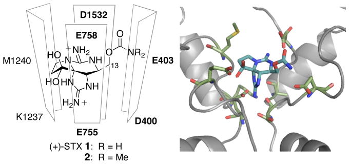
Figure 1.
A model of (+)-STX bound in the NaV channel pore.9b
The fully functional voltage-gated Na+ channel consists of a large heteromeric α-subunit (~260 kDa) and one or two auxiliary β-subunits (33–36 kDa).2 In the absence of protein crystallographic data, small molecule pharmacological probes together with protein mutagenesis and electrophysiology have provided much of the structural insights that currently exist for this family of macromolecules.8 These data together with X-ray structures of associated K+ ion channels, Kcsa and MthK, have made possible the construction of homology models of the NaV α-subunit.9 The outer mouth of the channel includes the ion selectivity filter and is considered to be the receptor site for STX and related guanidinium poisons (Figure 1).2 Five carboxylate residues line this pore region (D400, E755, E403, E758, D1532, NaV1.4 numbering); their presence is critical for high affinity STX binding, as shown by site-directed mutagenesis studies.8,9b,10 Computational models by Lipkind and Fozzard, Dudley, and Zhorov all posit that the 7,8,9-guanidine of STX points towards the ring of four amino acids that comprise the selectivity filter (also known as the DEKA loop).8,9 Specific contacts between the C13 carbamate unit, the C12-hydrated ketone, the 1,2,3-guanidinium moiety and the carboxylate residues of the outer vestibule loop (E403, E758, D1532) are also highlighted.
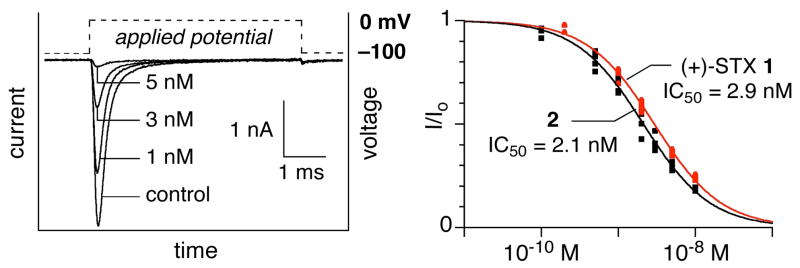
As a starting point for our investigations, we chose to examine the contribution of the C13 carbamate as a hydrogen-bond donor to the overall binding affinity of the toxin.8,9b Naturally occurring decarbamoyl STX (dc-STX) displays a modest reduction (< 10-fold) in potency relative to STX.6 Previous efforts to alter this functional group through semi-synthetic modification of dc-STX have been limited to a single succinate derivative.7a,c The availability of a de novo synthesis to STX (see below) remedies this problem. Accordingly, N,N-dimethyl-STX 2 has been prepared and evaluated for its ability to block Na+ current. Electrophysiology measurements are performed in a whole cell voltage-clamp format against the heterologously expressed α-subunit of the rat skeletal channel, NaV1.4 (CHO cell).11 Figure 2 shows current recordings following a 10 ms depolarizing pulse to 0 mV from a holding potential of −100 mV. Increasing concentrations of 2 are perfused into the external solution, resulting in decreased peak current. These data were fit to a Langmuir isotherm to give an IC50 of 2.1 ± 0.1 nM for 2, a value nearly equal to the IC50 recorded for our synthetic (+)-STX (2.9 ± 0.1 nM). This result seems to indicate that the role of the C13 carbamate in the natural product is not as a hydrogen-bond donor.9b Such a finding also provides the motivation for exploring further the steric environment of the protein pore in the vicinity of the carbamoyl residue.
Figure 2.
Current recordings at varying concentrations of 2 on rNaV1.4 expressed in CHO cells. Dose-response curves for (+)-STX 1 (red) and 2 (black).
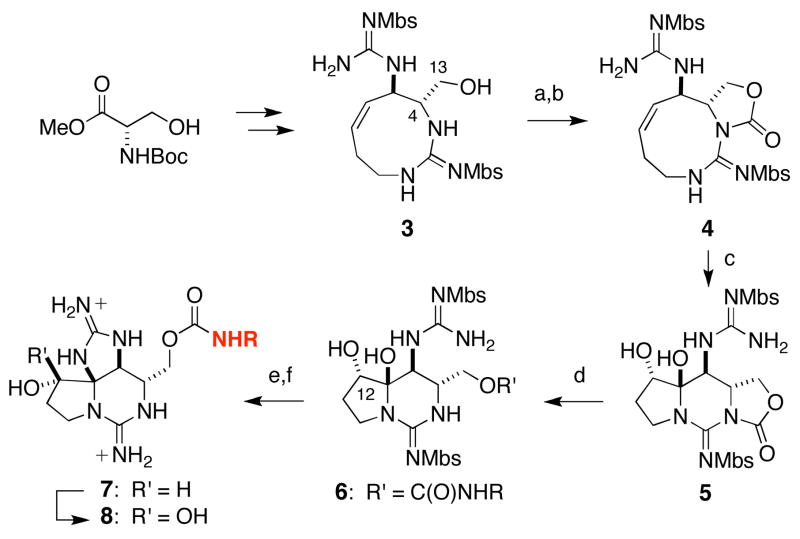
A strategic modification to one of our previously published routes to (+)-STX has been devised in order to prepare alternate C13 carbamate forms (Scheme 1).12 Tricyclic oxazolidinone 5 represents the cornerstone of this new synthetic plan, our assumption being that nucleophilic amines would open selectively this strained heterocycle. This structure can be fashioned in just three steps from the 9-membered ring guanidine 3, a material that we now routinely synthesize on >5 g scales. To access 5, sequential formation of the C13-Troc carbonate and ring closure has proven necessary; the use of other carbonylating agents (i.e., phosgene, carbonyldiimidazole) gives almost exclusively the C4–C13 alkene. Catalytic ketohydroxylation of 4 proceeds efficiently, albeit with modest selectivity, to generate hemiaminal 5.13 Oxazolidinone ring opening with a 1° amine at ambient temperature then furnishes the corresponding carbamate 6. Two subsequent transformations, which include Lewis acid-mediated guanidine ring closure and deprotection and C12 oxidation, complete the assembly of the tailored saxitoxins.
Scheme 1.
Synthetic route to C13-modified saxitoxins.a
a(a) Cl3CCH2C(O)Cl, C5H5N, 0 °C, 93%; (b) iPr2NEt, CH3CN, 86%; (c) 10 mol% OsCl3, Oxone, 44%; (d) RNH2, THF, 77–99%; (e) B(O2CCF3)3, 92%; (f) DCC, DMSO, C5H5NH+ −O2CCF3, 63%. Mbs = p-methoxybenzenesulfonyl.
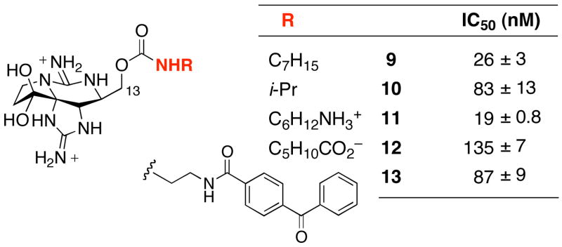
Inhibitory constants have been determined for STX C13-derivatives 9–13 against rNaV1.4 (Figure 3). In spite of the steric, electronic, and polar modifications to the C13 carbamate, all of these compounds exhibit channel blocking potency within 1–1.5 orders of magnitude of the parent toxin. The most severe loss in affinity is noted for acid 12, possibly the result of a charge interaction between the carboxylate residue and one of the guanidinium moieties.14 Other data, namely results from voltage-clamp recordings with 9 and 10, suggest that the carbamate appendage sits in a narrow gorge between protein domains. Future experiments will continue to test this hypothesis. Finally, we note that, to the best of our knowledge, the benzophenone-labeled toxin 13 represents the first of any such STX photoaffinity probes.15
Figure 3.
Recorded IC50 values for novel STX’s against rNaV1.4.
The power of de novo synthesis to provide novel forms of STX is further underscored with compounds such as 11 (Figure 4). The presence of two guanidinium groups notwithstanding, 11 undergoes selective coupling with an N-hydroxysuccinimide (NHS) benzoate ester to give the desired amide 14. This final-step, “post-synthetic” coupling reaction makes possible the attachment of structurally complex payloads to the STX core (i.e., fluorogenic groups, cofactors), side chain elements that might otherwise be incompatible with the chemistries employed for guanidine deprotection and/or C12 oxidation (see Scheme 1).
Figure 4.
A final-step ligation strategy for STX modification.
Access to (+)-STX through asymmetric total synthesis has empowered the development of unnatural analogues of this unique natural product. New saxitoxins have been evaluated for their efficacy in blocking NaV function using whole cell, voltage-clamp techniques. These findings have revealed opportunities for re-engineering the C13-carbamoyl unit of STX with any one of a number of different structural groups. Studies of this type together with the tools of molecular biology should allow us to map in greater detail the three-dimensional arrangement of the channel pore. We view these studies as a necessary step in a larger plan to utilize designed chemical tools to interrogate dynamic processes associated with NaV function.
Supplementary Material
Acknowledgments
B.M.A. is the recipient of graduate fellowships from Amgen and the ACS Division of Organic Chemistry, sponsored by Wyeth. We are grateful to Professors Richard Aldrich (University of Texas) and Merritt Maduke (Stanford University) for allowing use of their laboratory equipment and for many helpful discussions. This work has been supported by a grant from the NIH and with a gift from Pfizer.
Footnotes
Supporting information. General experimental protocols and characterization data for all new compounds. This material is available free of charge via the internet at http://pubs.acs.org
References
- 1.Hille B. Ion Channels of Excitable Membranes. 3. Sinauer; Sunderland, MA: 2001. [Google Scholar]
- 2.(a) Catterall WA, Yu FH. Genome Biology. 2003;4:207. doi: 10.1186/gb-2003-4-3-207. [DOI] [PMC free article] [PubMed] [Google Scholar]; (b) Catterall WA, Goldin AL, Waxman SG. Pharm Rev. 2005;57:397. doi: 10.1124/pr.57.4.4. [DOI] [PubMed] [Google Scholar]
- 3.(a) Novakovic SD, Eglen RM, Hunter JC. Trends in Neurosci. 2001;24:473. doi: 10.1016/s0166-2236(00)01884-1. [DOI] [PubMed] [Google Scholar]; (b) Lai HC, Jan LY. Nat Rev Neurosci. 2006;7:548. doi: 10.1038/nrn1938. [DOI] [PubMed] [Google Scholar]; (c) Rush AM, Cummins TR, Waxman SG. J Physiol. 2007;579:1. doi: 10.1113/jphysiol.2006.121483. [DOI] [PMC free article] [PubMed] [Google Scholar]
- 4.Termin A, Martinborough E, Wilson D. Ann Rep Med Chem. 2008;43:43. and references therein. [Google Scholar]
- 5.(a) Anger T, Madge DJ, Mulla M, Ridall D. J Med Chem. 2001;44:115. doi: 10.1021/jm000155h. [DOI] [PubMed] [Google Scholar]; (b) Priest BT, Kaczorowski GJ. Proc Nat Acad Sci. 2007;104:8205. doi: 10.1073/pnas.0703091104. [DOI] [PMC free article] [PubMed] [Google Scholar]
- 6.Llewellyn LE. Nat Prod Rep. 2006;23:200. doi: 10.1039/b501296c. [DOI] [PubMed] [Google Scholar]
- 7.For previous reports of STX derivatives, see: Koehn FE, Ghazarossian VE, Schantz EJ, Schnoes HK, Strong FM. Biorg Chem. 1981;10:412.Strichartz GR, Hall S, Magnani B, Hong CY, Kishi Y, Debin JA. Toxicon. 1995;33:723. doi: 10.1016/0041-0101(95)00031-g.Iwamoto O, Shinohara R, Nagasawa K. Chem Asian J. 2009;4:277. doi: 10.1002/asia.200800382.Robillot C, Kineavy D, Burnell J, Llewellyn LE. Toxicon. 2009;53:460. doi: 10.1016/j.toxicon.2008.12.020.
- 8.Choudhary G, Shang L, Li XF, Dudley SC. Biophys J. 2002;83:912. doi: 10.1016/S0006-3495(02)75217-X. and references therein. [DOI] [PMC free article] [PubMed] [Google Scholar]
- 9.Lipkind GM, Fozzard HA. Biochemistry. 2000;39:8161. doi: 10.1021/bi000486w.Tikhonov DB, Zhorov BS. Biophys J. 2005;88:184. doi: 10.1529/biophysj.104.048173.Also, see: Scheib H, McLay I, Guex N, Clare JJ, Flaney FE, Dale TJ, Tate SN, Robertson GM. J Mol Modeling. 2006;12:813. doi: 10.1007/s00894-005-0066-y.
- 10.An aromatic residue in the pore lining may also be important, see: Santarelli VP, Eastwood AL, Dougherty DA, Horn R, Ahern CA. J Biol Chem. 2007;282:8044. doi: 10.1074/jbc.M611334200.Also, see: Lee CH, Ruben PC. Channels. 2008;2:407. doi: 10.4161/chan.2.6.7429.
- 11.Moran O, Picollo A, Conti F. Biophys J. 2003;84:2999. doi: 10.1016/S0006-3495(03)70026-5. [DOI] [PMC free article] [PubMed] [Google Scholar]
- 12.Fleming JJ, McReynolds MD, Du Bois J. J Am Chem Soc. 2007;129:9964. doi: 10.1021/ja071501o. [DOI] [PubMed] [Google Scholar]
- 13.~40% of the regioisomeric α-hydroxyketone is also obtained.
- 14.This result is consistent with a previous report showing decreased in vivo potency of an STX C13-hemisuccinate derivative, see ref. 7a.
- 15.Tetrodotoxin-based photoaffinity probes have been prepared, see: Guillory RJ, Rayner MD, D’Arrigo JS. Science. 1977;196:883. doi: 10.1126/science.404708.Nakayama H, Hatanaka Y, Yoshida E, Oka K, Takanohashi M, Amano Y, Kanaoka Y. Biochem Biophys Res Commun. 1992;184:900. doi: 10.1016/0006-291x(92)90676-c.
Associated Data
This section collects any data citations, data availability statements, or supplementary materials included in this article.


