The evaluation of biomarkers as prognostic factors in lymphomas requires studies in the context of well designed clinical trials. In this study, the authors show the predictive value of immunoblastic morphology and loss of HLA-DR but not the cell of origin immunohistochemical classification in diffuse large B-cell lymphoma treated in large clinical trials.
Keywords: diffuse large B-cell lymphoma, prognosis, HLA-DR, immunoblastic lymphoma
Abstract
Background
Research on prognostically relevant immunohistochemical markers in diffuse large B-cell lymphomas has mostly been performed on retrospectively collected clinical data. This is also true for immunohistochemical classifiers that are thought to reflect the cell-of-origin subclassification of gene expression studies. In order to obtain deeper insight into the heterogeneous prognosis of diffuse large B-cell lymphomas and to validate a previously published immunohistochemical classifier, we analyzed data from a large set of cases from prospective clinical trials with long-term follow-up.
Design and Methods
We performed morphological and extensive immunohistochemical analyses in 414 cases of diffuse large B-cell lymphoma from two prospective randomized clinical trials (NHL-B1/B2, Germany). Classification into germinal center and non-germinal center subtypes of B-cell lymphoma was based on the expression pattern of CD10, BCL6, and IRF4. Multivariate analyses were performed adjusting for the factors in the International Prognostic Index.
Results
Analyzing 20 different epitopes on tissue microarrays, expression of HLA-DR, presence of CD23+ follicular dendritic cell meshworks, and monotypic light chain expression emerged as International Prognostic Index-independent markers of superior overall survival. Immunoblastic morphology was found to be related to poor event-free survival. The non-germinal center subtype, according to the three-epitope classifier (CD10, BCL6, and IRF4) did not have prognostic relevance when adjusted for International Prognostic Index factors (relative risk=1.2, p=0.328 for overall survival; and relative risk=1.1, p=0.644 for event-free survival).
Conclusions
The previously reported International Prognostic Index-independent prognostic value of stratification into germinal center/non-germinal center B-cell lymphoma using the expression pattern of CD10, BCL6, and IRF4 was not reproducible in our series. However, other markers and the morphological subtype appear to be of prognostic value.
Introduction
A milestone in the analysis of the heterogeneity of prognosis in patients with diffuse large B-cell lymphoma (DLBCL) was the development of the International Prognostic Index (IPI), which enables patients to be assigned to several clinical risk groups and for treatment strategies to be stratified accordingly.1 A second major step was the segregation of DLBCL into distinct subtypes based on gene expression profiling.2–4 Two main subgroups were identified: the germinal center B-cell (GCB)-like type showed a gene expression profile reminiscent of normal germinal center B cells, whereas the activated B-cell (ABC)-like type had a gene signature similar to that of activated peripheral blood B cells. A third intermediate group could not be allocated to either GCB or ABC type. The different gene expression patterns corresponded to different clinical outcomes.3,4
Several attempts have been made to substitute this gene expression-based classification by more routinely applicable methods. Hans et al.5 suggested that immunohistochemistry, using the expression pattern of CD10, BCL6, and IRF4, could assign DLBCL cases to a GCB or a non-GCB type. In their retrospective series, the non-GCB phenotype, defined according to the immunohistochemical-based subclassification, was an IPI-independent predictor of adverse overall survival.5
In order to validate these results in an independent large cohort of patients, we retrospectively analyzed 414 cases of DLBCL included in the randomized prospective clinical trials NHL-B1/B2 of the German High Grade Non-Hodgkin’s Lymphoma Study Group (DSHNHL),1 with tissue microarray data. Furthermore, we analyzed additional markers that might enable prognostic subgrouping independently from the IPI, which is still regarded the gold standard for risk stratification in DLBCL. Some of these additional markers, including HLA-DR and BCL2, have been investigated in several previous studies and were variably associated with prognosis.8–16
Design and Methods
Patients and samples
All patients were enrolled in the prospective randomized multicenter clinical trials NHL-B1/B2 of the DSHNHL.6,7 These trials were conducted in accordance with the Helsinki declaration. The protocols and the translational investigations had been approved by the ethical review board of all participating centers. All patients gave written informed consent.
The patients received six cycles of either CHOP (cyclophosphamide, doxorubicin, vincristine, prednisolon) or CHOEP (CHOP plus etoposide) chemotherapy, with the cycles being administered every 2 or 3 weeks, respectively. Rituximab was not included.
All cases were reviewed by expert hematopathologists in at least one of the reference centers for lymph node pathology in Germany (Berlin, Frankfurt, Kiel, Lübeck, Würzburg, Ulm) or Switzerland (St. Gallen) in order to obtain a standardized reference diagnosis. Morphological analysis was performed on conventional hematoxylin and eosin- and Giemsa-stained slides and subgrouping followed the 2001 WHO classification.17 According to this classification the centroblastic variant is characterized by medium-sized to large lymphoid cells with oval to round vesicular nuclei which contain two to four membrane-bound nucleoli. The immunoblastic variant of DLBCL consists predominantly (>90%) of immunoblasts with a single centrally located nucleolus and an appreciable amount of basophilic cytoplasm. Centroblasts represent less than 10% of the population in immunoblastic lymphomas.
Tissue microarray construction
Immunohistochemical profiling was performed on tissue microarrays constructed in cooperation with Euroimmun Medizinische Labordiagnostika AG (Lübeck, Germany).18,19 These tissue microarrays were constructed from conventional tissue sections, rather than the more widely used punch technique. Briefly, whole tissue sections were mounted on cover slip glass slides, which were cut into regular small squares of 3.24 mm2. Areas of interest were defined and indexed for robot-assisted transfer onto conventional glass slides. A single tissue microarray contained up to 96 squares. The main advantage of this alternative microarray technology over punch tissue microarrays is that the efficiency per case is optimum, since even almost exhausted paraffin blocks provide sufficient areas of tumor tissue for conventional sections. Furthermore, the remaining paraffin tissue is not perforated with this method, and shipping of blocks from one laboratory to another was avoided.
Immunohistochemical staining and scoring
The antibody panel included CD10, CD5, BCL2, BCL6, IRF4, κ and λ light chains, CD138, CD38, HLA-DR, CD30, Ki-67, IgM, IgD, IgA, IgG, Ki-B3, CD45RA, CD45RO, and CD23.
Since the Ki-B3, Ki-B5 (CD45RA) and OPD4 (CD45RO) antibodies are not commonly used, they are briefly described here. The Ki-B3 antibody predominantly recognizes B cells of follicular mantle and germinal center cells as well as plasma cells in normal lymphoid tissue; furthermore, it recognizes a subset of monocytic cells and some immature hematopoetic and lymphatic cell populations.20 Presumably this antibody detects a subtype of leukocyte common antigen, but has not been clustered.21 Ki-B5 shows a high specificity for the B-cell lineage including a minor subset of plasma cells, but also reacts with a subgroup of myelomonocytic leukemias and T-cell lymphomas.22 It detects the CD45RA molecule.21 The monoclonal antibody OPD4 recognizes the CD45RO molecule and preferentially reacts with normal and neoplastic post-thymic T cells including CD4 and CD8-positive subpopulations. The CD45RO molecule can also be detected by this antibody in a subset of B-cell neoplasms.23
Staining procedures followed a standard three-step protocol with diaminobenzidine as the chromogen. Primary antibodies, suppliers, and staining conditions are listed in Online Supplementary Table s1. All staining was performed in a single institution (Lübeck, Germany). Tissue microarrays were evaluated by expert hematopathologists of the German lymphoma reference panel. A given immunohistochemical marker was evaluated by two observers who had to reach a consensus decision on every case. If this was not possible, the case was excluded from the statistical analyses. To test for interobserver variation, selected markers were scored twice in different institutions and κ statistics were calculated.
With the exception of Ki-67 (MIB1) and immunoglobulin light chains, all stains were scored semiquantitatively estimating the percentage of positive tumor cells: less than 1% (score 0), 1–25% (score 1), 26–50% (score 2), 51–75% (score 3), and more than 75% (score 4). For Ki-67 (MIB1) the number of positive tumor cells was estimated and recorded in 10% increments. Restricted (monotypic) light chain expression by the tumor cells was noted as being present or not, regardless of the number of positive cells. Only cases in which both light chain stains were evaluable and in which only one chain was expressed were considered as having monotypic light chain restriction. With regards to the CD23 staining, not only were the tumor cells evaluated, but also the presence or absence of follicular dendritic cell (FDC) meshworks (referred to as CD23FDC: none = score 0, rare = score 1, moderate = score 2, abundant = score 3).
Statistical analyses
Event-free survival was defined as the time from the beginning of therapy to either disease progression under therapy, failure to achieve a confirmed or unconfirmed complete remission, initiation of additional (off-protocol) or salvage therapy, relapse or death. Overall survival was defined as the time from the beginning of therapy to death of any cause. Event-free survival and overall survival rates were estimated according to Kaplan and Meier.24
After extensive statistical pre-evaluation of results (for a detailed description see the Online Supplementary Data), we summarized the immunohistochemical score groups as follows: the cases were assigned as negative for the respective marker if less than 1% of the cells were stained, and positive if 1% or more of the tumor cells were positive. To allow for comparison with previously published data, we also performed analyses for some of the markers with different cut-offs (see below). For Ki67 (MIB1) the most suitable cut-off was at 80%. For CD23FDC we considered all cases with FDC (score groups 1 to 3) as one group (CD23FDC-positive) and compared these to the cases without any detectable FDC (CD23FDC-negative).
The assignment to the GCB and non-GCB groups was based on the expression of CD10, BCL6, and IRF4, as described by Hans and co-workers.5 Statistical tests were performed twice using cut-off levels of 1% and 25% for the included markers.
Log-rank tests were performed for univariate analyses. Immunohistochemical markers whose p-values were less than 0.100 for event-free survival or overall survival were included in the multivariate analysis.
For the selected markers proportional hazard models were fitted separately adjusted for the factors of the IPI. Prior to modeling, relevant correlations of the respective marker with IPI factors (i.e., Person’s correlation coefficient >0.7) were excluded. For markers that were not significant in the univariate analysis (p-values >0.1), we checked whether the marker was prognostically relevant after adjustment for the IPI factors. Relative risks with 95% confidence intervals (CI) are shown in a forest plot.
The χ2 test or, if required, Fisher’s exact text was used for the correlation of marker expression with qualitative data (age, GCB/non-GCB group, morphological subtypes). Because of the descriptive nature of these comparisons, the p-values were not adjusted for multiple comparisons and the significance level was p=0.05.
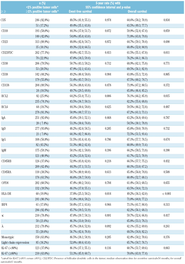
Cases that failed to give reliable staining results for a given marker were excluded from further analysis. Unreliable staining was assumed if there was a lack of an appropriate internal control and/or significant background staining preventing the identification of specific reactivity on tumor cells. Furthermore, in some of the cases tissue was lost after the staining procedure. Thus, the number of evaluable cases varied from one marker to another (Table 2). This also decreased the number of cases available for the GCB/non-GCB subgrouping and the Cox models that included more than one marker.
Table 2.
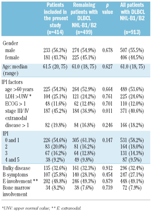
Patients’ characteristics and clinical parameters of the study cases compared to those of all patients with DLBCL in the NHL-B1/B2 trial of the German High-Grade Non-Hodgkin’s Lymphoma Study Group (DSHNHL).
To assess the concordance between different observers, κ statistics were performed. All statistical analyses were done with SPSS 15.0 software.
Results
Patients’ characteristics
The patients’ clinical characteristics are summarized in Table 2. The clinical features of the patients in the present study (n = 414) closely matched those of the entire study population of the NHL-B1/B2 trials (n = 913). According to the study protocol, patients 60 years of age or less were included only if their lactate dehydrogenase (LDH) values were within normal limits.7
The median age of the patients in the present analysis was 61.5 years (range, 20 to 75 years). Two hundred and twenty-five patients (54.3%) were over 60 years of age at randomization. The median observation time was 61 months for overall survival.
Morphological subtypes
Of the 414 cases of DLBCL, 75.5% (n=313) showed a centroblastic morphology. Immunoblastic B-cell lymphoma was seen in 11.4% (n=47), and the remainder were classified as not otherwise specified (NOS; 6.3%; n=26), anaplastic large cell variant, and T-cell/histiocyte rich B-cell lymphoma (3.4%; n=14 each).
Interobserver reliability
Staining for BCL2, HLA-DR, and IRF4 was scored independently by observers in two different institutions. Regarding positive (≥1% positive cells) and negative cases (<1% positive cells), the κ-values were 0.76, 0.84 and 0.75, respectively. If the missing values were included, the lowest κ value was still 0.68. Thus, a satisfactory interobserver reliability was achieved.
Staining results
Comparing the numbers of positive and negative cases for all the immunohistochemical markers in the two age groups of the IPI (≤ vs. >60 years), we found significantly more CD10-positive cases in younger patients (51.3% vs. 33.9%; p=0.001), and significantly more cases positive for BCL2, IRF4 and monotypic light chain expression (MLCE) in elderly patients (BCL2: 79.6% vs. 68.0%, p=0.018; IRF4: 86.8% vs. 77.1%, p=0.017; MLCE: 43.1% vs. 29.4%, p=0.029). Using the data only from patients without elevated LDH levels, the results were very similar (data not shown).
With respect to HLA-DR expression, we found a significantly higher number of negative cases among lymphomas that presented in the central nervous system, testes or ovaries (8/15 cases; 53.3%), which are considered as immunoprivileged sites, as compared to lymphomas in other sites (52/286 cases; 18.2%; Fisher’s exact test p=0.003).
Remarkably, the correlation between any pair of the 19 IHC markers investigated was generally weak with Pearson’s correlation coefficients not exceeding 0.35 (absolute value; correlation between κ, λ and MLCE not included) suggesting non-redundant information.
Univariate analyses of immunohistochemical markers
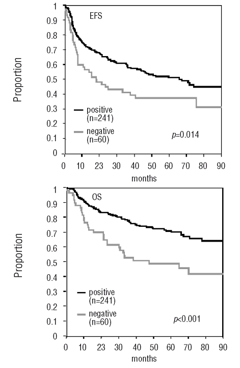
Expression of BCL2 was significantly associated with shorter event-free survival and overall survival (p=0.006 and 0.015, respectively). A similar adverse prognostic effect was seen for cases lacking HLA-DR expression (p=0.014 for event-free survival, and p<0.001 for overall survival; Figure 1). Furthermore, expression of CD10 was correlated, albeit not significantly, to longer event-free survival and overall survival (p=0.072 and 0.059, respectively). Interestingly, the presence of CD23-positive FDC meshworks (CD23FDC) within lymphoma infiltrates (observed in 78 of 340 cases; 22.9%) was significantly associated with superior overall survival (p=0.011), but not with event-free survival (p=0.413). Results of univariate analyses are summarized in Table 1.
Figure 1.
Univariate analysis for event-free survival (EFS) and overall survival (OS) HLA-DR-positive vs. HLA-DR-negative cases (positive = 1–100% stained tumor cells, negative = 0%).
Table 1.
Summary of univariate statistical test results for immunohistochemical markers.
Multivariate analyses
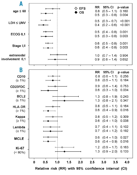
The IPI factors LDH, ECOG performance status, and stage were the strongest prognostic factors for event-free survival and overall survival: relative risks ranged from 1.6 to 2.7 (for LDH > normal, ECOG >1, and stage III/IV), and 0.6 to 0.4 (for LDH ≤ normal, ECOG ≤ 1, and stage <III), respectively.
In multivariate analyses adjusted for the IPI factors, we found significantly better overall survival if HLA-DR was positive (RR=0.6; p=0.016), or if remnants of CD23-positive FDC meshworks were present in the lymphoma (RR=0.6; p=0.034). Two additional markers were significant only in the multivariate analysis: a MLCE of the tumor was significantly correlated with superior event-free survival and overall survival (RR=0.6; p=0.027, and RR=0.6; p=0.016, respectively), and κ light chain expression was correlated with extended overall survival (RR=0.6; p=0.038). A summary of the results is given in Figure 2.
Figure 2.
Multivariate analysis for event-free (EFS) and overall survival (OS). (A) Cox model for IPI factors. (B) Cox model for each of the immunohistochemical markers adjusted for the IPI factors.
In multivariate analyses using the proportional hazard models to investigate the contribution of each individual marker if adjusted for IPI, only CD23FDC, HLA-DR and κ light chain expression for overall survival, and MLCE for both event-free and overall survival showed relative risks of a magnitude similar to those of the strongest IPI factors (Figure 2). By combining the three single independent markers CD23FDC, HLA-DR and MLCE (κ light chain expression is correlated to MLCE, and as such, not independent) in a Cox model adjusted for the IPI factors (n=192), HLA-DR remained significant for event-free survival (RR=0.6, p=0.019) and overall survival (RR=0.5, p=0.016) and MLCE for overall survival (RR=0.6, p=0.044), but not for event-free survival (RR=0.7, p=0.123). CD23FDC, however, was not significant in this model for either event-free survival or overall survival. Additional adjustment for treatment (14-day cycle, etoposide) did not change these results (data not shown).
Including only MLCE and HLA-DR in a further Cox model adjusted for the IPI factors (n=201), only HLA-DR remained significant for event-free survival (RR=0.6, 95%CI=0.4–0.9, p=0.028) and overall survival (RR= 0.5, 95%CI=0.3–0.9, p=0.016).
Formal testing for interactions between HLA-DR and the IPI groups (0–2 vs. 3–5) was not appropriate because the sample size was too small. However, we checked whether the relative risks were of a similar magnitude and they were found to be so for event-free survival [IPI 0–2: RR=0.7 (95%CI=0.4–1.2); IPI 3–5: RR=0.7 (95%CI=0.4–1.2)] as well as for overall survival [IPI 0–2: RR=0.4 (95%CI=0.2–0.7); IPI 3–5: RR=0.6 (95%CI=0.4–1.1)].
For BCL2 and BCL6 we performed repeated multivariate analyses adjusted for the IPI factors using additional cut-offs for positivity that more closely match those published in the literature (>25% and >50% positive tumor cells). However, neither BCL2 nor BCL6 qualified as an IPI-independent outcome predictor (data not shown)
Analyses of germinal center versus non-germinal center-like B-cell lymphomas
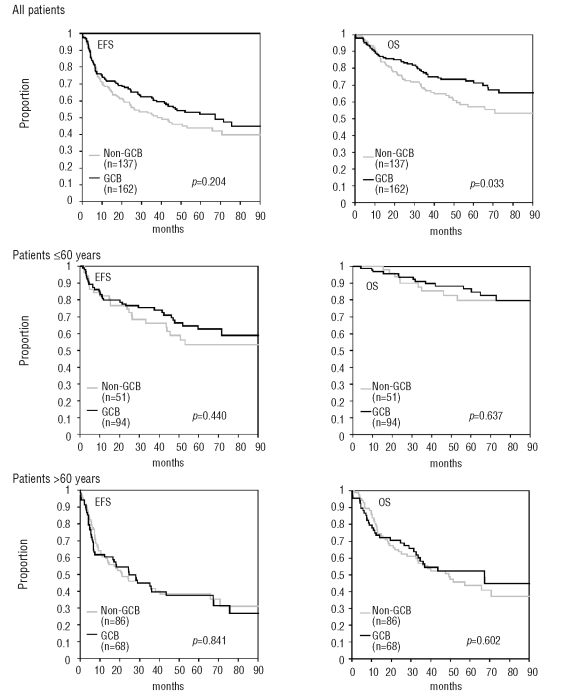
Cases were assigned to the GCB or non-GCB group as proposed by Hans and co-workers.5 In the first analysis, the cut-off level for CD10, BCL6, and IRF4 was 25%. Of 299 assessable cases, 162 lymphomas had a GCB-type pattern, and 137 cases were assigned to the non-GCB group. In the remaining cases, defaults for one or more of the immunohistochemical stains prevented further subclassification.
Comparing the two groups, the patients with GCB-type lymphoma turned out to be significantly younger than the non-GCB patients (58 vs. 64 years; p<0.001). Given the specific inclusion criteria for younger patients (see section Patients’ characteristics), this resulted in a significantly lower number of patients with elevated LDH levels in the GCB group (16.7% vs. 29.2%, p=0.010) as well as a higher number of patients within more favorable IPI risk groups (IPI 0,1: 65.4% vs. 48.9%; IPI 2: 16.0% vs. 23.4%; IPI 3: 11.7% vs. 16.8%; IPI 4, 5: 6.8% vs. 10.9%; p=0.039).
Looking for parameters potentially related to clinical outcome (Figure 3), GCB-type lymphomas were associated with an improved overall survival in the univariate analysis (p=0.033), whereas event-free survival was not statistically different between the GCB and non-GCB groups. Interestingly, when we compared GCB and non-GCB type lymphomas within the two age groups (≤60 and >60 years) separately, we could not find any differences for EFS and OS in univariate analysis (Figure 3).
Figure 3.
Univariate analysis for event-free survival (EFS) and overall survival (OS) – comparison of GCB and non-GCB type.
In multivariate analysis adjusted for the factors of the IPI, non-GCB immunophenotype did not emerge as an independent marker of inferior outcome (RR=1.2, 95%CI=0.8–1.8, p=0.328 for overall survival; and RR=1.1, 95%CI=0.8–1.5, p=0.644 for event-free survival).
In a separate analysis with a cut-off level of 1% for the markers included, our results did not substantially change. In the multivariate approach including the factors of the IPI, there was a trend to an association between non-GCB immunophenotype and reduced overall survival, but this was not statistically significant (RR=1.4, 95%CI=1.0–2.2, p=0.077). The same emerged when the possible effect of intensified treatment (14-day cycle, etoposide) was considered in the multivariate analysis (RR=1.4, 95%CI=0.9–2.1, p=0.093). With regards to event-free survival, the non-GCB profile did not qualify at all as an independent adverse prognostic marker (RR=1.2, 95%CI=0.9–1.7, p=0.231).
Analysis of morphological subtypes
Comparison of immunoblastic versus centroblastic lymphomas with regards to selected immunohistochemical markers (Online Supplementary Table S2) revealed a significantly higher rate of BCL6-positive cases among the centroblastic lymphomas (85.5% vs. 63.4%, p=0.001). In contrast, CD10 as a further germinal center cell associated marker was not differentially expressed in the two groups (p=0.420). The same held true for BCL2 (p=0.176), IgD (p=0.492), IgM (p=0.242), and IRF4 (p=0.211).
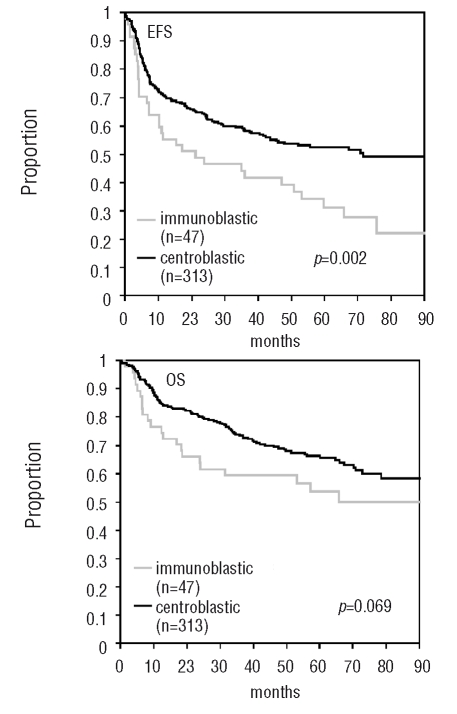
Interestingly, patients with centroblastic lymphomas showed a significantly superior event-free survival in both univariate analysis (3-year rate: 58.2% vs. 41.7%, p=0.002, Figure 4) and multivariate analysis. In other words, immunoblastic morphology turned out to be an adverse prognostic factor associated with shorter event-free survival, independently of the single IPI factors (RR1.7, 95%CI=1.2–2.5, p=0.005). The overall survival was not statistically different between patients with centroblastic or immunoblastic lymphomas, although a trend for shorter overall survival for those with immunoblastic lymphomas was apparent in both the univariate analysis (3-year rate: 73.4% vs. 59.3%, p=0.069, Figure 4) and the multivariate analysis (RR1.4, 95%CI=0.9–2.2, p=0.178).
Figure 4.
Univariate analysis for event-free survival (EFS) and overall survival (OS) – comparison of morphological subtypes.
Remarkably, the non-GCB immunophenotype was significantly more frequent among the immunoblastic lymphomas (64.9% in the immunoblastic group vs. 43.0% in the centroblastic group; p=0.013). However, this significant difference was only seen with the 25% cut-off, not with the 1% cut-off (p=0.227).
Discussion
Based on advances in the understanding of molecular mechanisms in lymphomagenesis, several investigators have subdivided DLBCL according to immunophenotype patterns that are thought to reflect the cell-of-origin subclassification determined by gene expression studies.
Hans et al.5 subclassified DLBCL into GCB and a non-GCB subtypes according to the expression pattern of CD10, BCL6, and IRF4. In their study, this classifier proved to be an independent predictor of clinical outcome in addition to the IPI by multivariate analysis. With similar, although not completely identical approaches, these results were confirmed by some,25–27 but not all studies.12,28,29 By applying the Hans classifier in our set of DLBCL, we found a longer overall survival for patients with GCB-type lymphomas in the univariate analysis; however, we failed to confirm this subclassification to be of independent prognostic impact if adjusted for the IPI in a multivariate analysis. The latter result is in line with the findings of Colomo et al.,12 DePaepe et al.,28 and Veelken et al.29 and challenges the value of the Hans classifier regarding prognosis. The difference in outcome in the univariate analysis in our series is obviously related to a higher number of patients with favorable prognostic attributes (lower age, normal LDH) in the GCB group.
The varying results among several immunohistochemical studies studies may be ascribed to a number of factors, a major one being the lack of standardization in the performance and evaluation of staining. However, as has recently been shown by the Lunenburg Lymphoma Biomarker Consortium,30 these problems can be overcome, at least in part, by harmonization of laboratory and evaluation procedures. In the present study, all stainings were carried out in a single laboratory in order to eliminate interlaboratory variations in antibody usage and staining processes as sources of artificial variations. All the pathologists who evaluated the stains are members of the German lymphoma reference pathology panel. Over the past decade, the panel members have met in regular workshop sessions to review lymphoma cases and this has led to a high level of harmonization in the evaluation of morphology and immunohistochemical stains. To further maximize consistency in the evaluation process, staining for a given antibody was evaluated by the same predetermined observers for the entire set of cases. For a selected subset of markers a second evaluation round with different observers was performed; the test of interobserver reliability showed satisfactory results.
Currently, there is no consensus across immunohistochemical studies regarding the number of stained tumor cells required to call a case positive. We, therefore, initially evaluated immunostains without a predetermined cut-off and scored them semiquantitatively for most of the antibodies by estimating the percentage of positive tumor cells in 25% increments. As described in detail in the Online Supplementary Data, a subsequent determination of formal cut-offs from Kaplan-Meier curves for the single score groups was unsuccessful. Further analysis revealed that a cut-off value of 1% in our series best discriminated groups of patients with different event-free and overall survivals. A separate analysis with additional cut-offs (>25%, >50% positive tumor cells) for some of the markers was done to allow comparison with results of previous studies.
The Hans classifier was tested twice with 25% and 1% cut-off levels. The first cut-off closely approximates that in the original report (30%)5 and hence enables direct comparison. The 1% cut-off was chosen in order to investigate, whether the protein expression by only a very small fraction of tumor cells might already enable a comparable subclassification.
The fact that we could not confirm an IPI-independent prognostic value of the Hans classifier highlights the difficulty in finding a reliable immunohistochemical surrogate for the gene expression profile. In an independent data set of the German research consortium Molecular Mechanisms of Malignant Lymphoma31 we found that the Hans classifier based on paraffin immunohistochemical is not perfectly correlated with the gene expression classification of GCB and ABC lymphoma subjects as determined according to Wright et al.32 (unpublished data). The prognostic value of gene expression-based subclassifications, on the other hand, has recently been confirmed also in DLBCL patients treated with additional rituximab immunotherapy (R-CHOP)33 underlining the necessity to refine immunohistochemical surrogates for existing gene expression profiles.
According to our results, loss of HLA-DR protein expression is a relevant and significant independent marker for worse overall survival. In fact, beside the factors of the IPI, loss of HLA-DR was the strongest outcome predictor in our series. This finding is well in line with previous reports and, thus, gives further support to the reliability of loss of HLA-DR as an adverse event in DLBCL.9–12 An association of high-grade malignancy in B-cell neoplasms with a lack of MHC class I and class II molecules on the tumor cells was already described in the 1980s.34,35 Correspondingly, gene expression studies on DLBCL showed that high expression of the MHC class II signature genes was correlated with a favorable outcome.4 As a member of the MHC class II family of proteins, HLA-DR is involved in cellular immune responses by participating in antigen presentation and stimulation of T cells.36,37 Hence, the lack of HLA-DR expression may constitute a mechanism by which tumor cells can escape immunosurveillance after chemotherapy. This in turn is reflected by significantly reduced numbers of tumor-infiltrating T-lymphocytes,11,38 decreased numbers of intratumoral CD1a-positive dendritic cells and, around the tumor edge, fewer S100-positive cells and CD45RO-positive T cells.8 More recently, a quantitative nuclease protection assay-based analysis on formalin-fixed, paraffin-embedded DLBCL samples identified low levels of HLA-DRB gene expression as an IPI-independent indicator of poor prognosis in rituximab-treated patients.39 This demonstrates that the prognostic impact of defective MHC class II expression can be reproduced using different detection technologies and at least to some extent is stable also with modern immunotherapy. The loss of MHC class II expression has been reported to be frequent in DLBCL arising in immunoprivileged sites such as the central nervous system, ovary and testis.40 Although in our series the number of cases with involvement of these sites was limited (n=15) we were able to confirm this observation: while about half of the cases from immunoprivileged sites were HLA-DR-negative, this was true for only 18% of lymphomas from other sites (p=0.003).
The presence of CD23-positive FDC meshworks was additionally significantly associated with superior overall survival in our study. This was a rather unexpected finding, although in an earlier investigation, the presence of FDC meshworks (demonstrated by CD21-staining) had already been shown to have clinical implications in terms of an increased rate of complete remissions in patients with poor-risk DLBCL.41 In this study, however, lymphomas with retained FDC meshworks had inferior disease-free survival and, contrasting to our findings, no differences in overall survival. Although it is tempting to speculate that FDC meshworks indicate a follicular lymphoma pathogenesis, there was no a correlation between the occurrence of FDC and expression of CD10 (Pearson’s correlation coefficient: −0.003) or between FDC and positivity for BCL2 or BCL6 in our series (Pearson’s correlation coefficient: −0.039 and −0.054, respectively). Moreover, follicular lymphomas were excluded from our study by reference pathology diagnosis.
The biological mechanisms that underlie a correlation between the presence of FDC and increased survival in de novo DLBCL are currently unknown. However, given the fact that FDC in this lymphoma entity most likely represent remnants of the pre-existing lymph node architecture, it might be a morphological manifestation of what has been termed the lymph node signature or stromal-1 signature in gene expression studies.4,33 This gene expression signature was correlated with a superior clinical outcome and linked to anti-tumor immune response. Alternatively, preservation of lymph node structures within the tumor infiltrate might indicate decreased destructive ability and, hence, typify, a biological attribute of the tumor itself.
MLCE of lymphoma cells was significantly associated with superior event-free survival and overall survival in our cohort. This was another unexpected finding that, to the best of our knowledge, has not been reported in DLBCL so far. In fact, some earlier reports showed that immunoglobulin-positive large cell lymphomas are associated with advanced stage and shorter overall survival42 or that immunoglobulin expression is more frequently seen in patients with unfavorable prognostic markers.43 However, in these earlier studies no separate correlation with light chain expression was provided and the stains had been performed on frozen sections. Thus, discussion of these results in the context of our findings is not really feasible.
The number of evaluable cases was too small to develop and validate a prognostic model based on the whole set of immunohistochemical markers used in our series; however, combining the three IPI-independent immunohistochemical markers, i.e., HLA-DR, CD23FDC, and MCLE in a Cox model, we found that HLA-DR was the strongest prognostic factor for event-free survival and overall survival. Morphological subgrouping into centroblastic and immunoblastic lymphomas in our series was associated with clinical outcome: immunoblastic morphology emerged as an adverse prognostic factor with shortened event-free survival, independently of by the IPI factors. The prognostic value of morphological subtyping is controversial.44–51 The difficulties, even among expert hematopathologists,52 in reliably identifying morphological subtypes of DLBCL may be caused, at least in part, by insufficient material (e.g. needle biopsies) or by suboptimal tissue processing, which hampers thorough cytomorphological analysis.
When we correlated the morphological subtype and the immunohistochemical classifier proposed by Hans et al., we found a significantly higher number of non-GCB type cases within the immunoblastic group than in the centroblastic group. However, since this correlation was not very strong, morphology apparently cannot be regarded as an adequate surrogate for immunophenotype, although it is obvious that both, morphology and immunophenotype, are indicators of a distinct tumor biology. This concept is also supported by gene expression studies in which a similar correlation between immunoblastic morphology and non-GCB molecular profile has been observed.4 Our findings demonstrate that careful morphological subtyping of DLBCL leads to a prognostically significant and thus relevant subclassification.
In the NHL-B1/B2 trials, intensified therapy led to a significant improvement of the outcome in comparison to that obtained with the standard CHOP regimen.6,7 This effect is not, however, likely to be sufficient to explain why we could not confirm the prognostic value of the Hans classifier or other previously reported immunohistochemical markers.14–16,53
The combination of the CHOP regimen with the monoclonal anti-CD20 antibody rituximab (R-CHOP) has considerably improved the outcome of patients with DLBCL.54,55 In this context, there is also some evidence to indicate that the impact of prognostic markers may be altered with the use of R-CHOP.56–58 The effect of rituximab does not, however, completely abolish the biological attributes of these lymphomas since at least some of the markers, for instance MHC class II gene expression, retain thier prognostic value although the observable effects may be diminished.39 Trials from the pre-rituximab era, such as the NHL-B1/B2 investigated here, are, therefore, suitable for identifying novel prognostic factors due to the high rate of events and concomitant high power. We will soon validate our results on data sets including patients treated with rituximab.
In conclusion, this is the first report on a large series of patients with DLBCL enrolled in a prospective clinical study with long-term follow-up. The results, on the one hand challenge the prognostic value of established immunohistochemical markers and the immunohistochemical-based GCB/non-GCB subtyping mostly determined from retrospective analyses and, on the other hand, suggest in part new markers and that morphological subgrouping can be indicative of prognosis independently of IPI factors.
Acknowledgments
we thank Ilona Schliephake, Katharina Vogel (both from Lübeck), Olivera Batic and Christiane Stange (both from Kiel) and Ulrike Schoenwiese (Leipzig) for their excellent technical assistance.
Footnotes
The online version of this article contains a supplementary appendix.
Authorship and Disclosures
HWB, CT, WK, HHW, MH, HS, MLH, GO, AR, HKMH, TFEB, PM, SBC, SH, and ACF performed morphological and immunohistochemical analyses; MZ and ML collected and analyzed the data; MP, NS and LT provided clinical data; HWB, MZ, ACF designed the research and wrote the manuscript. All authors approved the final version of the manuscript and declare that they have no conflicts of interest and no competing financial interests.
Funding: the NHL-B1 and NHL-B2 trials were supported by a grant from Deutsche Krebshilfe and unrestricted grants from Amgen and Bristol-Myers. SC was supported by the Swiss Group for Clinical Cancer Research (SAKK).
References
- 1.Shipp MA, Harrington DP, Anderson JR, Armitage JO, Bonadonna G, Brittinger G, et al. A predictive model for aggressive NHL: the International Non-Hodgkin’s Lymphoma Prognostic Factors Project. N Engl J Med. 1993;329:987. doi: 10.1056/NEJM199309303291402. [DOI] [PubMed] [Google Scholar]
- 2.Shipp MA, Ross KN, Tamayo P, Weng AP, Kutok JL, Aguiar RC, et al. Diffuse large B-cell lymphoma outcome prediction by gene-expression profiling and supervised machine learning. Nat Med. 2002;8:68–74. doi: 10.1038/nm0102-68. [DOI] [PubMed] [Google Scholar]
- 3.Alizadeh AA, Eisen MB, Davis RE, Ma C, Lossos IS, Rosenwald A, et al. Distinct types of diffuse large B-cell lymphoma identified by gene expression profiling. Nature. 2000;403:503–11. doi: 10.1038/35000501. [DOI] [PubMed] [Google Scholar]
- 4.Rosenwald A, Wright G, Chan WC, Connors JM, Campo E, Fisher RI, et al. Lymphoma/Leukemia Molecular Profiling Project. The use of molecular profiling to predict survival after chemotherapy for diffuse large-B-cell lymphoma. N Engl J Med. 2002;346:1937–47. doi: 10.1056/NEJMoa012914. [DOI] [PubMed] [Google Scholar]
- 5.Hans CP, Weisenburger DD, Greiner TC, Gascoyne RD, Delabie J, Ott G, et al. Confirmation of the molecular classification of diffuse large B-cell lymphoma by immunohistochemistry using a tissue microarray. Blood. 2004;103:275–82. doi: 10.1182/blood-2003-05-1545. [DOI] [PubMed] [Google Scholar]
- 6.Pfreundschuh M, Trümper L, Kloess M, Schmits R, Feller AC, Rübe C, et al. German High-Grade Non-Hodgkin’s Lymphoma Study Group. Two-weekly or 3-weekly CHOP chemotherapy with or without etoposide for the treatment of elderly patients with aggressive lymphomas: results of the NHL-B2 trial of the DSHNHL. Blood. 2004;104:634–41. doi: 10.1182/blood-2003-06-2095. [DOI] [PubMed] [Google Scholar]
- 7.Pfreundschuh M, Trümper L, Kloess M, Schmits R, Feller AC, Rudolph C, et al. German High-Grade Non-Hodgkin’s Lymphoma Study Group. Two-weekly or 3-weekly CHOP chemotherapy with or without etoposide for the treatment of young patients with good-prognosis (normal LDH) aggressive lymphomas: results of the NHL-B1 trial of the DSHNHL. Blood. 2004;104:626–33. doi: 10.1182/blood-2003-06-2094. [DOI] [PubMed] [Google Scholar]
- 8.Chang KC, Huang GC, Jones D, Lin YH. Distribution patterns of dendritic cells and T cells in diffuse large B-cell lymphomas correlate with prognoses. Clin Cancer Res. 2007;13:6666–72. doi: 10.1158/1078-0432.CCR-07-0504. [DOI] [PubMed] [Google Scholar]
- 9.Miller TP, Lippman SM, Spier CM, Slymen DJ, Grogan TM. HLA-DR (Ia) immune phenotype predicts outcome for patients with diffuse large cell lymphoma. J Clin Invest. 1988;82:370–2. doi: 10.1172/JCI113598. [DOI] [PMC free article] [PubMed] [Google Scholar]
- 10.Rybski JA, Spier CM, Miller TP, et al. Prediction of outcome in diffuse large cell lymphoma by the major histocompatibility complex class I (HLA-A, -B, -C) and class II (HLA-DR, -DP, -DQ) phenotype. Leuk Lymphoma. 1991;6:31–8. doi: 10.3109/10428199109064876. [DOI] [PubMed] [Google Scholar]
- 11.Rimsza LM, Roberts RA, Miller TP, Unger JM, LeBlanc M, Braziel RM, et al. Loss of MHC class II gene and protein expression in diffuse large B-cell lymphoma is related to decreased tumor immunosurveillance and poor patient survival regardless of other prognostic factors: a follow-up study from the Leukemia and Lymphoma Molecular Profiling Project. Blood. 2004;103:4251–8. doi: 10.1182/blood-2003-07-2365. [DOI] [PubMed] [Google Scholar]
- 12.Rimsza LM, Farinha P, Fuchs DA, Masoudi H, Connors JM, Gascoyne RD. HLA-DR protein status predicts survival in patients with diffuse large B-cell lymphoma treated on the MACOP-B chemotherapy regimen. Leuk Lymphoma. 2007;48:542–6. doi: 10.1080/10428190601078605. [DOI] [PubMed] [Google Scholar]
- 13.Colomo L, López-Guillermo A, Perales M, Rives S, Martínez A, Bosch F, et al. Clinical impact of the differentiation profile assessed by immunophenotyping in patients with diffuse large B-cell lymphoma. Blood. 2003;101:78–84. doi: 10.1182/blood-2002-04-1286. [DOI] [PubMed] [Google Scholar]
- 14.Gascoyne RD, Adomat SA, Krajewski S, Krajewska M, Horsman DE, Tolcher AW, et al. Prognostic significance of bcl-2 protein expression and bcl-2 gene rearrangement in diffuse aggressive non-Hodgkin’s lymphoma. Blood. 1997;90:244–51. [PubMed] [Google Scholar]
- 15.Hermine O, Haioun C, Lepage E, d’Agay MF, Briere J, Lavignac C, et al. Prognostic significance of bcl-2 protein expression in aggressive non-Hodgkin’s lymphoma. Blood. 1996;7:265–72. [PubMed] [Google Scholar]
- 16.Kramer MH, Hermans J, Parker J, Krol AD, Kluin-Nelemans JC, Haak HL, et al. Clinical significance of bcl2 and p53 protein expression in diffuse large B-cell lymphoma: a population-based study. J Clin Oncol. 1996;14:2131–8. doi: 10.1200/JCO.1996.14.7.2131. [DOI] [PubMed] [Google Scholar]
- 17.Gatter KC, Warnke RA. Diffuse large B-cell lymphoma. In: Jaffe ES, Harris NL, Stein H, Vardiman JW, editors. World Health Organization Classification of Tumours. Pathology and Genetics of Tumours of Haematopoietic and Lymphoid Tissues. Lyon, France: 2001. pp. 171–4. [Google Scholar]
- 18.Stocker K, Stocker W, Ritter-Frank Y, Scriba PC. Chemically activated glass slides for frozen sections and their use in autoantibody diagnosis. Acta Histochem. 1985;(Suppl 31):283–94. [PubMed] [Google Scholar]
- 19.Stocker W. Rational histochemistry using a new microanalytic technic. Acta Histochem. 1985;(Suppl 31):269–81. [PubMed] [Google Scholar]
- 20.Feller AC, Wacker HH, Moldenhauer G, Radzun HJ, Parwaresch MR. Monoclonal antibody Ki-B3 detects a formalin resistant antigen on normal and neoplastic B cells. Blood. 1987;70:629–36. [PubMed] [Google Scholar]
- 21.HLDA Database at: http://www99.mh-hannover.de/aktuelles/projekte/hlda7/hldabase/select.htm.
- 22.Hansmann ML, Wacker HH, Gralla J, Lumbeck H, Kossmahl M, Heidebrecht HJ, et al. Ki-B5: a monoclonal antibody unrelated to CD45 recognizes normal and neoplastic human B cells in routine paraffin sections. Blood. 1991;77:809–17. [PubMed] [Google Scholar]
- 23.Chadburn A, Husain S, Knowles DM. Monoclonal antibody OPD4 detects neoplastic T cells but does not distinguish between CD4 and CD8 neoplastic T cells in paraffin tissue sections. Hum Pathol. 1992;23:940–7. doi: 10.1016/0046-8177(92)90409-v. [DOI] [PubMed] [Google Scholar]
- 24.Kaplan EL, Meier P. Nonparametric estimation from incomplete observations. Journal of the American Statistical Association. 1958;53:457–481. [Google Scholar]
- 25.Chang CC, McClintock S, Cleveland RP, Trzpuc T, Vesole DH, Logan B, et al. Immunohistochemical expression patterns of germinal center and activation B-cell markers correlate with prognosis in diffuse large B-cell lymphoma. Am J Surg Pathol. 2004;28:464–70. doi: 10.1097/00000478-200404000-00005. [DOI] [PubMed] [Google Scholar]
- 26.Berglund M, Thunberg U, Amini RM, Book M, Roos G, Erlanson M, et al. Evaluation of immunophenotype in diffuse large B-cell lymphoma and its impact on prognosis. Mod Pathol. 2005;18:1113–20. doi: 10.1038/modpathol.3800396. [DOI] [PubMed] [Google Scholar]
- 27.Haarer CF, Roberts RA, Frutiger YM, Grogan TM, Rimsza LM. Immunohistochemical classification of de novo, transformed, and relapsed diffuse large B-cell lymphoma into germinal center B-cell and nongerminal center B-cell subtypes correlates with gene expression profile and patient survival. Arch Pathol Lab Med. 2006;130:1819–24. doi: 10.5858/2006-130-1819-ICODNT. [DOI] [PubMed] [Google Scholar]
- 28.De Paepe P, Achten R, Verhoef G, Wlodarska I, Stul M, Vanhentenrijk V, et al. Large cleaved and immunoblastic lymphoma may represent two distinct clinicopathologic entities within the group of diffuse large B-cell lymphomas. J Clin Oncol. 2005;23:7060–8. doi: 10.1200/JCO.2005.15.503. [DOI] [PubMed] [Google Scholar]
- 29.Veelken H, Vik Dannheim S, Schulte Moenting J, Martens UM, Finke J, Schmitt-Graeff A. Immunophenotype as prognostic factor for diffuse large B-cell lymphoma in patients undergoing clinical risk-adapted therapy. Ann Oncol. 2007;18:931–9. doi: 10.1093/annonc/mdm012. [DOI] [PubMed] [Google Scholar]
- 30.de Jong D, Rosenwald A, Chhanabhai M, Gaulard P, Klapper W, Lee A, et al. Lunenburg Lymphoma Biomarker Consortium. Immunohistochemical prognostic markers in diffuse large B-cell lymphoma: validation of tissue microarray as a prerequisite for broad clinical applications–a study from the Lunenburg Lymphoma Biomarker Consortium. J Clin Oncol. 2007;25:805–12. doi: 10.1200/JCO.2006.09.4490. [DOI] [PubMed] [Google Scholar]
- 31.Hummel M, Bentink S, Berger H, Klapper W, Wessendorf S, Barth TF, et al. Molecular Mechanisms in Malignant Lymphomas Network Project of the Deutsche Krebshilfe. A biologic definition of Burkitt’s lymphoma from transcriptional and genomic profiling. N Engl J Med. 2006;354:2419–30. doi: 10.1056/NEJMoa055351. [DOI] [PubMed] [Google Scholar]
- 32.Wright G, Tan B, Rosenwald A, Hurt EH, Wiestner A, Staudt LM. A gene expression-based method to diagnose clinically distinct subgroups of diffuse large B cell lymphoma. Proc Natl Acad Sci USA. 2003;100:9991–6. doi: 10.1073/pnas.1732008100. [DOI] [PMC free article] [PubMed] [Google Scholar]
- 33.Lenz G, Wright G, Dave SS, Xiao W, Powell J, Zhao H, et al. Lymphoma/Leukemia Molecular Profiling Project. Stromal gene signatures in large-B-cell lymphomas. N Engl J Med. 2008;359:2313–23. doi: 10.1056/NEJMoa0802885. [DOI] [PMC free article] [PubMed] [Google Scholar]
- 34.Möller P, Herrmann B, Moldenhauer G, Momburg F. Defective expression of MHC class I antigens is frequent in B-cell lymphomas of high-grade malignancy. Int J Cancer. 1987;40:32–9. doi: 10.1002/ijc.2910400107. [DOI] [PubMed] [Google Scholar]
- 35.Momburg F, Herrmann B, Moldenhauer G, Moller P. B-cell lymphomas of high-grade malignancy frequently lack HLA-DR, -DP and -DQ antigens and associated invariant chain. Int J Cancer. 1987;40:598–603. doi: 10.1002/ijc.2910400504. [DOI] [PubMed] [Google Scholar]
- 36.Issekutz T, Chu E, Geha RS. Antigen presentation by human B cells: T cell proliferation induced by Epstein Barr virus B lymphoblastoid cells. J Immunol. 1982;129:1446–50. [PubMed] [Google Scholar]
- 37.Santoli D, Radka SF, Igarashi M, Kreider BL, Ferrone S. Autologous B lymphoblastoid cell lines and long-term cultured T cells as stimulators in the mixed lymphocyte reaction: analysis of the role of HLA class II antigens as stimulatory molecules. J Immunol. 1986;137:400–7. [PubMed] [Google Scholar]
- 38.Stopeck AT, Gessner A, Miller TP, Hersh EM, Johnson CS, Cui H, et al. Loss of B7.2 (CD86) and intracellular adhesion molecule 1 (CD54) expression is associated with decreased tumor-infiltrating T lymphocytes in diffuse B-cell large-cell lymphoma. Clin Cancer Res. 2000;6:3904–9. [PubMed] [Google Scholar]
- 39.Rimsza LM, Leblanc ML, Unger JM, Miller TP, Grogan TM, Persky DO, et al. Gene expression predicts overall survival in paraffin-embedded tissues of diffuse large B-cell lymphoma treated with R-CHOP. Blood. 2008;112:3425–33. doi: 10.1182/blood-2008-02-137372. [DOI] [PMC free article] [PubMed] [Google Scholar]
- 40.Riemersma SA, Jordanova ES, Schop RF. Extensive genetic alterations of the HLA region, including homozygous deletions of HLA class II genes in B-cell lymphomas arising in immuneprivileged sites. Blood. 2000;96:3569–77. [PubMed] [Google Scholar]
- 41.van Imhoff GW, Boerma EJ, van der Holt B, Schuuring E, Verdonck LF, Kluin-Nelemans HC, et al. Prognostic impact of germinal center-associated proteins and chromosomal breakpoints in poor-risk diffuse large B-cell lymphoma. J Clin Oncol. 2006;24:4135–42. doi: 10.1200/JCO.2006.05.5897. [DOI] [PubMed] [Google Scholar]
- 42.Warnke R, Miller R, Grogan T, Pederson M, Dilley J, Levy R. Immunologic phenotype in 30 patients with diffuse large-cell lymphoma. N Engl J Med. 1980;303:293–300. doi: 10.1056/NEJM198008073030601. [DOI] [PubMed] [Google Scholar]
- 43.Horning SJ, Doggett RS, Warnke RA, Dorfman RF, Cox RS, Levy R. Clinical relevance of immunologic phenotype in diffuse large cell lymphoma. Blood. 1984;63:1209–15. [PubMed] [Google Scholar]
- 44.National Cancer Institute sponsored study of classifications of non-Hodgkin’s lymphomas: summary and description of a working formulation for clinical usage. The Non-Hodgkin’s Lymphoma Pathologic Classification Project. Cancer. 1982;49:2112–35. doi: 10.1002/1097-0142(19820515)49:10<2112::aid-cncr2820491024>3.0.co;2-2. [No authors listed] [DOI] [PubMed] [Google Scholar]
- 45.Brittinger G, Bartels H, Common H, Duhmke E, Fulle HH, Gunzer U, et al. Clinical and prognostic relevance of the Kiel classification of non-Hodgkin lymphomas: results of a prospective multicenter study by the Kiel Lymphoma Study Group. Hematol Oncol. 1984;2:269–306. doi: 10.1002/hon.2900020306. [DOI] [PubMed] [Google Scholar]
- 46.Engelhard M, Brittinger G, Huhn D, Gerhartz HH, Meusers P, Siegert W, et al. Subclassification of diffuse large B-cell lymphomas according to the Kiel classification: distinction of centroblastic and immunoblastic lymphomas is a significant prognostic risk factor. Blood. 1997;89:2291–7. [PubMed] [Google Scholar]
- 47.Vago JF, Weisenburger DD, Armitage JO. Do pathologic features predict prognosis in diffuse large B-cell lymphoma? Hematol Pathol. 1987;1:217–25. [PubMed] [Google Scholar]
- 48.Nathwani BN, Dixon DO, Jones SE, Hartsock RJ, Rebuck JW, Byrne GE, Jr, et al. The clinical significance of the morphological subdivision of diffuse “histocytic” lymphoma: a study of 162 patients treated by the Southwest Oncology Group. Blood. 1982;60:1068–74. [PubMed] [Google Scholar]
- 49.Diebold J, Anderson JR, Armitage JO, Connors JM, Maclennan KA, Muller-Hermelink HK, et al. Diffuse large B-cell lymphoma: a clinicopathologic analysis of 444 cases classified according to the updated Kiel classification. Leuk Lymphoma. 2002;43:97–104. doi: 10.1080/10428190210173. [DOI] [PubMed] [Google Scholar]
- 50.Kwak LW, Wilson M, Weiss LM, Horning SJ, Warnke RA, Dorfman RF. Clinical significance of morphologic subdivision in diffuse large cell lymphoma. Cancer. 1991;68:1988–93. doi: 10.1002/1097-0142(19911101)68:9<1988::aid-cncr2820680924>3.0.co;2-a. [DOI] [PubMed] [Google Scholar]
- 51.Camara DA, Stefanoff CG, Pires AR, Soares F, Biasoli I, Zalcberg I, et al. Immunoblastic morphology in diffuse large B-cell lymphoma is associated with a nongerminal center immunophenotypic profile. Leuk Lymphoma. 2007;48:892–6. doi: 10.1080/10428190701286470. [DOI] [PubMed] [Google Scholar]
- 52.Harris NL, Jaffe ES, Stein H, Banks PM, Chan JK, Cleary ML, et al. A revised European-American classification of lymphoid neoplasms: a proposal from the International Lymphoma Study Group. Blood. 1994;84:1361–92. [PubMed] [Google Scholar]
- 53.Lossos IS, Jones CD, Warnke R, Natkunam Y, Kaizer H, Zehnder JL, et al. Expression of a single gene, BCL-6, strongly predicts survival in patients with diffuse large B-cell lymphoma. Blood. 2001;98:945–51. doi: 10.1182/blood.v98.4.945. [DOI] [PubMed] [Google Scholar]
- 54.Coiffier B. State-of-the-art therapeutics: diffuse large B-cell lymphoma. J Clin Oncol. 2005;23:6387–93. doi: 10.1200/JCO.2005.05.015. [DOI] [PubMed] [Google Scholar]
- 55.Pfreundschuh M, Schubert J, Ziepert M, Schmits R, Mohren M, Lengfelder E, et al. German High-Grade Non-Hodgkin Lymphoma Study Group (DSHNHL) Six versus eight cycles of bi-weekly CHOP-14 with or without rituximab in elderly patients with aggressive CD20+ B-cell lymphomas: a randomised controlled trial (RICOVER-60) Lancet Oncol. 2008;9:105–16. doi: 10.1016/S1470-2045(08)70002-0. [DOI] [PubMed] [Google Scholar]
- 56.Nyman H, Adde M, Karjalainen-Lindsberg ML, Taskinen M, Berglund M, Amini RM, et al. Prognostic impact of immunohistochemically defined germinal center phenotype in diffuse large B-cell lymphoma patients treated with immunochemotherapy. Blood. 2007;109:4930–5. doi: 10.1182/blood-2006-09-047068. [DOI] [PubMed] [Google Scholar]
- 57.Winter JN, Weller EA, Horning SJ, Krajewska M, Variakojis D, Habermann TM, et al. Prognostic significance of Bcl-6 protein expression in DLBCL treated with CHOP or R-CHOP: a prospective correlative study. Blood. 2006;107:4207–13. doi: 10.1182/blood-2005-10-4222. [DOI] [PMC free article] [PubMed] [Google Scholar]
- 58.Mounier N, Briere J, Gisselbrecht C, Emile JF, Lederlin P, Sebban C, et al. Rituximab plus CHOP (R-CHOP) overcomes bcl-2--associated resistance to chemotherapy in elderly patients with diffuse large B-cell lymphoma (DLBCL) Blood. 2003;101:4279–84. doi: 10.1182/blood-2002-11-3442. [DOI] [PubMed] [Google Scholar]


