Approximately 30 hemoglobin (Hb) α-chain variants may entail chronic hemolytic anemia (CHA).1,2 In some, interaction between heme and globin is hampered, leading to an unstable Hb. In others, the change affects the domain which binds AHSP and associates with the β-chain partner.3 Variants underlying CHA may also result from in-frame deletion or insertion, leading to a shortened or elongated chain, or from frameshift (FS) deletions and insertions leading to a premature stop codon and a profoundly altered C-terminus.4
We herewith report 3 variants associating with CHA in the heterozygous state, a picture in contrast with that of many unstable α chain variants in which only borderline α thalassemia is displayed.4
Hb Sens was found in a French Caucasian patient (Table 1). The T>A heterozygous substitution at CD43(α2) changes Phe for Ile at CE1, one of the two invariable residues among all globin chains. This Phe maintains the heme in the proper position for interaction with the globin chain. Missense mutants at this position lead to severe unstable Hbs, with the anemia state aggravated by the consecutive shift of the heme towards the lower oxygen affinity conformation. Comparable variants are Hb Torino (Phe>Ser) and Hb Hirosaki (Phe>Leu) for the α chain, or Hb Hammersmith (Phe>Ser), Hb Sendagi (Phe>Val), and Hb Louisville (Phe>Leu) for the β chain (see HbVar for details).1 It is likely that these mutations do not impair the formation of tetramers but lead to unstable molecules which precipitate into Heinz bodies when submitted to oxidative stress.
Table 1.
Hematological and DNA sequence data from the 3 patients.*
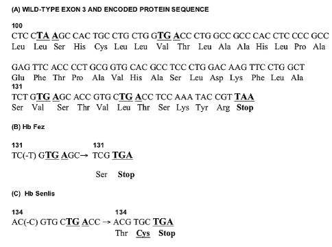
Hb Fez and Hb Senlis (Table 1) are specified by a single nucleotide deletion leading to FS and a premature stop codon. Hb Fez was identified in a Moroccan patient with Heinz bodies observed on the blood film, without any visible abnormal Hb. The (-T) deletion, within CDα1-131, leads to a synonymous change at CD132 (TCT>TCG, both encoding Ser), followed by TGA (Stop), and a resulting 131-residue long protein. Hb Senlis was identified in a French Caucasian patient without any apparent abnormal Hb. The (-C) deletion within CDα1-134 leads to FS with two novel residues (Thr and Cys) encoded by codons 134 and 135 (ACG and TGC, respectively), followed by a Stop codon (TGA), and a resulting 135-residue long protein.
Deletion within the 3rd exon has a different outcome whether affecting the α- or β globin-encoding genes. In the α genes, the meaningfulness of the FS is consequent upon its occurrence within the gene sequence (Figure 1). When the deletion involves a nucleotide located between CD100 and 106, a stop is met at position 101 or 107, leading to a protein where helix H is missing, and thus unable to interact with AHSP or the β chain to form α1β1 dimers. Such mutants are likely to be α-thalassemic. A nucleotide deletion occurring between CD107 and 132 (the next potential stop), will more or less significantly alter the structure of helix H, depending how early it occurs within the sequence. As for Hb Fez and Hb Senlis, with stop codons at positions 132 or 136, respectively, the deleted residues are located by the end of helix H, in a region that does not interact with either AHSP or the β-chain partner.6 Therefore, an abnormal, unstable, Hb tetramer may likely form and rapidly precipitate within the erythrocyte, accounting for the observed CHA. Furthermore, in Hb Senlis, a Cys residue occupies the C-terminus, allowing for possible interchain S-S bonds. When FS occurs more distal within an α2 gene sequence, the next stop is at position 147, leading to Hb Wayne,7 a relatively stable molecule. In the α1 gene, the nearest potential stop following codon 136 will occur only at position 173, a possibility unreported to this day.
Figure 1.
Nucleotide sequence of exon 3 in wild-type and mutated α globin genes, with resulting protein sequences. (A) Wild-type sequences of exon 3 in the α gene showing the potential stops (bold and underlined) that may result from FS. (B) In Hb Fez, the 3rd nucleotide of codon 131 is deleted, leading to FS and occurrence of a stop at CD132. (C) In Hb Senlis, deletion of a C within CD134 leads to a stop at CD136 and to a Cys as the outermost residue of the C-terminus.
In the β globin gene, deletion of 1 or 2 nucleotides within the 3rd exon leads to FS with occurrence of a stop codon at position 156. In such cases where FS occurs proximal within the sequence, helix H is drastically altered by incorporation of several hydrophobic residues (six Leu, two Ile, four Trp), leading to dominant β thalassemia syndrome.8 When the deletion is located at the very end of the 3rd exon, as in Hb Tak,9 the mere outcome is a 10-residue-long C-terminal tail leading to moderate instability and high oxygen affinity.
Unlike most other α gene defects reported, the mutations described here have a dominant effect. Thus, the biological consequences of those mutations, whether missense or FS, are highly dependent upon their occurrence within the gene sequence.
References
- 1.Hardison RC, Chui DHK, Giardine B, Riemer C, Patrinos GP, Anagnou N, et al. HbVar: a relational database of human hemoglobin variants and thalassemia mutations at the globin gene server. Hum Mutat. 2002;19:225–33. doi: 10.1002/humu.10044. [DOI] [PubMed] [Google Scholar]
- 2.Giardine B, van Baal S, Kaimakis P, Riemer C, Miller W, Samara M, et al. HbVar database of human hemoglobin variants and thalassemia mutations: 2007 update. Hum Mutat. 2007;28:206. doi: 10.1002/humu.9479. [DOI] [PubMed] [Google Scholar]
- 3.Vasseur C, Domingues-Hamdi E, Brillet T, Marden MC, Baudin-Creuza V. The α-hemoglobin stabilizing protein and expression of unstable a-Hb variants. Clin Biochem . 2009 May 29; doi: 10.1016/j.clinbiochem.2009.05.011. [Epub ahead of print] [DOI] [PubMed] [Google Scholar]
- 4.Wajcman H, Traeger-Synodinos J, Papassotiriou I, Giordano PC, Harteveld CL, Baudin-Creuza V, et al. Unstable and thalassemic α chain hemoglobin variants: a cause of Hb H disease and thalassemia intermedia. Hemoglobin. 2008;32:327–49. doi: 10.1080/03630260802173833. [DOI] [PubMed] [Google Scholar]
- 5.Moradkhani K, Mazurier E, Giordano PC, Wajcman H, Préhu C. An α 0-thalassemia-like mutation: Hb Suan-Dok [α109(G16)Leu_Arg] carried by a recombinant -α (3.7) gene. Hemoglobin. 2008;32:419–24. doi: 10.1080/03630260802173619. [DOI] [PubMed] [Google Scholar]
- 6.Feng L, Gell DA, Zhou S, Gu L, Kong Y, Li J, et al. Molecular mechanism of AHSP-mediated stabilization of α-hemoglobin. Cell. 2004;119:629–40. doi: 10.1016/j.cell.2004.11.025. [DOI] [PubMed] [Google Scholar]
- 7.Seid-Akhavan M, Winter WP, Abramson RK, Rucknagel DL. Hemoglobin Wayne: a frameshift mutation detected in human hemoglobin α chains. Proc Natl Acad Sci USA. 1976;73:882–6. doi: 10.1073/pnas.73.3.882. [DOI] [PMC free article] [PubMed] [Google Scholar]
- 8.Préhu C, Pissard S, Al-Sheikh M, Le Niger C, Bachir D, Galactéros F, et al. Two French Caucasian families with dominant thalassemia-like phenotypes due to hyper unstable hemoglobin variants: Hb Sainte Seve [codon 118 (-T)] and codon 127 [CAG_TAG (Gln_Stop]) Hemoglobin. 2005;29:229–33. doi: 10.1081/hem-200066335. [DOI] [PubMed] [Google Scholar]
- 9.Flatz G, Kinderlerer JL, Kilmartin JV, Lehmann H. Haemoglobin Tak: a variant with additional residues at the end of the β-chains. Lancet. 1971;1:732–3. doi: 10.1016/s0140-6736(71)91994-5. [DOI] [PubMed] [Google Scholar]


