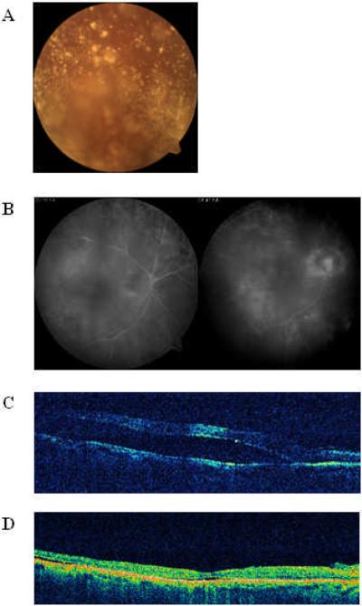
Figure 1.
Imaging Studies for Case 1. (A) Color photography demonstrates asteroid hyalosis light scatter (B) Angiographic evaluation displays a region of ill-defined inferotemporal hyperfluorescence (C) Preoperative OCT demonstrates displays a low-lying fovea-involving neurosensory detachment in the temporal macula (D) Postoperative OCT displays successful retinal detachment repair

