Abstract
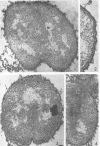
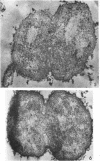
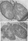
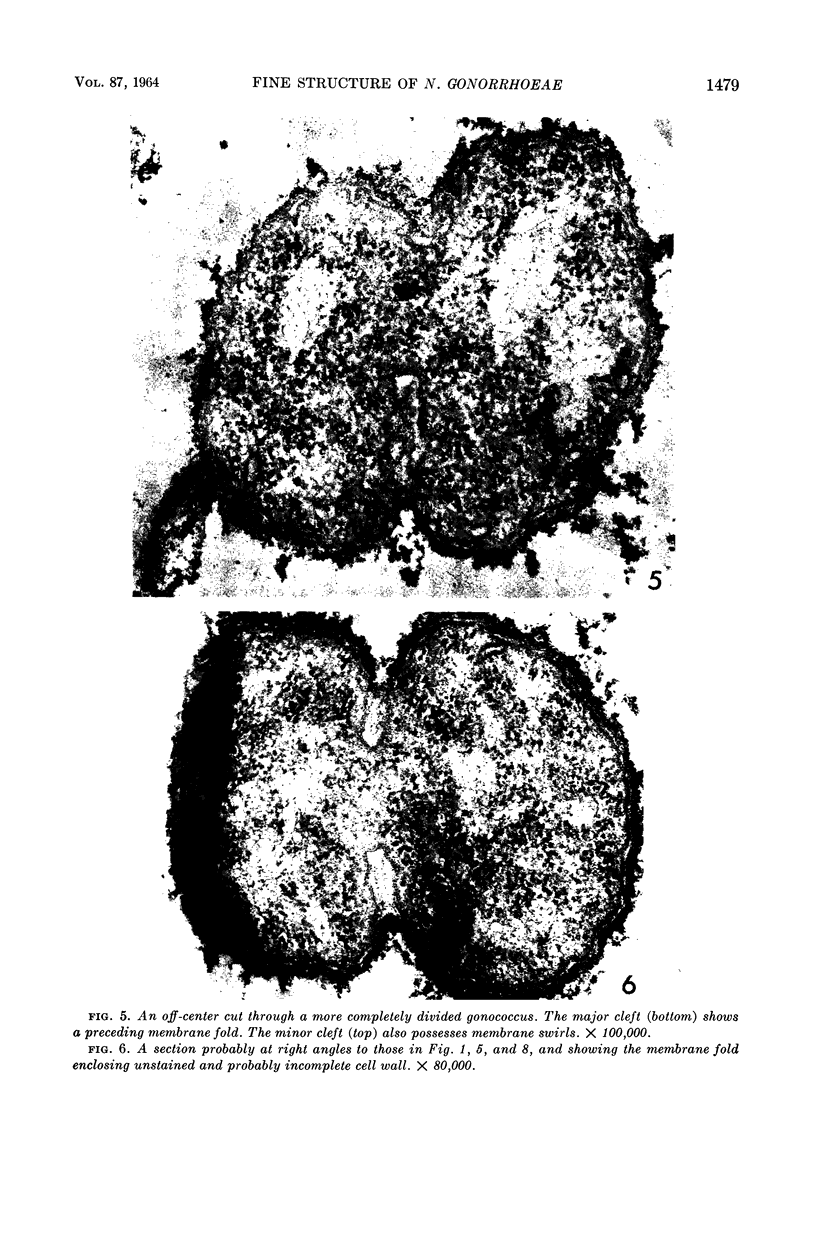
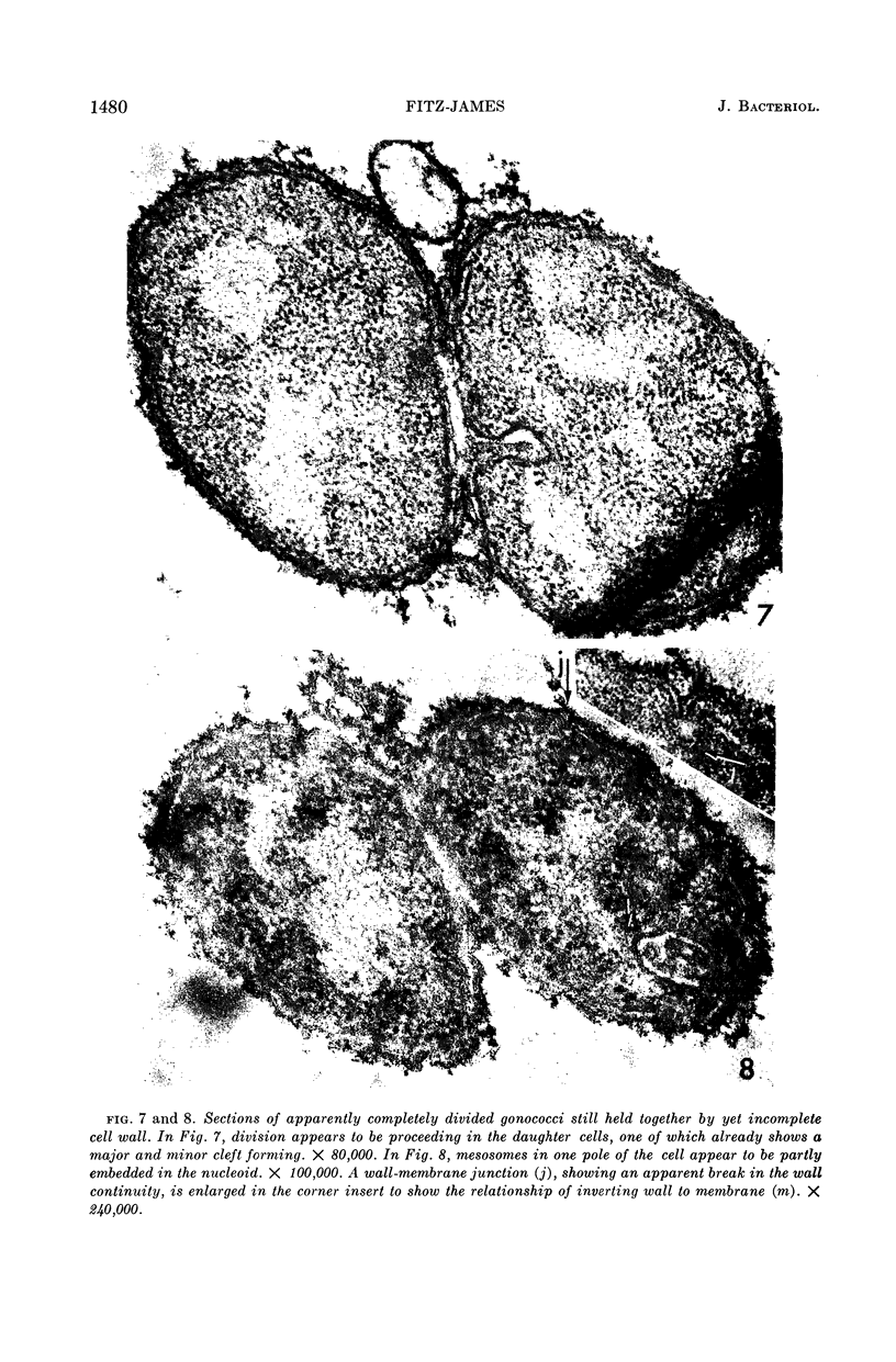
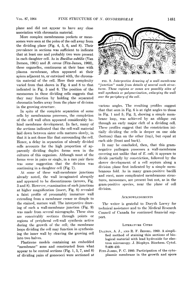
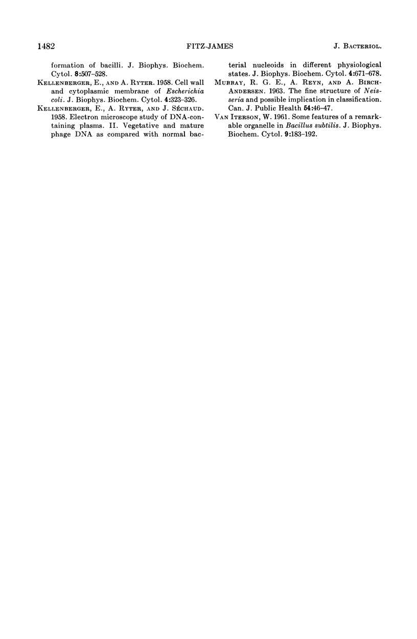
Fitz-James, Philip (University of Western Ontario, London, Ontario, Canada). Thin sections of dividing Neisseria gonorrhoeae. J. Bacteriol. 87:1477–1482. 1964.—The fine structure of the gram-negative pathogen Neisseria gonorrhoeae is described. Its cell-wall and membrane structures are not unlike those of Escherichia coli, possessing periodic points where wall and membrane are approximated. It appears to divide by a process of unequal constriction plus septum formation, preceded by a loop of membrane. Mesosomes are also seen but in the cell periphery away from the division plane. At some of the wall-membrane junctions, the cell wall appears to arise out of the membrane.
Full text
PDF

Images in this article
Selected References
These references are in PubMed. This may not be the complete list of references from this article.
- DALTON A. J., ZEIGEL R. F. A simplified method of staining thin sections of biolgical material with lead hydroxide for electron microscopy. J Biophys Biochem Cytol. 1960 Apr;7:409–410. doi: 10.1083/jcb.7.2.409. [DOI] [PMC free article] [PubMed] [Google Scholar]
- KELLENBERGER E., RYTER A. Cell wall and cytoplasmic membrane of Escherichia coli. J Biophys Biochem Cytol. 1958 May 25;4(3):323–326. doi: 10.1083/jcb.4.3.323. [DOI] [PMC free article] [PubMed] [Google Scholar]
- KELLENBERGER E., RYTER A., SECHAUD J. Electron microscope study of DNA-containing plasms. II. Vegetative and mature phage DNA as compared with normal bacterial nucleoids in different physiological states. J Biophys Biochem Cytol. 1958 Nov 25;4(6):671–678. doi: 10.1083/jcb.4.6.671. [DOI] [PMC free article] [PubMed] [Google Scholar]
- VAN ITERSON W. Some features of a remarkable organelle in Bacillus subtilis. J Biophys Biochem Cytol. 1961 Jan;9:183–192. doi: 10.1083/jcb.9.1.183. [DOI] [PMC free article] [PubMed] [Google Scholar]


