Abstract
Cystic echinococcosis (CE) is a widespread and severe zoonotic disease caused by infection with the larval stage of the eucestode Echinococcus granulosus sensu lato. The polymorphism exhibited by nuclear and mitochondrial markers conventionally used for the genotyping of different parasite species and strains does not reach the level necessary for the identification of genetic variants linked to restricted geographical areas. EmsB is a tandemly repeated multilocus microsatellite that proved its usefulness for the study of genetic polymorphisms within the species E. multilocularis, the causative agent of alveolar echinococcosis. In the present study, EmsB was used to characterize E. granulosus sensu lato samples collected from different host species (sheep, cattle, dromedaries, dogs, and human patients) originating from six different countries (Algeria, Mauritania, Romania, Serbia, Brazil, and the People's Republic of China). The conventional mitochondrial cox1 and nad1 markers identified genotypes G1, G3, G5, G6, and G7, which are clustered into three groups corresponding to the species E. granulosus sensu stricto, E. ortleppi, and E. canadensis. With the same samples, EmsB provided a higher degree of genetic discrimination and identified variations that correlated with the relatively small-scale geographic origins of the samples. In addition, one of the Brazilian single hydatid cysts presented a hybrid genotypic profile that suggested genetic exchanges between E. granulosus sensu stricto and E. ortleppi. In summary, the EmsB microsatellite exhibits an interesting potential for the elaboration of a detailed map of the distribution of genetic variants and therefore for the determination and tracking of the source of CE.
Cystic echinococcosis (CE) is a widespread and severe zoonosis caused by infection with the larval stage of the eucestode Echinococcus granulosus sensu lato. Classification of the organisms within this paraphyletic taxon has undergone and continues to undergo important changes. Mitochondrial DNA-based studies have shown that E. granulosus sensu lato is composed of 10 heterogeneous groups of variants, defined as strains (strains G1 to G10) (5, 6, 17). However, these strains are now reorganized within distinct species (21, 25). E. granulosus sensu stricto encompasses strains G1, G2, and G3; E. equinus corresponds to strain G4; and E. ortleppi comprises strain G5. Strains G6, G7, G8, G9, and G10 have been also classified under a well-supported monophyletic species, E. canadensis (16, 19, 21). Recently, the lion strain has been characterized as another new species, E. felidis (11).
Currently, mitochondrial and nuclear markers are not sufficiently polymorphic for use for the identification of genetic variations that could reflect geographically based peculiarities. The use of sensitive tools such as microsatellites may provide more information about the polymorphism of the parasite and the spatial-temporal characteristics of its patterns of transmission between foci. However, to date, only four single-locus microsatellites have been used to investigate E. granulosus sensu lato isolates: U1snRNA, EgmSca 1, EgmSca 2, and EgmSga 1. The U1snRNA gene exhibited 11 distinct profiles: 8 for E. granulosus sensu stricto (G1/G2), 2 for E. ortleppi (G5), and 1 for E. canadensis (G6) (22). However, no spatial correlation with the geographic origin of the isolates was observed. Among the three microsatellites described by Bartholomei-Santos et al. (4), only EgmSca 1 correlated the genotypes with the origins of the samples.
Recently, Bart et al. (3) developed EmsB, a tandemly repeated multilocus microsatellite, for the genotyping of E. multilocularis. This marker showed a higher level of intraspecific variability compared with that shown by any previously published marker, as well as a very high degree of sensitivity (7, 13-15). It is composed of an array of 800-bp DNA fragments containing a variable combination of CA and GA repeats. The use of such a microsatellite could contribute to the better identification of the spatial-temporal characteristics of the E. granulosus transmission patterns, as is the case for E. multilocularis. Indeed, using this new tool, Knapp et al. managed to perform efficient genetic tracking of E. multilocularis isolates in different foci of alveolar echinococcosis (13-15). Furthermore, because of its localization within the nuclear genome, cross-fertilization processes may modify EmsB patterns. Therefore, this microsatellite may be an interesting marker for use for both assessment of the genetic polymorphism of E. granulosus sensu lato and detection of the genetic exchange events between the variants.
In the present work, we tackled its variability using a panel of 127 E. granulosus sensu lato samples collected in six countries where CE is endemic.
MATERIALS AND METHODS
Isolates.
One hundred twenty-seven unilocular hydatid cysts (n = 125) and adult worms (n = 2) were collected from different intermediate or definitive hosts (their characteristics are reported in Table 1) in six different countries where CE is endemic. The cysts from animals were isolated in slaughterhouses by local veterinarians. The cysts from human patients were surgically resected in hospitals for the treatment of CE and were then given anonymously to the Department of Parasitology and Mycology, UMR6249, for scientific research. The two adult worms were isolated by local veterinarians from the intestines of two dogs captured and euthanatized during a campaign to regulate the stray dog population in Qinghai Province, People's Republic of China.
TABLE 1.
Characteristics of the 66 clusters of samples showing the same profile with the EmsB multilocus microsatellite (E1 to E66)
| EmsB profile | Characteristics of samples showing the EmsB profiles |
|||||
|---|---|---|---|---|---|---|
| No. of samples | Origin | Host | Mitochondrial polymorphisma: |
Species | ||
| cox1 | nad1 | |||||
| E1 | 2 | China | Fox | M1 | M1 | E. multilocularis |
| E2 | 3 | China | Dog, cattle, and human | G1 and G1 (246G → T) | G1 (282C → T) | E. granulosus sensu stricto |
| E3 | 2 | Algeria | Sheep | G1 and G1 (56 → T) | G1 (282C → T) | E. granulosus sensu stricto |
| E4 | 1 | Algeria | Cattle | G1 | G1 (282C → T) | E. granulosus sensu stricto |
| E5 | 1 | Brazil | Cattle | G1 | G1 (282C → T) | E. granulosus sensu stricto |
| E6 | 1 | Serbia | Human | G1 (154A → G, 211A → C) | G1 (282C → T) | E. granulosus sensu stricto |
| E7 | 1 | Algeria | Sheep | G1 | G1 (282C → T) | E. granulosus sensu stricto |
| E8 | 1 | Romania | Cattle | G1 | G1 (282C → T) | E. granulosus sensu stricto |
| E9 | 2 | Romania | Sheep | G1 | G1 (282C → T) | E. granulosus sensu stricto |
| E10 | 2 | Algeria | Sheep | G1 and G3 | G1 (282C → T) and G2/G3 (282C → T) | E. granulosus sensu stricto |
| E11 | 1 | Algeria | Cattle | G1 (56C → T) | G1 (150T → C, 282C → T) | E. granulosus sensu stricto |
| E12 | 1 | Algeria | Dromedary | G1 | G1 (282C → T) | E. granulosus sensu stricto |
| E13 | 1 | China | Human | G1 | G1 (282C → T) | E. granulosus sensu stricto |
| E14 | 2 | China | Human | G1 | G1 (282C → T) | E. granulosus sensu stricto |
| E15 | 1 | China | Human | G1 | G1 (282C → T) | E. granulosus sensu stricto |
| E16 | 1 | China | Human | G1 | G1 (282C → T) | E. granulosus sensu stricto |
| E17 | 1 | China | Cattle | G1 (123T → C) | G1 (282C → T) | E. granulosus sensu stricto |
| E18 | 4 | China | Cattle and human | G1, G1 (123T → C), and G3 | G1 (282C → T) and G2/G3 (282C → T) | E. granulosus sensu stricto |
| E19 | 1 | China | Human | G1 | G1 (282C → T, 426A → G) | E. granulosus sensu stricto |
| E20 | 2 | China | Human | G3 | G2/G3 (282C → T) | E. granulosus sensu stricto |
| E21 | 2 | China | Human | G1 | G1 (282C → T) | E. granulosus sensu stricto |
| E22 | 2 | China | Human | G1 | G1 (282C → T) | E. granulosus sensu stricto |
| E23 | 4 | China | Sheep | G1 (246G → T) and G3 | G1 (282C → T) and G2/G3 (282C → T) | E. granulosus sensu stricto |
| E24 | 1 | China | Dog | G1 | G1 (282C → T) | E. granulosus sensu stricto |
| E25 | 2 | China | Sheep | G1 and G1 (246G → T) | G1 (282C → T) | E. granulosus sensu stricto |
| E26 | 2 | Algeria | Cattle and human | G1 (66C → T) and G1 (108T → C, 111C → T) | G1 (282C → T) | E. granulosus sensu stricto |
| E27 | 4 | Algeria and Serbia | Cattle and human | G1 and G1 (220A → G) | G1 (282C → T) | E. granulosus sensu stricto |
| E28 | 1 | Algeria | Cattle | G1 | G1 (282C → T) | E. granulosus sensu stricto |
| E29 | 2 | Algeria | Sheep and cattle | G1 and G1 (56C → T) | G1 (282C → T) and G1 (121A → G, 237G → A, 282C → T) | E. granulosus sensu stricto |
| E30 | 1 | Romania | Pig | G3 | G2/G3 (282C → T) | E. granulosus sensu stricto |
| E31 | 2 | Algeria | Sheep and human | G1 | G1 (282C → T) and G1 (66C → T, 121A → G, 282C → T) | E. granulosus sensu stricto |
| E32 | 3 | Algeria and Romania | Sheep and cattle | G1 | G1 (282C → T) | E. granulosus sensu stricto |
| E33 | 2 | Algeria and Romania | Sheep and cattle | G1 | G1 (282C → T) | E. granulosus sensu stricto |
| E34 | 3 | Algeria | Cattle | G1 | G1 (282C → T | E. granulosus sensu stricto |
| E35 | 4 | Algeria and Romania | Sheep and cattle | G1, G1 (312G → A), G1 (56C → T, 325A → G), and G3 | G1 (282C → T) and G2/G3 (237T → A, 282C → T) | E. granulosus sensu stricto |
| E36 | 4 | Algeria, Romania and Brazil | Cattle and human | G1 and G1 (73T → C) | G1 (282C → T and G1 (208T → G, 282C → T) | E. granulosus sensu stricto |
| E37 | 2 | Romania and Brazil | Cattle | G1 | G1 (282C → T) and G1 (28C → T, 181T → C, 282C → T) | E. granulosus sensu stricto |
| E37 | Sample B395b | Brazil | Cattle | G1 | G1 (282C → T) | E. granulosus sensu stricto/E. ortleppi |
| E38 | 7 | Algeria and Romania | Sheep, cattle and human | G1 and G3 (228A → G) | G1 (282C → T) and G2/G3 (282C → T) | E. granulosus sensu stricto |
| E39 | 3 | Algeria and Romania | Sheep and cattle | G1 (56C → T, 111C → T), G1 (204T → G), and G3 | G1 (282C → T) and G2/G3 (282C → T) | E. granulosus sensu stricto |
| E40 | 2 | Algeria | Cattle | G1 | G1 (282C → T) | E. granulosus sensu stricto |
| E41 | 2 | Romania | Cattle | G3 | G2/G3 (282C → T) | E. granulosus sensu stricto |
| E42 | 2 | Romania | Sheep and cattle | G1 (66C → T, 228A → G) and G3 | G2/G3 (282C → T) | E. granulosus sensu stricto |
| E43 | 1 | Romania | Sheep | G1 | G1 (282C → T) | E. granulosus sensu stricto |
| E44 | 1 | China | Human | G1 | G1 (228T → C, 282C → T) | E. granulosus sensu stricto |
| E45 | 10 | China | Human | G1 and G3 | G1 (282C → T), G1 (228T → C, 282C → T), and G2/G3 (282C → T) | E. granulosus sensu stricto |
| E46 | 3 | Brazil | Cattle | G5 | G5 | E. ortleppi |
| E46 | Sample B395b | Brazil | Cattle | G1 | G1 (282C → T) | E. granulosus sensu stricto/E. ortleppi |
| E47 | 1 | Mauritania | Human | G6 (129T → C, 269T → C) | G6 | E. canadensis |
| E48 | 1 | Mauritania | Dromedary | G6 | G6 | E. canadensis |
| E49 | 1 | Mauritania | Dromedary | G6 | G6 | E. canadensis |
| E50 | 2 | Mauritania | Dromedary, Human | G6 | G6 and G6 (165T → C) | E. canadensis |
| E51 | 1 | Mauritania | Dromedary | G6 | G6 | E. canadensis |
| E52 | 2c | Mauritania | Dromedary | G6 | G6 | E. canadensis |
| E53 | 3 | Algeria | Dromedary | G6 | G6 | E. canadensis |
| E54 | 1 | Algeria | Dromedary | G6 | G6 | E. canadensis |
| E55 | 1 | Algeria | Dromedary | G6 | G6 | E. canadensis |
| E56 | 1 | Serbia | Human | G7 (195T → C) | G7 | E. canadensis |
| E57 | 1 | Mauritania | Dromedary | G6 | G6 | E. canadensis |
| E58 | 1 | Mauritania | Dromedary | G6 | G6 | E. canadensis |
| E59 | 1 | Serbia | Human | G7 | G7 | E. canadensis |
| E60 | 1 | Romania | Pig | G7 | G7 | E. canadensis |
| E61 | 2 | Romania | Pig | G7 | G7 | E. canadensis |
| E62 | 1 | Romania | Pig | G7 | G7 | E. canadensis |
| E63 | 1 | Romania | Pig | G7 | G7 | E. canadensis |
| E64 | 1 | Algeria | Dromedary | G6 | G6 (165T → C) | E. canadensis |
| E65 | 1 | Algeria | Dromedary | G6 | G6 | E. canadensis |
| E66 | 1 | Algeria | Dromedary | G6 | G6 | E. canadensis |
The mitochondrial cox1 and nad1 sequences obtained from the 129 samples of this study were compared with the sequences reported by Bowles et al. (5, 6); their E. granulosus sensu lato genotypes G1, G2, G3, G5, G6, and G7 and E. multilocularis genotype M1 were used here as reference strains. With the marker nad1, G2 and G3 could not be distinguished (G2/G3). When a sequence showed additional mutations when it was compared to the sequence of the reference genotype, these mutations and their positions in this genotype are indicated in parentheses.
B395, E. granulosus sensu stricto/E. ortleppi hybrid Brazilian sample.
One of the two samples showing this EmsB profile was selected to study the repeatability of the EmsB profiles and determine the threshold used for hierarchical classification.
Among the 45 samples from Algeria, 24 samples (from sheep, cattle, and human patients) originated from the urban region along the Mediterranean coast of northern Algeria, while the other samples (from sheep, cattle, and dromedaries) came from the pastoral Ouargla/Touggourt region near the Sahara Desert. Ten samples (from human patients or cattle) came from the region of Nouakchott, the capital of Mauritania. Seven samples were isolated from cattle in the Brazilian region of Rio Grande do Sul. Thirty-six cysts (from sheep, cattle, and human patients) were collected in five different prefectures of Qinghai Province. Twenty-three samples (from sheep, cattle, and pigs) were collected from four different places in Romania, and four samples were taken from human patients in the hospital of Belgrade, Serbia. Two adult worms were collected from foxes killed by hunters in Qinghai Province.
DNA extraction, PCR amplification, and sequencing.
For each isolate, genomic DNA was extracted by using a High Pure PCR preparation kit (Roche Diagnostics, Mannheim, Germany).
A portion of the cox1 mitochondrial gene, which codes for subunit 1 of the cytochrome c oxidase and a portion of the nad1 mitochondrial gene, which codes for subunit 1 of the NADH dehydrogenase, were amplified by PCR (the primers have been described by Bart et al. [3]). PCR amplification was performed as previously reported by Maillard et al. (18). The presence of amplicons was checked by electrophoresis in a 1.5% (wt/vol) Tris-acetate-EDTA agarose gel. The sequencing protocols and the subsequent ethanol purification of the fragments were also the same as those reported by Maillard et al. (18).
Genotype identification and phylogenetic reconstruction.
The cox1 and nad1 mitochondrial gene sequences were aligned by using Bioedit software, version 7.0.5.3 (10). These sequences were compared to those reported by Bowles and colleagues (5, 6). The genetic distances were calculated in accordance with the Kimura two-parameter model of nucleotide substitution (12), and the samples were clustered by the neighbor-joining method (23). The phylogenic reconstructions were performed with MEGA software (24). The dendrograms were rooted with the sequences obtained from the two E. multilocularis samples, and a bootstrap test with 1,000 repetitions was done to evaluate the robustness of each dichotomy (8).
The EmsB tandemly repeated multilocus microsatellite.
PCR amplification of the EmsB fragments was carried out in a reaction mixture with a final volume of 10 μl containing 1 μl genomic DNA, 150 μM of each deoxynucleoside triphosphate (GeneAmp; Applied Biosystems), 0.5 μM of fluorescent forward primer/1 μM of nonlabeled reverse primer (the primers used have been described by Bart et al. [3]), and 0.5 U of Red Taq DNA polymerase in 1.5 μl 10× reaction buffer (Sigma-Aldrich, St. Louis, MO). The reaction was performed as follows: 30 cycles of denaturation at 94°C for 30 s, annealing at 60°C for 30 s, and extension at 72°C for 1 min. The presence of amplicons was determined by electrophoresis in 1.5% (wt/vol) Tris-acetate-EDTA agarose gels.
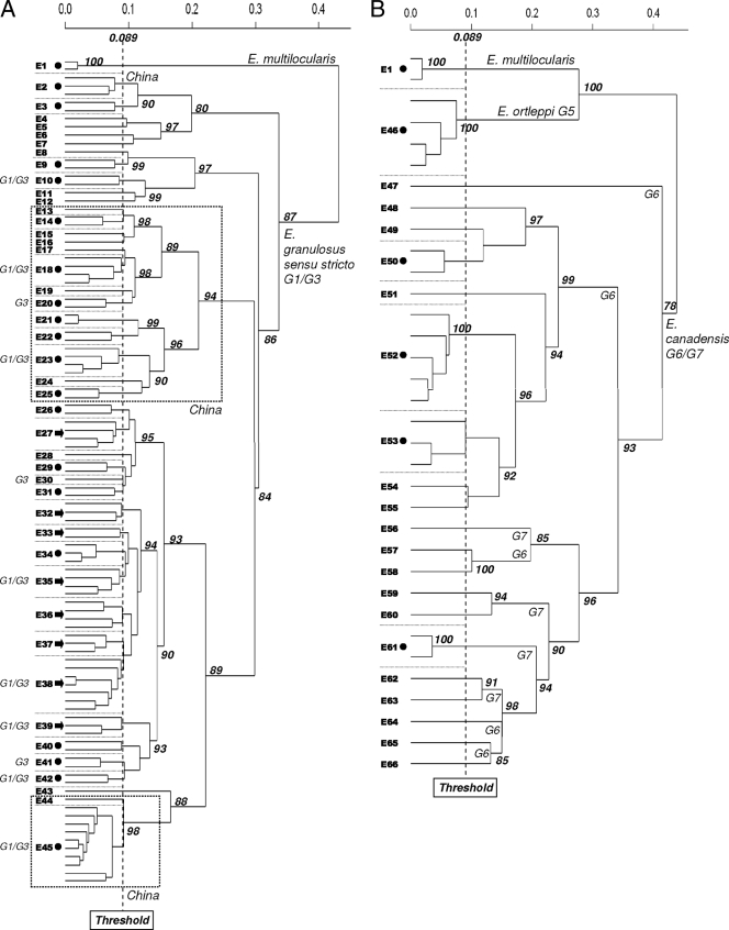
Fragment analysis was performed with an automatic sequencer (ABI Prism 3130; Applied Biosystems). For each sample, we obtained a profile composed of several peaks; the time of migration of each fragment is on the abscissa, and the intensity of the fluorescence is on the ordinate. These parameters were directly related to the size of each locus and the number of repetitions of the locus in the genome (2). For the mathematical study of these profiles, we followed the procedures reported by Knapp et al. (13-15). By using R software and the pvclust library (20), the Euclidean distances between these profiles were calculated and a hierarchical clustering analysis was performed with an average linkage algorithm (see Fig. 2). The robustness of each dichotomy was calculated with a bootstrap test of 1,000 iterations (8). Knapp et al. tested the reproducibility and repeatability of EmsB microsatellite analyses by comparing the profiles obtained from several experiments performed with the same isolate maintained in vivo for several months and calculated a genetic threshold for identification of the isolate clusters (14). In order to adapt this threshold to the experimental conditions of the present work and to ascertain the clonality of the protoscoleces for the EmsB microsatellite, one cyst (cyst M110) was selected from the panel, and the protocol was performed four times: four distinct tubes, each of which contained 25 mg fragment of the germinal layer and a pool of protoscoleces, were investigated. The tubes were then used separately for DNA extraction, PCR amplification, and EmsB fragment analysis. The Euclidean distances between the four profiles were calculated, and a threshold was determined by using the same process described by Knapp et al. (14). Below this limit, the profiles were considered identical.
FIG. 2.
Hierarchical clustering analysis tree constructed with the 129 profiles provided by the EmsB multilocus microsatellite. The Euclidean distances calculated between the profiles are represented on the abscissa. A bootstrap test was done to assess the robustness of the branches. All mathematical analyses were performed with R software (20). Two dendrograms are drawn: the first one included all 99 E. granulosus sensu stricto G1/G3 samples (A), while the second one included the 25 E. canadensis G6/G7 samples and the 3 E. orlteppi G5 samples (B). A threshold was calculated with the four profiles obtained with different pools of protoscoleces and fragments of the germinal layer of the same cyst (cyst M110). Under this threshold, the EmsB profiles are considered to be identical. By this method, E1 to E66 represent clusters of samples showing the same profile. The characteristics of these clusters are detailed in Table 1. When a profile was found in more than one sample, a symbol was added: circles, all the samples showing this profile were collected from the same area; arrows, all the samples were collected from different areas.
RESULTS
Mitochondrial polymorphism.
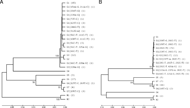
The cox1 and nad1 sequences were compared to those reported by Bowles and colleagues (5, 6). The genotypes identified in this study and detailed in Table 1 were used to draw two mitochondrial dendrograms, represented in Fig. 1. The two adult worms collected from foxes belonged to the species E. multilocularis and were used as the outgroup control.
FIG. 1.
Dendrograms constructed with the cox1 (A) and nad1 (B) mitochondrial gene sequences obtained from the 129 samples evaluated in this study. MEGA software was used (24). A bootstrap test with 1,000 repetitions was performed, and values above 75% are indicated. The sequences were compared to the sequences of E. granulosus sensu lato genotypes G1, G3, G6, and G7 and E. multilocularis genotypes M1 and M2 reported by Bowles et al. (5, 6). When a sequence showed mutations compared to the sequence of the reference genotype, these mutations and their positions in this genotype are indicated in brackets. For each sequence, the corresponding number of samples is reported in parentheses.
The EmsB tandemly repeated multilocus microsatellite.
All of the fragments produced by the amplification of EmsB were between 250 bp and 310 bp for samples from E. granulosus sensu stricto and E. canadensis and between 220 bp and 240 bp for samples from E. otleppi and E. multilocularis. The four profiles obtained by the analysis of sample M110 four times were extremely similar. The threshold calculated from these profiles was equal to 0.089. At this limit, 44 EmsB profiles were discriminated within E. granulosus sensu stricto samples (samples E2 to E45): 20 within E. canadensis samples (samples E47 to E66), 1 within E. ortleppi samples (sample E46), and 1 within E. multilocularis samples (the outgroup control, sample E1). In a global hierarchical classification of all the samples based on the similarities of the EmsB profiles, no E. granulosus sensu stricto profile clustered with the E. canadensis profiles. To obtain more robust ramifications and better visibility, the classification was subdivided into two dendrograms (Table 1 and Fig. 2). The first dendrogram included the E. granulosus sensu stricto samples, and the second contained the E. canadensis and the E. ortleppi samples. In the first dendrogram, 44 distinct EmsB profiles were distinguished. Among those, 27 profiles included more than one sample. Nineteen of them grouped samples collected in the same area, and eight corresponded to samples collected from different foci. The hierarchical clustering analysis showed that 34 of the 37 Chinese samples were regrouped into two specific groups. The most important was an assemblage of 13 profiles found in 24 samples (samples E13 to E25 in Table 1). The second assemblage was formed by two profiles (named E44 and E45 in Table 1) that were weakly separated, and 1 and 10 samples, respectively, had those two profiles. The 13 G3 samples provided 11 different EmsB profiles, but 8 of them were also identified in G1 samples.
In the second dendrogram, the three E. ortleppi samples showed the same profile. Only 4 of the 20 E. canadensis profiles reassembled more than one sample. Each of these four profiles corresponded to a given geographical location. For the seven G7 samples, the EmsB microsatellite produced six different profiles. Even though we found no profile that was shared by the G6 and G7 samples, the hierarchical clustering analysis failed to separate the G6 profile from the G7 profile, and all of them were mixed in a monophyletic cluster of profiles (Fig. 2).
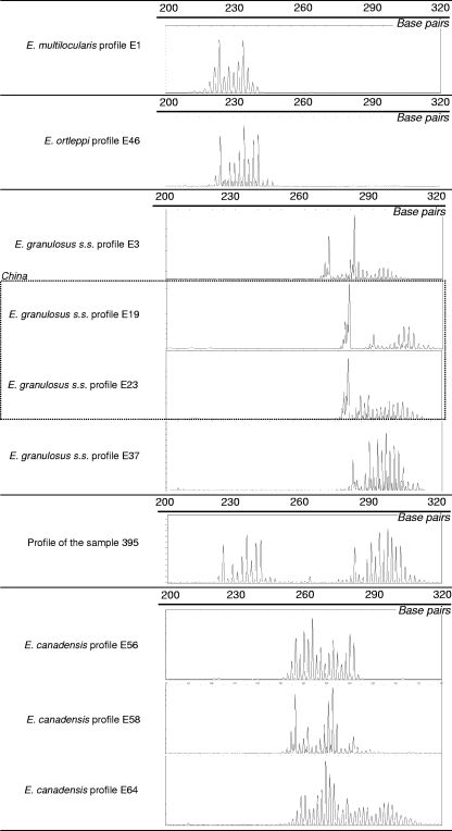
Using the nuclear marker EmsB, we also obtained a hybrid profile for a Brazilian isolate (Fig. 3). This profile was composed of two ranges of peaks: the first range was between 220 bp and 240 bp (13 peaks, sample E46), and the other was between 250 bp and 310 bp (21 peaks, sample E36). When they were added to the hierarchical clustering analysis of the EmsB profiles, the first range strongly clustered with the Brazilian E. ortleppi samples and the second range clustered with the Brazilian E. granulosus sensu stricto samples. The cox1 and nad1 sequences obtained from this sample were clearly G1, which allowed us to completely exclude the hypothesis that a mixture of two samples (one G1 and one G5) was present.
FIG. 3.
Examples of the profiles obtained with the EmsB multilocus microsatellite. The lengths of the EmsB fragments are indicated on the abscissa, and the measure of fluorescence is indicated on the ordinate. For Brazilian sample B395, EmsB showed a mixed E. granulosus sensu stricto (s.s.) and E. ortleppi profile.
DISCUSSION
The main objective of this study was to estimate the value of the EmsB microsatellite for the identification and the geographical tracking of variants within E. granulosus parasite populations (strains and other groups). By using two mitochondrial markers of reference, cox1 and nad1, and the EmsB multilocus microsatellite, we assessed the polymorphisms of 127 samples collected from six different hosts originating from six areas of endemicity: Algeria, Mauritania, Serbia, Romania, the People's Republic of China, and Brazil.
Mitochondrial markers could be discriminated into three groups, corresponding to samples from E. granulosus sensu stricto (99 samples), E. canadensis (25 samples), and E. ortleppi (3 samples). The results corroborated the regrouping of genotypes G1 and G3 into a monophyletic and robust species, E. granulosus sensu stricto, and the regrouping of G6 and G7 into another monophyletic and robust species, E. canadensis, while E. ortleppi could be clearly individualized. On a continental scale, mitochondrial sequences of samples belonging to the same species could sometimes specify the geographical origins of the samples. For instance, the E. canadensis G6 samples from North Africa differed slightly from the E. canadensis G7 samples from Europe. In this study, genotype G6 was found in dromedaries and human patients in Algeria and Mauritania, while genotype G7 was found in pigs and human patients in Romania and Serbia. Similarly, some specific mutations could be used to identify Chinese E. granulosus sensu stricto isolates. However, within a given species, mitochondrial markers failed to exhibit a polymorphism that could pinpoint the geographical origin of samples at the subcontinental scale.
On the other hand, the EmsB multilocus microsatellite showed a higher degree of polymorphism and discriminatory power than the mitochondrial markers. However, because we found an overlap between the profiles for E. granulosus sensu stricto and E. canadensis, as well as between the profiles for E. ortleppi and E. multilocularis, we do not recommend the use of this marker to identify a sample to the species level. At such a level, mitochondrial and nuclear DNAs are polymorphic enough to allow the species identification of isolates by sequencing of the PCR products. As previously reported for E. multilocularis, EmsB is useful for investigation of the genetic variability at the subspecies level, especially for epidemiological purposes (7, 13-15). The present study showed that the EmsB microsatellite was sufficiently polymorphic for assessment of populations of parasites at the scale of their foci. The two profiles showed by almost all of the Chinese samples suggest a local and isolated population of parasites and an absence of interactions with the other foci studied. Conversely, some G1 profiles were found in several foci and were able to spread from one continent to another. For example, the E36 profile clustered the genotype G1 samples collected from Algeria, Romania, and Brazil. Owing to the nature of the hosts involved in the developmental cycle of E. granulosus sensu lato, the circulation of genotypes may be attributed to worldwide commercial exchanges. With regard to the genotype G3 samples, the EmsB microsatellite provided eight profiles that had also been identified in G1 samples. This result corroborated the weak pertinence of this genetic discrimination and the regrouping of the G1 and G3 strains into a monophyletic species, E. granulosus sensu stricto. The G6 and G7 samples showed distinct profiles, but these profiles did not form specific groups in the hierarchical clustering analysis. The EmsB microsatellite distinguished the G6 and G7 samples, but despite the high degree of polymorphism provided by this tool, this distinction stays weak and could corroborate the regrouping of G6 and G7 into the monophyletic species E. canadensis. Furthermore, with the microsatellite, one of the Brazilian samples showed a G1/G5 hybrid profile. Such a pattern cannot be found in a pure clonal context. This result proved that cross-fertilization processes between E. granulosus sensu stricto and E. orlteppi have certainly occurred at least once. The observation of such a hybrid genotype could suggest the absence of a strongly installed reproductive barrier between these two species (1). The observation of such a heterozygote is very rare in the genus Echinococcus (9).
Overall, the EmsB microsatellite may potentially be useful for assessment of the genetic diversity within E. granulosus: both genotypes with a restricted distribution and genotypes with a widespread distribution were revealed in the field, and this might potentially have been driven by the trading of the hosts. Furthermore, EmsB genetic exchanges between distinct G1 and G5 populations of parasites circulating in the same focus may occur. As such, EmsB may be useful for assessment of the genetic polymorphism of E. granulosus and of its spatial distribution in detail. Knapp and colleagues earlier showed that this microsatellite enriched a powerful panel of markers that may be used to study the temporal and spatial development of E. multilocularis (7, 13-15). In the present study, we demonstrated that this observation also holds true for E. granulosus sensu lato. The next challenge will be to enlarge the panel of E. granulosus isolates and genotypes and to superimpose the genetic distribution map on ecoepidemiological data. The principal interest in such studies is the determination, at the local scale, of the genotypes that circulate in different foci of E. granulosus. The establishment of a detailed geographical map of genotypes could be essential to determine and track the source of CE. In addition, the accurate identification of the paths of circulation used by the parasite could be useful for isolation of the source and could thus increase the efficiency and the stability in time of the actions engaged against CE.
Footnotes
Published ahead of print on 9 September 2009.
REFERENCES
- 1.Badaraco, J. L., F. J. Ayala, J. M. Bart, B. Gottstein, and K. L. Haag. 2008. Using mitochondrial and nuclear markers to evaluate the degree of genetic cohesion among Echinococcus populations. Exp. Parasitol. 119:453-459. [DOI] [PubMed] [Google Scholar]
- 2.Bart, J. M., J. Knapp, B. Gottstein, F. El-Garch, P. Giraudoux, M. L. Glowatzki, H. Berthoud, S. Maillard, and R. Piarroux. 2006. EmsB, a tandem repeated multi-loci microsatellite, new tool to investigate the genetic diversity of Echinococcus multilocularis. Infect. Genet. Evol. 6:390-400. [DOI] [PubMed] [Google Scholar]
- 3.Bart, J. M., S. Morariu, J. Knapp, M. S. Ilie, M. Pitulescu, A. Anghel, I. Cosoroaba, and R. Piarroux. 2006. Genetic typing of Echinococcus granulosus in Romania. Parasitol. Res. 98:130-137. [DOI] [PubMed] [Google Scholar]
- 4.Bartholomei-Santos, M. L., L. S. Heinzelmann, R. P. Oliveira, G. Chemale, A. M. Gutierrez, L. Kamenetzky, K. L. Haag, and A. Zaha. 2003. Isolation and characterization of microsatellites from the tapeworm Echinococcus granulosus. Parasitology 126(Pt 6):599-605. [PubMed] [Google Scholar]
- 5.Bowles, J., D. Blair, and D. P. McManus. 1992. Genetic variants within the genus Echinococcus identified by mitochondrial DNA sequencing. Mol. Biochem. Parasitol. 54:165-173. [DOI] [PubMed] [Google Scholar]
- 6.Bowles, J., and D. P. McManus. 1993. NADH dehydrogenase 1 gene sequences compared for species and strains of the genus Echinococcus. Int. J. Parasitol. 23:969-972. [DOI] [PubMed] [Google Scholar]
- 7.Casulli, A., J. M. Bart, J. Knapp, G. La Rosa, G. Dusher, B. Gottstein, A. Di Cerbo, M. T. Manfredi, C. Genchi, R. Piarroux, and E. Pozio. 2009. Multi-locus microsatellite analysis supports the hypothesis of an autochthonous focus of Echinococcus multilocularis in northern Italy. Int. J. Parasitol. 39:837-842. [DOI] [PubMed] [Google Scholar]
- 8.Felsenstein, J. 1985. Confidence limits on phylogenies: an approach using the bootstrap. Evolution 39:783-791. [DOI] [PubMed] [Google Scholar]
- 9.Haag, K. L., A. M. Araujo, B. Gottstein, M. Siles-Lucas, R. C. Thompson, and A. Zaha. 1999. Breeding systems in Echinococcus granulosus (Cestoda; Taeniidae): selfing or outcrossing? Parasitology 118(Pt 1):63-71. [DOI] [PubMed] [Google Scholar]
- 10.Hall, T. A. 1999. BioEdit: a user-friendly biological sequence alignment editor and analysis program for Windows 95/98/NT. Nucleic Acids Symp. Ser. 41:95-98. [Google Scholar]
- 11.Huttner, M., M. Nakao, T. Wassermann, L. Siefert, J. D. Boomker, A. Dinkel, Y. Sako, U. Mackenstedt, T. Romig, and A. Ito. 2007. Genetic characterization and phylogenetic position of Echinococcus felidis (Cestoda: Taeniidae) from the African lion. Int. J. Parasitol. 38:861-868. [DOI] [PubMed] [Google Scholar]
- 12.Kimura, M. 1980. Average time until fixation of a mutant allele in a finite population under continued mutation pressure: studies by analytical, numerical, and pseudo-sampling methods. Proc. Natl. Acad. Sci. USA 77:522-526. [DOI] [PMC free article] [PubMed] [Google Scholar]
- 13.Knapp, J., J.-M. Bart, P. Giraudoux, M. L. Glowatzki, I. Breyer, F. Raoul, P. Deplazes, G. Dusher, K. Martinek, P. Dubinsky, M. H. Guislain, F. Cliquet, T. Romig, P. Malczewski, B. Gottstein, and R. Piarroux. 2009. Genetic diversity of the cestode Echinococcus multilocularis in red foxes at a continental scale in Europe. PLoS Negl. Trop. Dis. 3:e452. [DOI] [PMC free article] [PubMed] [Google Scholar]
- 14.Knapp, J., J. M. Bart, M. L. Glowatzki, A. Ito, S. Gerard, S. Maillard, R. Piarroux, and B. Gottstein. 2007. Assessment of use of microsatellite polymorphism analysis for improving spatial distribution tracking of Echinococcus multilocularis. J. Clin. Microbiol. 45:2943-2950. [DOI] [PMC free article] [PubMed] [Google Scholar]
- 15.Knapp, J., M. H. Guislain, J. M. Bart, F. Raoul, B. Gottstein, P. Giraudoux, and R. Piarroux. 2008. Genetic diversity of Echinococcus multilocularis on a local scale. Infect. Genet. Evol. 8:367-373. [DOI] [PubMed] [Google Scholar]
- 16.Lavikainen, A., M. J. Lehtinen, S. Laaksonen, E. Agren, A. Oksanen, and S. Meri. 2006. Molecular characterization of Echinococcus isolates of cervid origin from Finland and Sweden. Parasitology 133(Pt 5):565-570. [DOI] [PubMed] [Google Scholar]
- 17.Lavikainen, A., M. J. Lehtinen, T. Meri, V. Hirvela-Koski, and S. Meri. 2003. Molecular genetic characterization of the Fennoscandian cervid strain, a new genotypic group (G10) of Echinococcus granulosus. Parasitology 127(Pt 3):207-215. [DOI] [PubMed] [Google Scholar]
- 18.Maillard, S., M. C. Benchikh-Elfegoun, J. Knapp, J. M. Bart, P. Koskei, B. Gottstein, and R. Piarroux. 2007. Taxonomic position and geographical distribution of the common sheep G1 and camel G6 strains of Echinococcus granulosus in three African countries. Parasitol. Res. 100:495-503. [DOI] [PubMed] [Google Scholar]
- 19.Nakao, M., D. P. McManus, P. M. Schantz, P. S. Craig, and A. Ito. 2006. A molecular phylogeny of the genus Echinococcus inferred from complete mitochondrial genomes. Parasitology 134:713-722. [DOI] [PubMed] [Google Scholar]
- 20.R Development Core Team. 2005. R: a language and environment for statistical computing. R Foundation for Statistical Computing, Vienna, Austria.
- 21.Romig, T., A. Dinkel, and U. Mackenstedt. 2006. The present situation of echinococcosis in Europe. Parasitol. Int. 55(Suppl.):S187-S191. [DOI] [PubMed] [Google Scholar]
- 22.Roratto, P. A., M. L. Bartholomei-Santos, A. M. Gutierrez, L. Kamenetzky, M. C. Rosenzvit, and A. Zaha. 2006. Detection of genetic polymorphism among and within Echinococcus granulosus strains by heteroduplex analysis of a microsatellite from the U1 snRNA genes. Genet. Mol. Res. 5:542-552. [PubMed] [Google Scholar]
- 23.Saitou, N., and M. Nei. 1987. The neighbor-joining method: a new method for reconstructing phylogenetic trees. Mol. Biol. Evol. 4:406-425. [DOI] [PubMed] [Google Scholar]
- 24.Tamura, K., J. Dudley, M. Nei, and S. Kumar. 2007. MEGA4: Molecular Evolutionary Genetics Analysis (MEGA) software version 4.0. Mol. Biol. Evol. 24:1596-1599. [DOI] [PubMed] [Google Scholar]
- 25.Thompson, R. C., and D. P. McManus. 2002. Towards a taxonomic revision of the genus Echinococcus. Trends Parasitol. 18:452-457. [DOI] [PubMed] [Google Scholar]


