

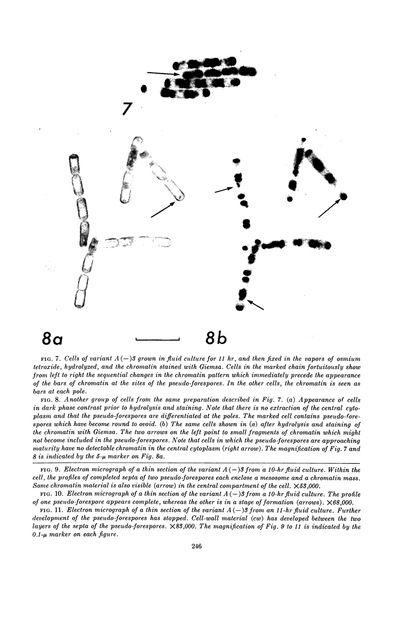
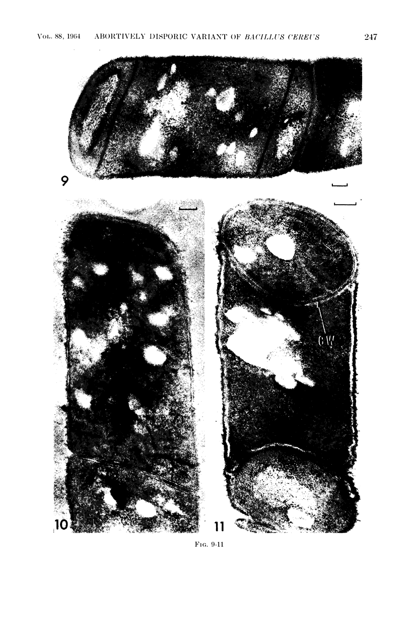
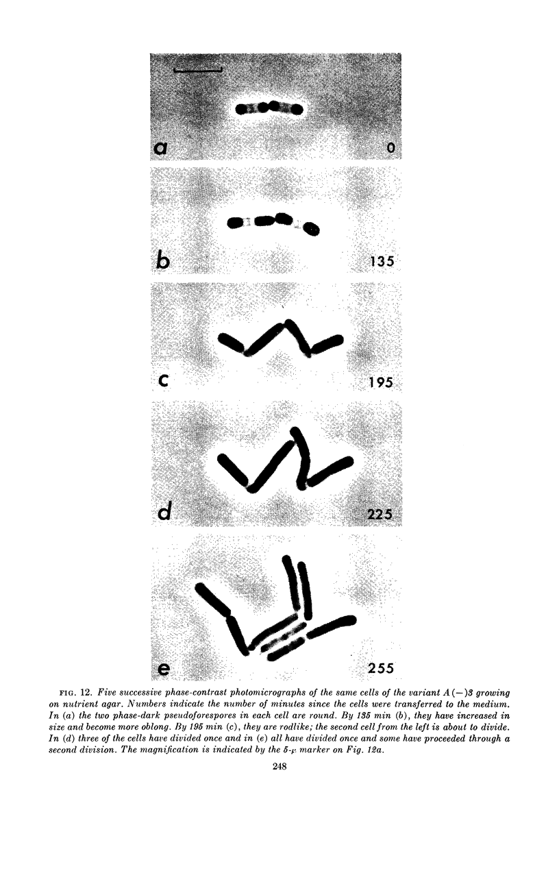
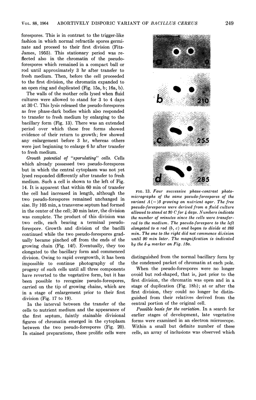
Abstract
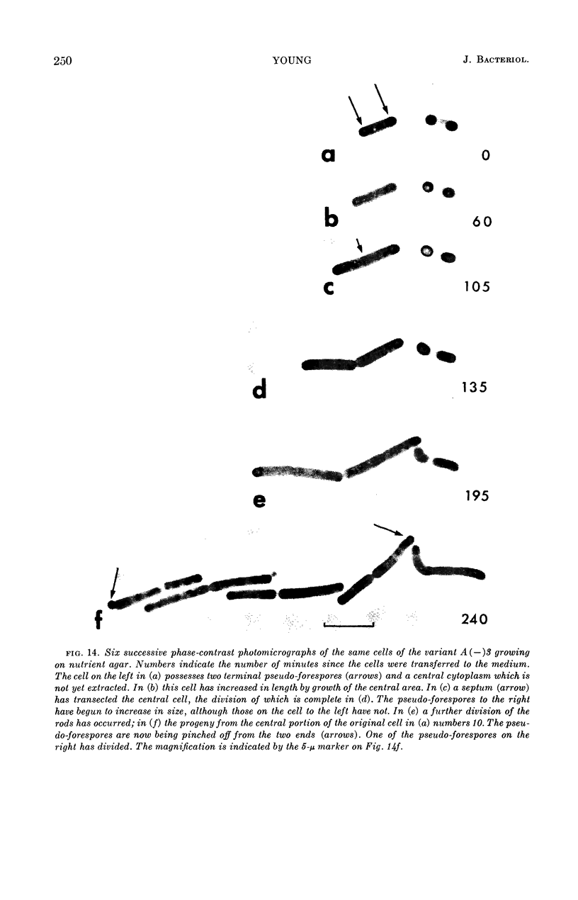
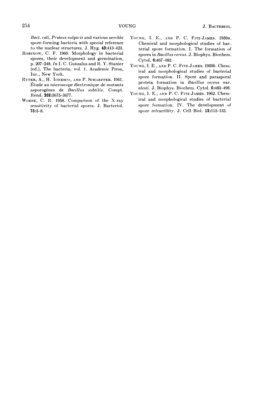
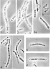
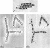
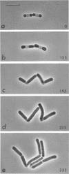
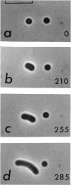
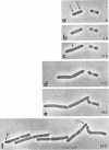
Young, I. Elizabeth (University of Alberta, Edmonton, Alberta, Canada). Characteristics of an abortively disporic variant of Bacillus cereus. J. Bacteriol. 88:242–254. 1964.—A variant [A(−)3] of the Bacillus cereus group has been isolated which appears to begin the formation of a spore at each pole of the cell. These pseudo-forespores, like conventional forespores, are initially formed by invagination of the plasma membrane. However, their maturation does not continue the course of normal spore development. There is no further proliferation of the membrane and no development of the peripheral layers, the cortex, spore coat, or exosporium; there is deposition of apparent cell-wall material between the layers of the septa. Each pseudo-forespore receives approximately one-half of the cell's chromatin. With a supply of fresh nutrients, each is able to resume division after elongation to the bacillary form. Furthermore, the portion of the cell lying between the two pseudo-forespores, containing at most a fragment of the total chromatin, is able to resume growth and division. This growth potential has been interpreted as evidence that the chromatin body of the variant contains more than one set of the genetic instructions characteristic of the organism. A single growth cycle in the presence of dipicolinic acid (thought to cure some Bacillus species of phage) results in approximately 40% of the cells producing a single spore. These spores resemble those formed by the organism from which A(−)3 was isolated. The possibility that the abortively disporic state is associated with the presence of an infecting phage is discussed.
Full text
PDF
Images in this article
Selected References
These references are in PubMed. This may not be the complete list of references from this article.
- Bayne-Jones S., Petrilli A. Cytological Changes during the Formation of the Endospore in Bacillus megatherium. J Bacteriol. 1933 Mar;25(3):261–275. doi: 10.1128/jb.25.3.261-275.1933. [DOI] [PMC free article] [PubMed] [Google Scholar]
- FITZ-JAMES P. C. Participation of the cytoplasmic membrane in the growth and spore fromation of bacilli. J Biophys Biochem Cytol. 1960 Oct;8:507–528. doi: 10.1083/jcb.8.2.507. [DOI] [PMC free article] [PubMed] [Google Scholar]
- FITZ-JAMES P. C. The phosphorus fractions of Bacillus cereus and Bacillus megaterium. II. A correlation of the chemical with the cytological changes occurring during spore germination. Can J Microbiol. 1955 Aug;1(7):525–548. doi: 10.1139/m55-066. [DOI] [PubMed] [Google Scholar]
- FITZ-JAMES P. C., YOUNG I. E. Comparison of species and yarieties of the genus Bacillus. Structure and nucleic acid content of spores. J Bacteriol. 1959 Dec;78:743–754. doi: 10.1128/jb.78.6.743-754.1959. [DOI] [PMC free article] [PubMed] [Google Scholar]
- WOESE C. R. Comparison of the x-ray sensitivity of bacterial spores. J Bacteriol. 1958 Jan;75(1):5–8. doi: 10.1128/jb.75.1.5-8.1958. [DOI] [PMC free article] [PubMed] [Google Scholar]
- YOUNG I. E., FITZ-JAMES P. C. Chemical and morphological studies of bacterial spore formation. II. Spore and parasporal protein formation in Bacillus cereus var. alesti. J Biophys Biochem Cytol. 1959 Dec;6:483–498. doi: 10.1083/jcb.6.3.483. [DOI] [PMC free article] [PubMed] [Google Scholar]
- YOUNG I. E., JAMES P. C. Chemical and morphological studies of bacterial spore formation. IV. The development of spore refractility. J Cell Biol. 1962 Jan;12:115–133. doi: 10.1083/jcb.12.1.115. [DOI] [PMC free article] [PubMed] [Google Scholar]