

Abstract
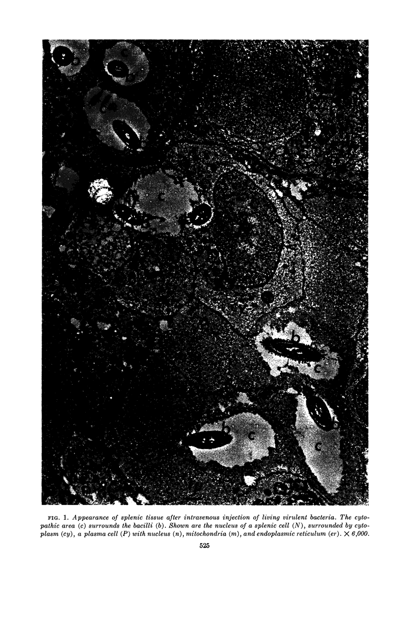
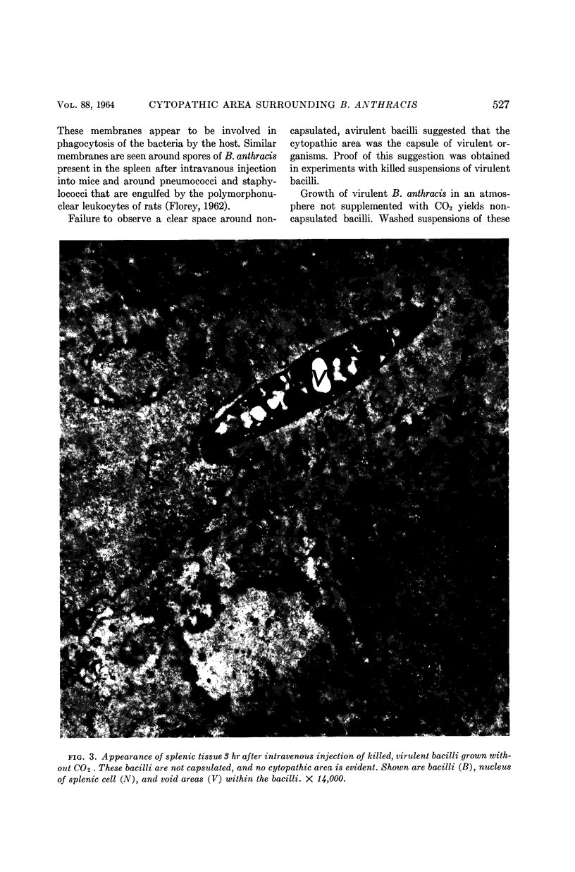
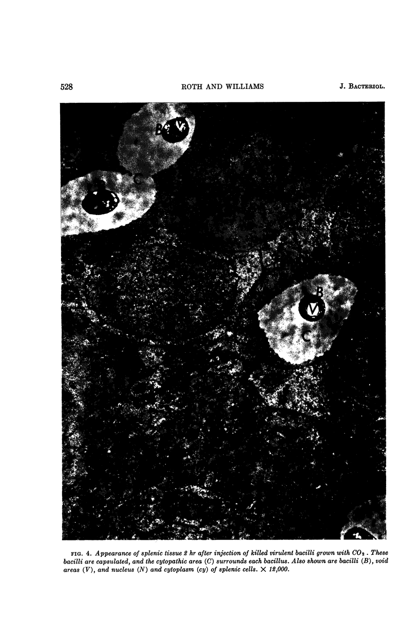
Roth, Ivan L. (Baylor University College of Medicine, Houston, Tex.), and Robert P. Williams. Nature of the cytopathic area surrounding virulent cells of Bacillus anthracis in mouse spleen. J. Bacteriol. 88:523–530. 1964.—Virulent anthrax bacilli in splenic tissue of mice were seen by electron microscopy to be surrounded by a clear zone that was designated the cytopathic area. Previous experiments did not establish the nature of the area. In the present experiments, virulent anthrax bacilli were grown in a system enriched with CO2. The bacilli were capsulated and retained the capsule after they were killed by autoclaving. When grown without CO2, these virulent bacilli were not capsulated. Also used was an avirulent strain of Bacillus anthracis that did not form capsules in either the presence or absence of CO2. Mice were inoculated intravenously with suspensions containing 5 × 10⁸ per 0.5 ml of (i) living or (ii) killed, capsulated virulent bacilli; (iii) killed, noncapsulated virulent bacilli; and (iv) living, avirulent bacilli. Bacteria were found in the spleen 1 hr after injection. Electron microscopy revealed that both living and dead, capsulated virulent bacilli were surrounded by a clear area. No clear area was found around killed, noncapsulated virulent bacilli. Clear areas were never seen around avirulent bacilli. Light microscopy of smears prepared from splenic tissue and stained for the presence of bacterial capsules corroborated the electron microscopic findings. These results established that the cytopathic area surrounding virulent bacilli was capsule.
Full text
PDF
Images in this article
Selected References
These references are in PubMed. This may not be the complete list of references from this article.
- CHU H. P. Variation of Bacillus anthracis with special reference to the non-capsulated avirulent variant. J Hyg (Lond) 1952 Dec;50(4):433–444. doi: 10.1017/s0022172400019720. [DOI] [PMC free article] [PubMed] [Google Scholar]
- ROTH I. L., LEWIS C. W., Jr, WILLIAMS R. P. Electron microscope study of Bacillus anthracis in mouse spleen. J Bacteriol. 1960 Dec;80:772–782. doi: 10.1128/jb.80.6.772-782.1960. [DOI] [PMC free article] [PubMed] [Google Scholar]
- ROTH I. L., WILLIAMS R. P. COMPARISON OF THE FINE STRUCTURE OF VIRULENT AND AVIRULENT SPORES OF BACILLUS ANTHRACIS. Tex Rep Biol Med. 1963;21:394–399. [PubMed] [Google Scholar]
- THORNE C. B. Biochemical properties of virulent and avirulent strains of Bacillus anthracis. Ann N Y Acad Sci. 1960 Nov 21;88:1024–1033. doi: 10.1111/j.1749-6632.1960.tb20094.x. [DOI] [PubMed] [Google Scholar]