Abstract
Context
2-[18F]fluoro-3-(2(S)-azetidinylmethoxy) pyridine (2-F-A-85380, abbreviated as 2-FA) is a recently developed radioligand that allows for visualization of brain α4β2* nicotinic acetylcholine receptors (nAChRs) with positron emission tomography (PET) scanning in humans.
Objective
To determine the effect of cigarette smoking on α4β2* nAChR occupancy in tobacco-dependent smokers.
Design
Fourteen 2-FA PET scanning sessions were performed. During the PET scanning sessions, subjects smoked 1 of 5 amounts (none, 1 puff, 3 puffs, 1 full cigarette, or to satiety [2½ to 3 cigarettes]).
Setting
Academic brain imaging center.
Participants
Eleven tobacco-dependent smokers (paid volunteers).
Main Outcome Measure
Dose-dependent effect of smoking on occupancy of α4β2* nAChRs, as measured with 2-FA and PET in nAChR-rich brain regions.
Results
Smoking 0.13 (1 to 2 puffs) of a cigarette resulted in 50% occupancy of α4β2* nAChRs for 3.1 hours after smoking. Smoking a full cigarette (or more) resulted in more than 88% receptor occupancy and was accompanied by a reduction in cigarette craving. A venous plasma nicotine concentration of 0.87 ng/mL (roughly of the level achieved in typical daily smokers) was associated with 50% occupancy of α4β2* nAChRs.
Conclusions
Cigarette smoking in amounts used by typical daily smokers leads to nearly complete occupancy of α4β2* nAChRs, indicating that tobacco-dependent smokers maintain α4β2* nAChR saturation throughout the day. Because prolonged binding of nicotine to α4β2* nAChRs is associated with desensitization of these receptors, the extent of receptor occupancy found herein suggests that smoking may lead to withdrawal alleviation by maintaining nAChRs in the desensitized state.
TOBACCO DEPENDENCE (PRImarily through cigarette smoking) is a major risk factor for death and disability worldwide.1,2 This condition is relatively resistant to treatment, as evidenced by the fact that most smokers endorse a desire to quit3 but very few are able to do so on their own4 and fewer than half are able to quit long-term even with comprehensive treatment.3,5 A greater understanding of the mechanisms that underlie tobacco dependence may aid in the development of improved treatments for this condition.
Of the thousands of components of tobacco smoke, nicotine is the one that is most closely linked to tobacco dependence.6 Nicotine administration provides positive reinforcement7,8 and ameliorates a range of behavioral states that accompany smoking abstinence, including irritability,9 anxiety,9 and deficits in cognitive performance.10-12 Extensive animal research demonstrates that the interaction of nicotine with nicotinic acetylcho-line receptors (nAChRs)13 (along with associated actions of nicotine) activates dopamine pathways projecting to the nucleus accumbens,14-19 leading to positive reinforcement.8,20
The nAChR containing α4 and β2 subunits (α4β2* subtype) is the predominant receptor subtype in the mammalian brain, and the α421 and β222-25 subunits have been linked to the positive-reinforcing (and cognitive function–enhancing) effects of nicotine. Nicotine has high affinity for α4β2* receptors, and therefore, these receptors are considered as primary targets for the actions of nicotine during cigarette smoking. Studies with engineered mutant mice suggest that α4β2* nAChRs are necessary and sufficient to exhibit in vivo effects of smoking, such as tolerance and sensitization.21
The affinity of nicotine for the α4β2* nAChR measured in vitro is in the range of 0.5 to 14 nmol,26 which is equal to a concentration of 0.01 to 2.3 ng/mL. Typical human smokers have venous plasma nicotine concentrations of 10 to 50 ng/mL during the day.27 Based on these reports, we hypothesized that human cigarette smoking results in nearly complete saturation of α4β2* nAChRs.
2-[18F]fluoro-3-(2(S)azetidinylmethoxy) pyridine (2-F-A-85380, abbreviated as 2-FA) is a radiotracer recently developed for the in vivo imaging of α4β2* nAChRs with positron emission tomography (PET).28-30 Positron emission tomography studies in nonhuman primates demonstrate that receptor binding of 2-FA31 (and another PET radiotracer for α4β2* nAChRs32) can be decreased by administration of nicotine or inhalation of tobacco smoke. Recent studies have demonstrated the safety of this radiotracer for use in humans.30,33-35 Using 2-FA and PET, we sought to determine the effect of cigarette smoking on brain α4β2* nAChR occupancy in tobacco-dependent smokers.
METHODS
SUBJECTS
Eleven tobacco-dependent smokers (>20 cigarettes per day), who were recruited through advertisements in local newspapers and the Internet, participated in the study. Eight subjects were scanned only once in an experimental (smoking) condition, while 3 subjects underwent 2 scanning sessions, 1 in an experimental (smoking) condition and 1 in a control (no smoking) condition, as described later, so that a total of 14 PET scanning sessions were performed for this study. Subjects met DSM-IV criteria for nicotine dependence but were otherwise healthy.
Initial screening consisted of an anonymous telephone interview in which medical, psychiatric, and substance abuse histories were obtained. Qualified subjects were then assessed in person using screening questions from the Structured Clinical Interview for DSM-IV36 2 days prior to PET scanning. The central inclusion criterion was the DSM-IV diagnosis of nicotine dependence, while any history of an Axis I psychiatric or substance abuse/dependence diagnosis other than nicotine dependence was exclusionary. Other exclusion criteria were pregnancy and current use of medications or any history of a medical condition that might affect the central nervous system at the time of scanning (eg, current treatment with a β-blocker or analgesic medication or history of head trauma with loss of consciousness or epilepsy). Women of childbearing potential had a urine pregnancy test. Subjects who occasionally used alcohol, caffeine, or other drugs, but did not meet criteria for abuse or dependence, were allowed to participate in the study but were instructed to abstain for the 2 days prior to PET scanning. Subjects who drank more than the equivalent of 2 cups of coffee per day (300 mg of caffeine per day) were also excluded, as were subjects who experienced caffeine withdrawal symptoms (such as irritability, flushing, or headache) temporally associated with caffeine ingestion. After complete description of the study to subjects, written informed consent was obtained using forms approved by the local institutional review board.
During the initial visit, additional screening data were obtained, including the smoker's profile form (which includes smoking history data, such as current smoking level, years smoked, brand of cigarette smoked, and quit periods) and scores on the Fagerström Test for Nicotine Dependence,37,38 the Beck Depression Inventory,39 the Spielberger State-Trait Anxiety Inventory,40 and the Shiffman-Jarvik Withdrawal Scale.41 An exhaled carbon monoxide (CO) level was obtained using the MicroSmokerlyzer (Bedfont Scientific Ltd, Kent, England) at the initial visit to verify smoking status (subjects were considered to be active smokers if a CO level of >8 ppm was obtained).
GENERAL STUDY DESIGN
Participants underwent the following sequence of procedures (described in greater detail later): abstinence from cigarettes and nicotine-containing products for 2 days, a bolus-plus–continuous infusion 2-FA PET scanning session including smoking between 0 and 3.5 cigarettes, blood sampling for plasma nicotine levels and withdrawal symptom monitoring during the PET scanning session, and structural magnetic resonance imaging (MRI) of the brain within 1 week of PET scanning to aid in localization of regions of interest on PET scans.
ABSTINENCE PERIOD
After the initial screening, participants were instructed to begin smoking/nicotine abstinence at 6 pm 2 nights prior to PET scanning. They reported to our laboratory at 1 pm the day after initiating abstinence. At that time, an exhaled CO level was measured and a brief clinical interview was performed. Participants were deemed to be compliant with study protocol if they reported no smoking since 6 pm the previous night and had an exhaled CO measurement of 8 ppm or less. Subjects were then seen the following day for PET scanning and were required to report continuous abstinence since 2 nights previously and have an exhaled CO level of 3 ppm or less to undergo PET scanning.
PET PROTOCOL
At noon on the day of PET scanning, subjects arrived at the Greater Los Angeles Veterans Affairs Healthcare System PET Center, and abstinence was verified as described earlier. Each participant then had an intravenous catheter placed at 12:45 pm in a room adjacent to the PET scanner. At 1 pm, bolus-plus–continuous infusion 2-FA was initiated. In this study, the amount of 2-FA administered as a bolus was equal to the amount infused over 500 minutes (K =500bolus minutes).42,43 Consistent with this paradigm, 144 MBq (mean±SD, 3.88±0.16 mCi) of 2-FA was administered as an intravenous bolus, followed by continuous infusion of 138 MBq (mean±SD, 3.72±0.15 mCi) in 57.6 mL of saline over the next 480 minutes (7.2 mL per hour) for a total effective dose of radioactivity of 187.5 MBq (mean±SD, 5.08±0.21 mCi). All PET scans were obtained as series of 10-minute frames.
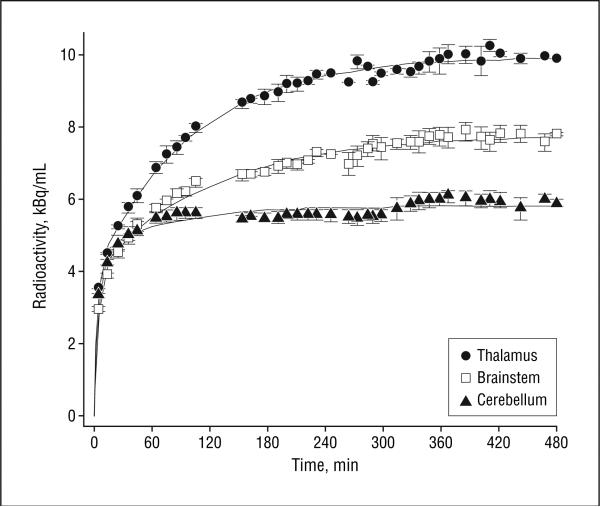
Three PET scanning sessions were performed without smoking, as control sessions. For 2 of these sessions, subjects were positioned in the scanner before administration of 2-FA, and PET scanning began at the time of bolus injection and continued for 8 hours with 7 scheduled breaks and no smoking to demonstrate the full time-activity curves for the 2-FA method used herein (Figure 1). For the third session, a subject preferred not to be scanned for the full 8 hours and was scanned without smoking, using the shorter (5 hours) scanning protocol as in the experimental sessions described next.
Figure 1.
Time-activity curves (mean±SEM) for the 2 subjects undergoing full (8 hours) scanning with no smoking. The graphs demonstrate that 3 to 4 hours from the beginning of radioligand infusion are needed to reach a near steady state in the primary regions of interest for the bolus/infusion paradigm used herein. The average increase in radioactivity starting 3.5 hours after initiation of 2-[18F]fluoro-3-(2(S)-azetidinylmethoxy) pyridine (2-F-A-85380) administration was 3.2%, 3.2%, and 2.6% per hour for the thalamus, brainstem, and cerebellum, respectively.
For the 11 experimental sessions with smoking, subjects received the bolus injection of 2-FA in a room adjacent to the PET scanner. They then remained seated in this room for the next 3 hours to allow the radiotracer to reach a relatively steady state. At 4 pm, brain scanning commenced and continued for 60 minutes. At 5 pm (4 hours after the initiation of 2-FA administration), subjects had a 10-minute break in scanning, during which they smoked between 0 and 3 cigarettes of their favorite brand (because we were interested in studying α4β2* nAChR occupancy from typical smoking conditions). All subjects smoked regular (not light) cigarettes with similar nicotine yields (range, 1.2-1.4 mg).44 The 5 smoking levels for this study were no smoking (n=3); a single puff, which included only lighting a cigarette and inhaling (measured as roughly one twelfth of a cigarette) (n=2); 3 puffs of a cigarette (measured as approximately one quarter of a cigarette) (n=3); a full cigarette (n=3); and satiety (2½-3 cigarettes) (n=3). Subjects were then scanned for 3 hours 50 minutes more with 3 scheduled 10- or 15-minute breaks. Scanning ended at 9 pm.
Blood samples (5 mL) for assay of venous plasma nicotine levels were drawn immediately before and at 10 and 185 minutes following the smoke break from a dual port in the intravenous catheter placed for 2-FA infusion. Two subjects who smoked a full cigarette had a full series of venous plasma nicotine levels drawn prior to the smoke break and at 2, 10, 30, 65, and 185 minutes after the smoke break. Samples were centrifuged, and concentrations of venous plasma nicotine were determined in the laboratory of Peyton Jacob III, PhD, at the University of California, San Francisco, by gas chromatography with nitrogen-phosphorus detection,45 using 5-methyl-nicotine as an internal standard. The lower limit of quantitation was 1 ng/mL.
Cigarette craving was monitored with the Urge to Smoke Scale,46,47 an analog scale (range, 0-6) with 10 craving-related questions. This scale was filled out at the beginning of the first break in scanning (prior to smoking), at the end of the first break (after smoking), and during each of the 3 remaining breaks in scanning.
The PET scans were obtained on a General Electric Advance NXi scanner (General Electric Medical Systems, Milwaukee, Wis) with 35 slices in 3-dimensional mode, transaxial resolution full width at half maximum 5.2 to 7.7 mm.48 Scans were acquired as series of 10-minute frames. Attenuation correction scanning was performed with the germanium rotating rod source built into the General Electric scanner for 5 minutes at the end of the scanning session, and this attenuation correction was applied to all scans from the session. F 18 was prepared with the CP-45 variable proton energy negative ion cyclotron (The Cyclotron Corporation) and 2-FA was prepared by a published method.49 Specific activities ranged from 122 to 241 GBq/μmol (range, 3.3-6.5 Ci/μmol).
MAGNETIC RESONANCE IMAGING
An MRI of the brain was obtained within a week of PET scanning to aid in localization of brain regions on the PET scans, with the following specifications: 3-dimensional Fourier-transform spoiled gradient-recalled acquisition with repetition time=30 milliseconds, echo time=7 milliseconds, 30° angle, 2 acquisitions, and 256×192 view matrix. The acquired volume was reconstructed as roughly 90 contiguous 1.5-mm-thick transaxial sections.
REGION OF INTEREST PLACEMENT
Magnetic resonance imaging to PET coregistration was performed using an automated image registration method.50 Regions of interest were drawn on MRI and transferred to coregistered PET scans. Regions of interest included the thalamus, brainstem, cerebellum, prefrontal cortex, and corpus callosum. The thalamus, brainstem, and cerebellum were analyzed as whole structures while representative sections of the prefrontal cortex (middle frontal gyrus parallel to the body of cingulate) and genu of the corpus callosum (on sagittal images) were drawn. These brain regions were chosen based on prior reports indicating a range of nAChR densities in these regions from highest (thalamus) to lowest (corpus callosum).51-54 Mean±SD volumes for the thalamus, brainstem, and cerebellum were 4.97±0.22, 18.37±3.41, and 95.71±13.77 cm3, respectively. The expected ratio of specific to nonspecific binding for the thalamus based on a study of nonhuman primates using 2-FA PET was roughly 2:1.29
DERIVATIONS OF RECEPTOR OCCUPANCY PARAMETERS
The total concentration of tissue radioactivity (CT) without smoking can be expressed as follows: CT=CSB+CF+CNB, where CSB is the concentration of specifically bound radioligand, CF is the concentration of free radioligand, and CNB is the concentration of nonspecifically bound radioligand. After smoking, C′T = C′SB + CF + CNB. In subjects who smoked, C′SB<CSB because nicotine, its metabolites, or perhaps other components of tobacco smoke displace some of the specifically bound radiotracer.
At equilibrium conditions and assuming that CF and CNB are unchanged as a result of smoking, the fractional displacement (FD) of radiotracer can be expressed as the following:
| (1) |
where
| (2) |
is the fraction of the tissue radioactivity (before smoking) that represents specifically bound radioligand and is therefore the maximum fraction of radiotracer that can be displaced.
At a tracer dose of radioligand (CSB<Bmax), where Bmax is the receptor concentration, we can derive the following equation from equation 1 (again assuming equilibrium):
| (3) |
where
| (4) |
[Ni] and [Ach] are the tissue concentrations of nicotine and acetylcholine, respectively, and K[Ni] and K[ACh] are the equilibrium dissociation constants for nicotine and acetylcholine, respectively.
The tissue nicotine concentration is assumed to be directly proportional to the amount of cigarette smoked and to the venous plasma nicotine concentration. Therefore, equation 3 can be expressed as:
| (5) |
| (6) |
where ED50 and [D] are in units of cigarettes smoked (or fractions thereof) and EC50 (plasma) (simply called EC50 in the rest of this article) and plasma nicotine concentration [Ni] are in units of nanograms per milliliter or nanomole (nM).
Maximum fractional displacement (R) is also related to the commonly used parameter binding potential (BP*), which is defined as the ratio of VDSB to VDNDR,55 where VDSB and VDNDR are the volumes of distribution for the specific binding and nondisplaceable radioactivity compartments, respectively. Consistent with equation 3, R is defined by the equation:
| (7) |
and BP* can be determined from R, using the equation:
| (8) |
DATA ANALYSIS
For all drawn regions of interest, displacement of radiotracer was found from before to after smoking (even in the lowest nAChR density region, the corpus callosum). This observation limited using the reference region approach for calculations of specific binding. In addition, this evaluation revealed that the highest levels of specific binding were in the thalamus, brainstem, and cerebellum, so that these regions were used for analysis herein, and each of the 3 regions was analyzed independently. For calculation of the dose-dependent effects of smoking on receptor occupancy, we calculated fractional displacement of total radioactivity for each subject and each smoking condition.
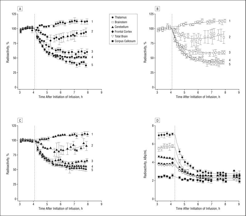
CT and C′T values were determined by averaging data from the six 10-minute frames (1 hour) prior to the smoking break (180-240 minutes [mean, 3.5 hours] after bolus injection) and from the last seven 10-minute frames of the PET scanning session (395-480 minutes, including a 15-minute break, [mean, 7.3 hours] after bolus injection) (Figure 2 and Figure 3).
Figure 2.
Time-activity curves for the 5 smoking levels for the primary regions of interest. A, Thalamus. B, Brainstem. C, Cerebellum. Radioactivity is expressed as percentage of baseline value (mean radioactivity for the hour before the smoking break±standard errors of the mean between subjects). 1 indicates 0 cigarettes; 2 indicates 1 puff; 3 indicates 3 puffs; 4 indicates 1 full cigarette; and 5 indicates satiety (2.8 cigarettes). Dotted lines indicate the time of the smoking break. D, Displacement of radioactivity in the whole brain and regions of interest in a subject who smoked to satiety (2.5 cigarettes) during the break (mean±SE in volume of interest).
Figure 3.
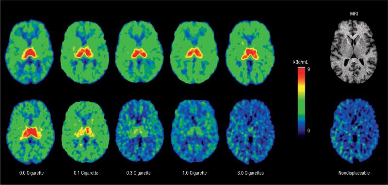
2-[18F]fluoro-3-(2(S)-azetidinylmethoxy) pyridine (2-F-A-85380) positron emission tomography (PET) images before (top row) and 3.1 hours after (bottom row) cigarette smoking. Images were obtained by averaging the six 10-minute frames over the 1 hour prior to the smoking break and by averaging the seven 10-minute scans from a mean of 3.1 hours after smoking the cigarette amount listed. The far right column shows a magnetic resonance image (MRI) of the brain and a PET image of nondisplaceable radioactivity distribution (calculated). All PET images were aligned to the level shown on the MRI.
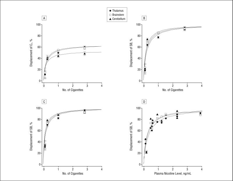
The displacement of total radioactivity (CT minus C′T) for each cigarette dose or venous plasma nicotine level was expressed as a percentage of CT and plotted as a function of amount of cigarette smoked (Figure 4A) or venous plasma nicotine concentration. In these plots, the asymptotic portion of the curve represents the maximum fractional displacement of radioactivity. The plotted data were fitted to equations 5 and 6 mentioned earlier, using nonlinear regression with SigmaStat software (Systat Software, Inc, Richmond, Calif) to determine ED50 (percentage of a cigarette needed to displace 50% of the radio-tracer), EC50 (venous plasma nicotine concentration that was associated with 50% radiotracer displacement), and R (maximum fractional displacement). Percentage of receptor occupancy was also calculated by dividing the fractional displacement (as determined in equation 1) by R.
Figure 4.
Effects of variable smoking (number of cigarettes) and venous plasma nicotine levels on radiotracer displacement. A, Percentage of displacement of total radioactivity (Ct) in regions of interest for the range of smoking levels from 0 to 2.8 cigarettes, based on the ratio of displaced Ct by each cigarette dose to prebreak Ct, without correction for the imperfect steady state of the radiotracer found in control scans. B and C, Displacement of specific binding (SB) (a measure that takes into account nondisplaceable radioactivity derived from the asymptotic portion of the saturation curve in part A) by the varying smoking levels without and with correction for the imperfect steady state, respectively. D, Specific binding displacement as a function of venous plasma nicotine levels, corrected.
We performed these calculations for the raw CT value data and for data corrected for the imperfect steady state found in the no-smoking (control) scans (see “Results” section, paragraphs 2 and 4). The BP* values were calculated based on the R value for each region using equation 8.
RESULTS
Subjects were adults (mean±SD age, 36.6±11.8 years; 8 men, 3 women) who smoked 22.2±2.8 (mean±SD) regular cigarettes per day and had been smoking for a mean±SD of 17.5±10.5 years. Six subjects were African American, 4 subjects were white, and 1 subject was Asian American. One participant's longest previous quit period was 2 years, while the others had longest quit periods ranging from a few days to 1 year. Mean±SD total scores for the Fagerström Test for Nicotine Dependence (5.9±2.2) and the Beck Depression Inventory (3.1±2.5) and mean±SD per item scores for the Spielberger State-Trait Anxiety Index (1.8 ± 0.5) and the Shiffman-Jarvik Withdrawal Scale (4.2±0.7) indicated that subjects were moderately dependent on tobacco and had low levels of depression and anxiety during the study. At the time of screening, subjects had exhaled CO levels consistent with tobacco dependence (mean±SD, 18.1±5.7 ppm), while at the time of scanning, subjects had low exhaled CO levels (mean±SD, 1.9±1.0 ppm), consistent with their compliance with the abstinence protocol of the study. For all subjects, venous plasma nicotine levels were lower than the level of detection of the laboratory for this study (<1 ng/mL) prior to the smoking break in scanning and were the highest in the first sample after smoking, with a half-life of 145 minutes.
Time-activity curves for the control (no-smoking) scans (Figure 1 and Figure 2A-C) demonstrate that the distribution of radioligand reached a near equilibrium state 3.5 hours after initiation of 2-FA administration, with tissue concentrations maintained in a near steady state for the remainder of the scanning sessions. The percentage change per hour in the no-smoking group was calculated by determining the difference in average radioactivity for each region of interest between the period at which 2-FA reached an approximate steady state (180-240 minutes after infusion initiation) and the final 70 minutes of scanning (395-480 minutes after infusion initiation, including a 15-minute break) and dividing by the number of hours (3.8) between the midpoint of these 2 periods. This calculation resulted in average increases in radioactivity for the thalamus, brainstem, and cerebellum of 12%, 12%, and 10%, respectively (3.2%, 3.2%, and 2.6% per hour, respectively). For the last 70 minutes of scanning (last 85 minutes of the scanning session, including a 15-minute break), an almost complete steady state (indicating equilibrium between plasma and brain tissue) was reached, with change in radioactivity in the thalamus, brainstem, and cerebellum of only 0.0±1.0% (mean±SE), 1.0±1.0%(mean±SE), and 1.0±1.0%(±mean±SE), respectively (determined by linear regression).
For the scans with cigarette smoking, decreases in total radioactivity for the 3 studied brain regions with the highest radioactivity accumulation (thalamus, brainstem, and cerebellum) were clearly dose dependent (Figure 2 and Figure 3). A single puff of a cigarette was the only amount of smoking that was followed by recovery of receptor availability within the 3-hour 50-minute time frame after smoking (Figure 2A-C), while the medium to high levels of smoking (one quarter of a cigarette to satiety) resulted in new steady-state conditions at 3 hours after smoking (Figure 2 and Figure 3). Smoking to satiety resulted in a profound decrease of radioactivity in all brain regions studied (Figure 2D). The dose-response curves for displacement of total and specifically bound radioactivities are shown in Figure 4A-C.
The mean±SE effective dose of a cigarette required to occupy 50% of receptors (ED50) for the 3 primary regions of interest at 3.1 hours after smoking was 0.19±0.05 (Table 1). After correcting for the imperfect steady state found in the control group that did not smoke (by multiplying the prebreak mean total radioactivities for the thalamus, brainstem, and cerebellum by 1.12, 1.12, and 1.10, respectively), ED50 was calculated to be 0.13±0.03 (mean±SE) of a cigarette (Table 1). The mean±SE venous plasma nicotine concentration associated with 50% occupancy of receptors (EC50) was determined from the dose response curve (Figure 4D) to be 0.87±0.16 ng/mL (5.3±1.0 nM) (Table 1). Apparent binding potential (BP*) values for the thalamus (Table 1) were similar to those in another published report using 2-FA.29
Table 1.
Effective Dose of a Cigarette and Effective Concentration of Venous Plasma Nicotine Needed to Occupy 50% of α4β2* nAChRs During Positron Emission Tomography Scanning for the 3 Primary Regions of Interesta
| No Correction (Cigarette) |
With Correction (Cigarette) |
With Correction (Plasma Nicotine Level) |
|||||||
|---|---|---|---|---|---|---|---|---|---|
| ED50, Cigarette | R, % | BP* | ED50, Cigarette | R, % | BP* | EC50, ng/mL | R, % | BP* | |
| Thalamus | 0.22 ± 0.11 | 61 ± 8 | 1.8 | 0.15 ± 0.05 | 67 ± 5 | 2.0 | 1.03 ± 0.26 | 71 ± 6 | 2.4 |
| Brainstem | 0.19 ± 0.07 | 64 ± 6 | 1.8 | 0.13 ± 0.03 | 67 ± 4 | 2.0 | 0.81 ± 0.27 | 70 ± 6 | 2.3 |
| Cerebellum | 0.16 ± 0.09 | 52 ± 7 | 1.1 | 0.11 ± 0.05 | 56 ± 5 | 1.3 | 0.73 ± 0.31 | 58 ± 6 | 1.4 |
| Avg ± SD | 0.19 ± 0.05 | 0.13 ± 0.03 | 0 87 ± 0.16 | ||||||
Abbreviations: 2-FA, 2-[18F]fluoro-3-(2(S)-azetidinylmethoxy) pyridine; Avg, weighted average for all 3 brain regions listed; BP*, binding potential, defined as the ratio VDSB/VDNDR, where VDSB and VDNDR are the volumes of distribution for specific binding and nondisplaceable radioactivity, respectively, calculated as R/(1-R); EC50, effective concentration of venous plasma nicotine needed for 50% displacement of bound 2-FA; ED50, effective dose of a cigarette needed for 50% displacement of bound 2-FA; nAChR, nicotinic acetylcholine receptor; R, maximum fractional displacement of the total radioactivity.
Values are expressed as mean ± SE unless otherwise indicated. Correction was based on an imperfect steady state of 2-FA observed after 3.5 hours for subjects scanned in the control (no smoking) condition, as described in the “Results” section, paragraphs 2 and 4.
The mean receptor fractional occupancies at 3.1 hours after smoking 1 puff, 3 puffs, 1 full cigarette, and to satiety were 33%, 75%, 88%, and 95%, respectively (Figure 4) (Table 2). Earlier than 3.1 hours after smoking, we would hypothesize that the fractional occupancy of nAChRs would be even higher. For example, assuming a t1/2 (half-life) of venous plasma nicotine of 2.5 hours,27 we would expect that smoking 1 cigarette would result in venous plasma nicotine levels at 1 and 2.5 hours after smoking to be roughly 1.8 and 1.2 times higher than the levels at 3.1 hours. These higher nicotine levels would lead to estimated receptor occupancies at 1 and 2.5 hours of 93% and 90%, respectively.
Table 2.
Cigarette Dose, Venous Plasma Nicotine Concentration, and Percentage Change in Occupancy of Nicotinic Acetylcholine Receptors in Regions of Interest From Before Smoking to 3.1 Hours After Smokinga
| Dose, Cigarette | Nicotine Level, ng/mL | Without Correction, % |
With Correction,% |
||||||
|---|---|---|---|---|---|---|---|---|---|
| Thalamus | Brainstem | Cerebellum | Average | Thalamus | Brainstem | Cerebellum | Average | ||
| 0.08 | 0.5 ± 0.1b | 17 ± 7 | 21 ± 9 | 20 ± 8 | 19 ± 1 | 30 ± 6 | 34 ± 8 | 34 ± 7 | 33 ± 1 |
| 0.25 | 1.7 ± 0.1 | 66 ± 3 | 66 ± 3 | 74 ± 3 | 69 ± 2 | 72 ± 3 | 74 ± 3 | 80 ± 2 | 75 ± 2 |
| 1.00 | 3.8 ± 0.5 | 81 ± 2 | 85 ± 1 | 85 ± 2 | 83 ± 1 | 84 ± 2 | 90 ± 1 | 89 ± 2 | 88 ± 2 |
| 2.83 | 8.8 ± 1.7 | 98 ± 2 | 91 ± 2 | 92 ± 4 | 93 ± 2 | 98 ± 2 | 93 ± 1 | 93 ± 2 | 95 ± 2 |
Values are expressed as mean±SE. Data are presented both without and with correction for the imperfect steady state from the bolus-plus-infusion method used herein as described in the “Results” section, paragraph 4.
At 3.1 hours after smoking, the concentration was lower than the detection limit, so values were calculated based on the concentrations measured 10 minutes after smoking (1.2 ± 0.2 ng/mL) and assuming that the half-life for nicotine was t1/2= 145 minutes.
Craving was only alleviated with higher smoking levels (1 full cigarette or satiety). For the 5 smoking levels (none, 1 puff, 3 puffs, 1 cigarette, and satiety), changes in mean±SD Urge to Smoke Scale scores (0- to 6-point scale) from before to after smoking were –0.8 ± 0.7, 0.2 ± 0.2, 0.1 ± 0.4, –4.6 ± 1.2, and –4.8 ± 1.1, respectively. Craving relief was temporary and elevated craving levels returned later during the scanning session. For example, a mean ± SD change of –2.3 ± 1.6 in Urge to Smoke Scale score was found from before to 2.5 hours after smoking for the group that smoked 1 full cigarette.
COMMENT
The central findings of this study indicate that typical daily smokers have nearly complete saturation of brain α4β2* nAChRs throughout the day. The ED50 (0.13 cigarette) is very small compared with smoking levels (1 to 2 cigarettes per hour) found in tobacco-dependent smokers. The EC50 of venous plasma nicotine (0.87 ng/mL or 5.4 nM) found herein is very low compared with venous plasma nicotine levels (which range from 10-37, 10-50, and 19-50 ng/mL for trough, afternoon, and peak levels,respectively)27,56 found in daily smokers. This EC50 estimate indicates that 96% to 98% of α4β2* nAChRs are occupied during the day in tobacco-dependent smokers. Additionally, the EC50 found herein is in the range of concentrations causing desensitization of 50% of α4β2* nAChRs. As demonstrated recently, 100 seconds of coincubation of cells expressing α4β2* nAChRs with 10 nM of nicotine (1.6 ng/mL) resulted in 70% receptor desensitization. Thus, results of this study in the context of other α4β2* nAChR research indicate a nearly complete shift from other potential nAChR states (such as resting and intermediate desensitized)58 to the high-affinity desensitization state during the day in tobacco-dependent smokers. Such prolonged desensitization may be responsible for the up-regulation of these receptors found in smokers.59,60
In our study, craving was only reduced with near total occupancy of nAChRs, with up to one quarter of a cigarette doing little to alleviate craving, despite up to 75% receptor occupancy, whereas smoking 1 full cigarette or to satiety resulted in significant craving reductions and at least 88% and 95% receptor occupancy, respectively. Similarly, though roughly 50% of the presmoking craving level returned in the 1 full cigarette group 2.5 hours after smoking, the calculated receptor occupancy by nicotine for that time was roughly 90% (as described earlier).
Continued smoking (in tobacco-dependent smokers) despite near total α4β2* nAChR occupancy/desensitization may be explained in several ways. First, smokers may continue to smoke to avoid having free unbound receptors. In this situation, free (nondesensitized) receptors would beresponsible for cigarette craving, and nicotine binding and desensitization of these nAChRs may alleviate craving. This action of nicotine may provide positive reinforcement and craving alleviation through frequency-sensitive dopamine release61,62 and/or decreased γ-aminobutyric acid tone.19Second, it is possible that positive reinforcement from smoking is due to activation of other α4β2* nAChR subtypes that are not desensitized by high concentrations of nicotine and that are not labeled by 2-FA because of a smaller number of receptors or low affinity. Therefore, we cannot exclude the possibility that activation of other α4β2* nAChRs could be involved in tobacco dependence, and it may be important to identify such receptors in future research. And third, factors other than nicotine binding to nAChRs may be at least partly responsible for craving alleviation and positive reinforcement (which is supported by studies demonstrating that denicotinized cigarettes partially alleviate craving63). In any case, because α4β2* nAChRs are the predominant receptor subtype in humans and 2-FA labels almost the entire population of these receptors,64 the conclusion from our study still indicates that near complete desensitization of nAChRs has an important role in the pathophysiology of tobacco dependence.
This study should be interpreted in the context of severallimitations. First, cigarette smokers vary in their rate and depth of inhalation of cigarettes, and it is recognized that these interindividual differences could have affected theED50 calculation (though, as noted earlier, all subjects smoked cigarettes with similar nicotine contents and the total amount smokedwas measured). Second, test-retest data on the bolus-plus-infusion method used have not yet been published, although there was relatively little variability within study subgroups (based on smoking dosage) compared with the robust effects of cigarette smoking and the ED50 and EC50 determined herein remained relatively stable from region to region. Third, repeated breaks in scanning (which were necessary for subject comfort) may have led to slight shifts in overall radioactivity determinations due to small differences in position from before to after each scanning break, though (as noted earlier) a head holder and laser light positioning were used to attempt to accurately reposition subjects. And fourth, venous rather than arterial nicotine levels were used for study calculations, and arterial levels are reported to be higher than venous at the time of smoking.65 However,study calculations were performed on venousnicotine levels 3.1 hours after smoking, a time at which venous and arterial levels would be expected to be similar,65 so that our results should provide a reasonable approximation of EC50 for arterial, as well as venous, nicotine levels.
Finally, the association between cigarette smoking and high receptor occupancy found herein has implications for the study of indirect cigarette/nicotine exposures. Studies of fetal exposure to maternal smoking,66 neonate exposure to breast milk of mothers who smoke,67 and prolonged environmental tobacco smoke exposure68,69 all show evidence of venous plasma nicotine levels higher than 1 ng/mL in those who are indirectly exposed, with fetal levels being even higher than maternal levels. Our study indicates that even low levels of exposure result in substantial occupancy of brain α4β2* nAChRs, suggesting significant occupancy and desensitization of α4β2* nAChRs in subjects indirectly exposed to nicotine and pointing to the need for further research to address this issue.
Acknowledgment
We thank the laboratory of Peyton Jacob III, PhD, for determining venous plasma nicotine levels and Josephine Ribe, BS, and Michael Clark, BS, for performing positron emission tomography and magnetic resonance imaging, respectively.
Funding/Support: This study was supported by National Institute on Drug Abuse (NIDA) grants RO1 DA15059 and DA20872 (Dr Brody) and RO1 DA14093 (Dr London), a Veterans Affairs Type I Merit Review Award (Dr Brody), Tobacco-Related Disease Research Program grants 11RT-0024 (Dr Brody) and 10RT-0091 (Dr London), Office of National Drug Control Policy contract DABT63-00-C-1003 (Dr London), and the NIDA intramural research program (Drs Chefer and Mukhin).
REFERENCES
- 1.Michaud CM, Murray CJ, Bloom BR. Burden of disease—implications for future research. JAMA. 2001;285:535–539. doi: 10.1001/jama.285.5.535. [DOI] [PubMed] [Google Scholar]
- 2.Ezzati M, Lopez AD. Estimates of global mortality attributable to smoking in 2000. Lancet. 2003;362:847–852. doi: 10.1016/S0140-6736(03)14338-3. [DOI] [PubMed] [Google Scholar]
- 3.Fiore MC, Bailey WC, Cohen SJ, Dorfman SF, Goldstein MG, Gritz ER, Heyman RB, Jaén CR, Kottke TE, Lando HA, Mecklenburg RE, Mullen PD, Nett LM, Robinson L, Stitzer ML, Tommasello AC, Villejo L, Wewers ME. Treating Tobacco Use and Dependence: Clinical Practice Guideline. US Dept of Health and Human Services, Public Health Service; Rockville, Md: 2000. [Google Scholar]
- 4.Ashenden R, Silagy C, Weller D. A systematic review of the effectiveness of promoting lifestyle change in general practice. Fam Pract. 1997;14:160–176. doi: 10.1093/fampra/14.2.160. [DOI] [PubMed] [Google Scholar]
- 5.Jorenby DE, Leischow SJ, Nides MA, Rennard SI, Johnston JA, Hughes AR, Smith SS, Muramoto ML, Daughton DM, Doan K, Fiore MC, Baker TB. A controlled trial of sustained-release bupropion, a nicotine patch, or both for smoking cessation. N Engl J Med. 1999;340:685–691. doi: 10.1056/NEJM199903043400903. [DOI] [PubMed] [Google Scholar]
- 6.Henningfield JE, Fant RV. Tobacco use as drug addiction: the scientific foundation. Nicotine Tob Res. 1999;1(suppl 2):S31–S35. doi: 10.1080/14622299050011781. [DOI] [PubMed] [Google Scholar]
- 7.Cohen C, Pickworth WB, Henningfield JE. Cigarette smoking and addiction. Clin Chest Med. 1991;12:701–710. [PubMed] [Google Scholar]
- 8.Koob GF. Drugs of abuse: anatomy, pharmacology and function of reward pathways. Trends Pharmacol Sci. 1992;13:177–184. doi: 10.1016/0165-6147(92)90060-j. [DOI] [PubMed] [Google Scholar]
- 9.West R, Shiffman S. Effect of oral nicotine dosing forms on cigarette withdrawal symptoms and craving. Psychopharmacology (Berl) 2001;155:115–122. doi: 10.1007/s002130100712. [DOI] [PubMed] [Google Scholar]
- 10.Newhouse PA, Potter A, Singh A. Effects of nicotinic stimulation on cognitive performance. Curr Opin Pharmacol. 2004;4:36–46. doi: 10.1016/j.coph.2003.11.001. [DOI] [PubMed] [Google Scholar]
- 11.Ernst M, Heishman SJ, Spurgeon L, London ED. Smoking history and nicotine effects on cognitive performance. Neuropsychopharmacology. 2001;25:313–319. doi: 10.1016/S0893-133X(01)00257-3. [DOI] [PubMed] [Google Scholar]
- 12.Mendrek A, Monterosso J, Simon SL, Jarvik M, Brody A, Olmstead R, Domier CP, Cohen MS, Ernst M, London ED. Working memory in cigarette smokers: comparison to non-smokers and effects of abstinence. Addict Behav. 2006;31:833–844. doi: 10.1016/j.addbeh.2005.06.009. [DOI] [PMC free article] [PubMed] [Google Scholar]
- 13.Lukas RJ, Changeux JP, Le Novere N, Albuquerque EX, Balfour DJ, Berg DK, Bertrand D, Chiappinelli VA, Clarke PB, Collins AC, Dani JA, Grady SR, Kellar KJ, Lindstrom JM, Marks MJ, Quik M, Taylor PW, Wonnacott S. International Union of Pharmacology, XX: current status of the nomenclature for nicotinic acetylcholine receptors and their subunits. Pharmacol Rev. 1999;51:397–401. [PubMed] [Google Scholar]
- 14.Di Chiara G, Imperato A. Drugs abused by humans preferentially increase synaptic dopamine concentrations in the mesolimbic system of freely moving rats. Proc Natl Acad Sci U S A. 1988;85:5274–5278. doi: 10.1073/pnas.85.14.5274. [DOI] [PMC free article] [PubMed] [Google Scholar]
- 15.Pontieri FE, Tanda G, Orzi F, Di Chiara G. Effects of nicotine on the nucleus accumbens and similarity to those of addictive drugs. Nature. 1996;382:255–257. doi: 10.1038/382255a0. [DOI] [PubMed] [Google Scholar]
- 16.Sziraki, Lipovac MN, Hashim A, Sershen H, Allen D, Cooper T, Czobor P, Lajtha A. Differences in nicotine-induced dopamine release and nicotine pharmacokinetics between Lewis and Fischer 344 rats. Neurochem Res. 2001;26:609–617. doi: 10.1023/a:1010979018217. [DOI] [PubMed] [Google Scholar]
- 17.Damsma G, Day J, Fibiger HC. Lack of tolerance to nicotine-induced dopamine release in the nucleus accumbens. Eur J Pharmacol. 1989;168:363–368. doi: 10.1016/0014-2999(89)90798-x. [DOI] [PubMed] [Google Scholar]
- 18.Corrigall WA, Coen KM, Adamson KL. Self-administered nicotine activates the mesolimbic dopamine system through the ventral tegmental area. Brain Res. 1994;653:278–284. doi: 10.1016/0006-8993(94)90401-4. [DOI] [PubMed] [Google Scholar]
- 19.Mansvelder HD, Keath JR, McGehee DS. Synaptic mechanisms underlie nicotine-induced excitability of brain reward areas. Neuron. 2002;33:905–919. doi: 10.1016/s0896-6273(02)00625-6. [DOI] [PubMed] [Google Scholar]
- 20.Leshner AI, Koob GF. Drugs of abuse and the brain. Proc Assoc Am Physicians. 1999;111:99–108. doi: 10.1046/j.1525-1381.1999.09218.x. [DOI] [PubMed] [Google Scholar]
- 21.Tapper AR, McKinney SL, Nashmi R, Schwarz J, Deshpande P, Labarca C, Whiteaker P, Marks MJ, Collins AC, Lester HA. Nicotine activation of alpha4* receptors: sufficient for reward, tolerance, and sensitization. Science. 2004;306:1029–1032. doi: 10.1126/science.1099420. [DOI] [PubMed] [Google Scholar]
- 22.Picciotto MR, Zoli M, Changeux JP. Use of knock-out mice to determine the molecular basis for the actions of nicotine. Nicotine Tob Res. 1999;1(suppl 2):S121–S125. doi: 10.1080/14622299050011931. [DOI] [PubMed] [Google Scholar]
- 23.Salminen O, Murphy KL, McIntosh JM, Drago J, Marks MJ, Collins AC, Grady SR. Subunit composition and pharmacology of two classes of striatal presynaptic nicotinic acetylcholine receptors mediating dopamine release in mice. Mol Pharmacol. 2004;65:1526–1535. doi: 10.1124/mol.65.6.1526. [DOI] [PubMed] [Google Scholar]
- 24.Liu X, Koren AO, Yee SK, Pechnick RN, Poland RE, London ED. Self-administration of 5-iodo-A-85380, a beta 2-selective nicotinic receptor ligand, by operantly trained rats. Neuroreport. 2003;14:1503–1505. doi: 10.1097/00001756-200308060-00020. [DOI] [PubMed] [Google Scholar]
- 25.Maskos U, Molles BE, Pons S, Besson M, Guiard BP, Guilloux JP, Evrard A, Cazala P, Cormier A, Mameli-Engvall M, Dufour N, Cloez-Tayarani I, Bemelmans AP, Mallet J, Gardier AM, David V, Faure P, Granon S, Changeux JP. Nicotine reinforcement and cognition restored by targeted expression of nicotinic receptors. Nature. 2005;436:103–107. doi: 10.1038/nature03694. [DOI] [PubMed] [Google Scholar]
- 26.Sihver W, Nordberg A, Langstrom B, Mukhin AG, Koren AO, Kimes AS, London ED. Development of ligands for in vivo imaging of cerebral nicotinic receptors. Behav Brain Res. 2000;113:143–157. doi: 10.1016/s0166-4328(00)00209-6. [DOI] [PubMed] [Google Scholar]
- 27.Benowitz NL, Porchet H, Jacob PI. Pharmacokinetics, metabolism, and pharmacodynamics of nicotine. In: Wonnacott S, Russell MAH, Stolerman IP, editors. Nicotine Psychopharmacology: Molecular, Cellular, and Behavioural Aspects. Oxford University Press; Oxford, England: 1990. pp. 112–157. [Google Scholar]
- 28.Koren AO, Horti AG, Mukhin AG, Gundisch D, Kimes AS, Dannals RF, London ED. 2-, 5-, and 6-halo-3-(2(S)-azetidinylmethoxy)pyridines. J Med Chem. 1998;41:3690–3698. doi: 10.1021/jm980170a. [DOI] [PubMed] [Google Scholar]
- 29.Chefer SI, London ED, Koren AO, Pavlova OA, Kurian V, Kimes AS, Horti AG, Mukhin AG. Graphical analysis of 2-[F-18]FA binding to nicotinic acetylcholine receptors in rhesus monkey brain. Synapse. 2003;48:25–34. doi: 10.1002/syn.10180. [DOI] [PubMed] [Google Scholar]
- 30.Gallezot JD, Bottlaender M, Gregoire MC, Roumenov D, Deverre JR, Coulon C, Ottaviani M, Dolle F, Syrota A, Valette H. In vivo imaging of human cerebral nicotinic acetylcholine receptors with 2-18F-fluoro-A-85380 and PET. J Nucl Med. 2005;46:240–247. [PubMed] [Google Scholar]
- 31.Valette H, Bottlaender M, Dolle F, Coulon C, Ottaviani M, Syrota A. Long-lasting occupancy of central nicotinic acetylcholine receptors after smoking: a PET study in monkeys. J Neurochem. 2003;84:105–111. doi: 10.1046/j.1471-4159.2003.01502.x. [DOI] [PubMed] [Google Scholar]
- 32.Ding YS, Volkow ND, Logan J, Garza V, Pappas N, King P, Fowler JS. Occupancy of brain nicotinic acetylcholine receptors by nicotine doses equivalent to those obtained when smoking a cigarette. Synapse. 2000;35:234–237. doi: 10.1002/(SICI)1098-2396(20000301)35:3<234::AID-SYN9>3.0.CO;2-7. [DOI] [PubMed] [Google Scholar]
- 33.Obrzut SL, Koren AO, Mandelkern MA, Brody AL, Hoh CK, London ED. Whole-body radiation dosimetry of 2-[18F]fluoro-A-85380 in human PET imaging studies. Nucl Med Biol. 2005;32:869–874. doi: 10.1016/j.nucmedbio.2005.06.005. [DOI] [PubMed] [Google Scholar]
- 34.Mitkovski S, Villemagne VL, Novakovic KE, O'Keefe G, Tochon-Danguy H, Mulligan RS, Dickinson KL, Saunder T, Gregoire MC, Bottlaender M, Dolle F, Rowe CC. Simplified quantification of nicotinic acetylcholine receptors with 2[18F]FA-85380 PET. Nucl Med Biol. 2005;32:585–591. doi: 10.1016/j.nucmedbio.2005.04.013. [DOI] [PubMed] [Google Scholar]
- 35.Kimes AS, Horti AG, London ED, Chefer SI, Contoreggi C, Ernst M, Friello P, Koren AO, Kurian V, Matochik JA, Pavlova O, Vaupel DB, Mukhin AG. 2-[18F]FA-85380: PET imaging of brain nicotinic acetylcholine receptors and whole body distribution in humans. FASEB J. 2003;17:1331–1333. doi: 10.1096/fj.02-0492fje. [DOI] [PubMed] [Google Scholar]
- 36.First MB, Spitzer RL, Gibbon M, Williams JBW. Structured Clinical Interview for DSM-IV Axis I Disorders–Patient Edition (SCID-I/P, version 2.0). Biometrics Research Department, New York State Psychiatric Institute; New York: 1995. [Google Scholar]
- 37.Fagerström KO. Measuring the degree of physical dependence to tobacco smoking with reference to individualization of treatment. Addict Behav. 1978;3:235–241. doi: 10.1016/0306-4603(78)90024-2. [DOI] [PubMed] [Google Scholar]
- 38.Heatherton TF, Kozlowski LT, Frecker RC, Fagerström KO. The Fagerström Test for Nicotine Dependence: a revision of the Fagerström Tolerance Questionnaire. Br J Addict. 1991;86:1119–1127. doi: 10.1111/j.1360-0443.1991.tb01879.x. [DOI] [PubMed] [Google Scholar]
- 39.Beck AT, Ward CH, Mendelson M, Mock J, Erbaugh J. An inventory for measuring depression. Arch Gen Psychiatry. 1961;4:561–571. doi: 10.1001/archpsyc.1961.01710120031004. [DOI] [PubMed] [Google Scholar]
- 40.Spielberger C. Manual for the State-Trait Anxiety Inventory. Consulting Psychologists Press; Palo Alto, Calif: 1983. [Google Scholar]
- 41.Shiffman SM, Jarvik ME. Smoking withdrawal symptoms in two weeks of abstinence. Psychopharmacology (Berl) 1976;50:35–39. doi: 10.1007/BF00634151. [DOI] [PubMed] [Google Scholar]
- 42.Carson RE. PET physiological measurements using constant infusion. Nucl Med Biol. 2000;27:657–660. doi: 10.1016/s0969-8051(00)00138-4. [DOI] [PubMed] [Google Scholar]
- 43.Carson RE, Channing MA, Blasberg RG, Dunn BB, Cohen RM, Rice KC, Herscovitch P. Comparison of bolus and infusion methods for receptor quantitation. J Cereb Blood Flow Metab. 1993;13:24–42. doi: 10.1038/jcbfm.1993.6. [DOI] [PubMed] [Google Scholar]
- 44.US Federal Trade Commission . “Tar,” Nicotine, and Carbon Monoxide of the Smoke of 1294 Varieties of Domestic Cigarettes for the Year 1998. US Federal Trade Commission; Washington, DC: 2000. [Google Scholar]
- 45.Jacob P, III, Wilson M, Benowitz NL. Improved gas chromatographic method for the determination of nicotine and cotinine in biologic fluids. J Chromatogr. 1981;222:61–70. doi: 10.1016/s0378-4347(00)81033-6. [DOI] [PubMed] [Google Scholar]
- 46.Jarvik ME, Madsen DC, Olmstead RE, Iwamoto-Schaap PN, Elins JL, Benowitz NL. Nicotine blood levels and subjective craving for cigarettes. Pharmacol Biochem Behav. 2000;66:553–558. doi: 10.1016/s0091-3057(00)00261-6. [DOI] [PubMed] [Google Scholar]
- 47.Brody AL, Mandelkern MA, London ED, Childress AR, Lee GS, Bota RG, Ho ML, Saxena S, Baxter LR, Jr, Madsen D, Jarvik ME. Brain metabolic changes during cigarette craving. Arch Gen Psychiatry. 2002;59:1162–1172. doi: 10.1001/archpsyc.59.12.1162. [DOI] [PubMed] [Google Scholar]
- 48.Dhawan V, Kazumata K, Robeson W, Belakhlef A, Margouleff C, Chaly T, Nakamura T, Dahl R, Margouleff D, Eidelberg D. Quantitative brain PET: comparison of 2D and 3D acquisitions on the GE Advance Scanner. Clin Positron Imaging. 1998;1:135–144. doi: 10.1016/s1095-0397(98)00009-0. [DOI] [PubMed] [Google Scholar]
- 49.Doll F, Dolci L, Valette H, Hinnen F, Vaufrey F, Guenther I, Fuseau C, Coulon C, Bottlaender M, Crouzel C. Synthesis and nicotinic acetylcholine receptor in vivo binding properties of 2-fluoro-3-[2(S)-2-azetidinylmethoxypyridine. J Med Chem. 1999;42:2251–2299. doi: 10.1021/jm9910223. [DOI] [PubMed] [Google Scholar]
- 50.Woods RP, Mazziotta JC, Cherry SR. MRI-PET registration with automated algorithm. J Comput Assist Tomogr. 1993;17:536–546. doi: 10.1097/00004728-199307000-00004. [DOI] [PubMed] [Google Scholar]
- 51.London ED, Waller SB, Wamsley JK. Autoradiographic localization of [3H]nicotine binding sites in the rat brain. Neurosci Lett. 1985;53:179–184. doi: 10.1016/0304-3940(85)90182-x. [DOI] [PubMed] [Google Scholar]
- 52.Villemagne VL, Horti A, Scheffel U, Ravert HT, Finley P, Clough DJ, London ED, Wagner HN, Jr, Dannals RF. Imaging nicotinic acetylcholine receptors with fluorine-18-FPH, an epibatidine analog. J Nucl Med. 1997;38:1737–1741. [PubMed] [Google Scholar]
- 53.Fujita M, Ichise M, van Dyck CH, Zoghbi SS, Tamagnan G, Mukhin AG, Bozkurt A, Seneca N, Tipre D, DeNucci CC, Iida H, Vaupel DB, Horti AG, Koren AO, Kimes AS, London ED, Seibyl JP, Baldwin RM, Innis RB. Quantification of nicotinic acetylcholine receptors in human brain using [I-123]5-I-A-85380 SPET. Eur J Nucl Med Mol Imaging. 2003;30:1620–1629. doi: 10.1007/s00259-003-1320-0. [DOI] [PubMed] [Google Scholar]
- 54.Valette H, Bottlaender M, Dolle F, Guenther I, Coulon C, Hinnen F, Fuseau C, Ottaviani M, Crouzel C. Characterization of the nicotinic ligand 2-[F-18]fluoro-3-[2(S)-2-azetidinylmethoxy]pyridine in vivo. Life Sci. 1999;64:PL93–PL97. doi: 10.1016/s0024-3205(98)00573-6. [DOI] [PubMed] [Google Scholar]
- 55.Ichise M, Meyer JH, Yonekura Y. An introduction to PET and SPECT neuroreceptor quantification models. J Nucl Med. 2001;42:755–763. [PubMed] [Google Scholar]
- 56.Hukkanen J, Jacob P, III, Benowitz NL. Metabolism and disposition kinetics of nicotine. Pharmacol Rev. 2005;57:79–115. doi: 10.1124/pr.57.1.3. [DOI] [PubMed] [Google Scholar]
- 57.Paradiso KG, Steinbach JH. Nicotine is highly effective at producing desensitization of rat alpha4beta2 neuronal nicotinic receptors. J Physiol. 2003;553:857–871. doi: 10.1113/jphysiol.2003.053447. [DOI] [PMC free article] [PubMed] [Google Scholar]
- 58.Quick MW, Lester RA. Desensitization of neuronal nicotinic receptors. J Neurobiol. 2002;53:457–478. doi: 10.1002/neu.10109. [DOI] [PubMed] [Google Scholar]
- 59.Benwell ME, Balfour DJK, Anderson JM. Evidence that tobacco smoking increases the density of (-)-[3H]nicotine binding sites in human brain. J Neurochem. 1988;50:1243–1247. doi: 10.1111/j.1471-4159.1988.tb10600.x. [DOI] [PubMed] [Google Scholar]
- 60.Breese CR, Marks MJ, Logel J, Adams CE, Sullivan B, Collins AC, Leonard S. Effect of smoking history on [3H]nicotine binding in human postmortem brain. J Pharmacol Exp Ther. 1997;282:7–13. [PubMed] [Google Scholar]
- 61.Rice ME, Cragg SJ. Nicotine amplifies reward-related dopamine signals in striatum. Nat Neurosci. 2004;7:583–584. doi: 10.1038/nn1244. [DOI] [PubMed] [Google Scholar]
- 62.Zhang H, Sulzer D. Frequency-dependent modulation of dopamine release by nicotine. Nat Neurosci. 2004;7:581–582. doi: 10.1038/nn1243. [DOI] [PubMed] [Google Scholar]
- 63.Robinson ML, Houtsmuller EJ, Moolchan ET, Pickworth WB. Placebo cigarettes in smoking research. Exp Clin Psychopharmacol. 2000;8:326–332. doi: 10.1037//1064-1297.8.3.326. [DOI] [PubMed] [Google Scholar]
- 64.Chefer SI, Mukhin AG, Horti AG, Koren AD, Pavlova OI, Kurian V, Stratton M, Kimes AS, London ED. 2-[18F]-fluoro-a-85380.. Proceedings of the International Conference on Mathematics and Engineering Techniques in Medicine and Biology..2000. pp. 409–415. [Google Scholar]
- 65.Henningfield JE, Stapleton JM, Benowitz NL, Grayson RF, London ED. Higher levels of nicotine in arterial than in venous blood after cigarette smoking. Drug Alcohol Depend. 1993;33:23–29. doi: 10.1016/0376-8716(93)90030-t. [DOI] [PubMed] [Google Scholar]
- 66.Luck W, Nau H, Hansen R, Steldinger R. Extent of nicotine and cotinine transfer to the human fetus, placenta and amniotic fluid of smoking mothers. Dev Pharmacol Ther. 1985;8:384–395. doi: 10.1159/000457063. [DOI] [PubMed] [Google Scholar]
- 67.Becker AB, Manfreda J, Ferguson AC, Dimich-Ward H, Watson WT, Chan-Yeung M. Breast-feeding and environmental tobacco smoke exposure. Arch Pediatr Adolesc Med. 1999;153:689–691. doi: 10.1001/archpedi.153.7.689. [DOI] [PubMed] [Google Scholar]
- 68.Jarvis MJ, Russell MA, Feyerabend C. Absorption of nicotine and carbon monoxide from passive smoking under natural conditions of exposure. Thorax. 1983;38:829–833. doi: 10.1136/thx.38.11.829. [DOI] [PMC free article] [PubMed] [Google Scholar]
- 69.Jarvis MJ. Uptake of environmental tobacco smoke. IARC Sci Publ. 1987;(87):43–58. [PubMed] [Google Scholar]


