

Abstract
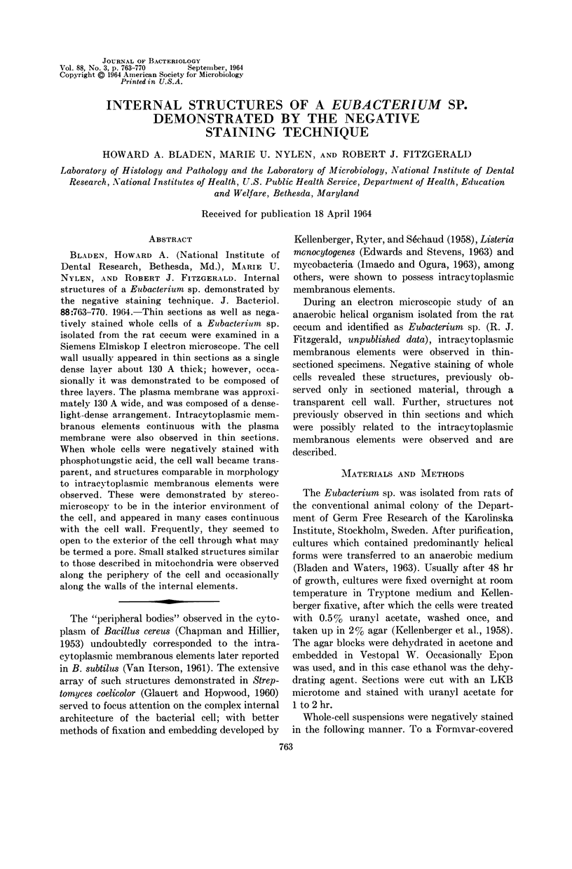
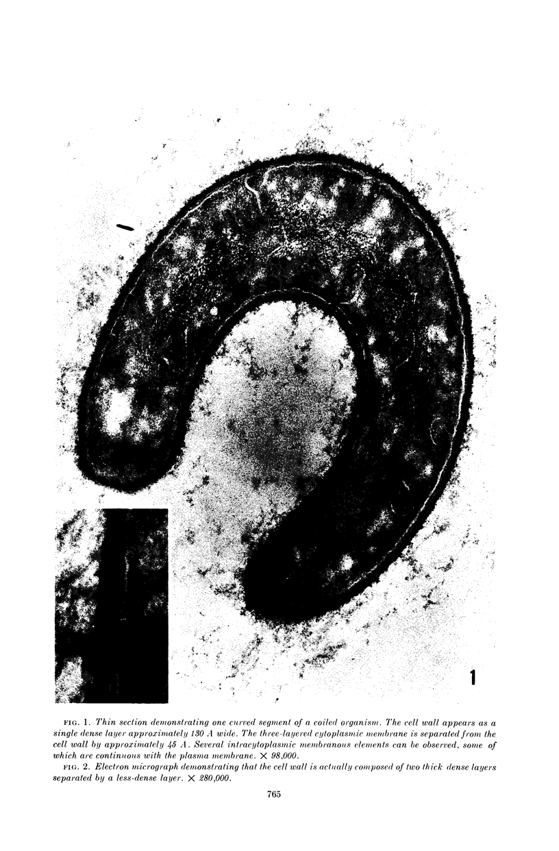
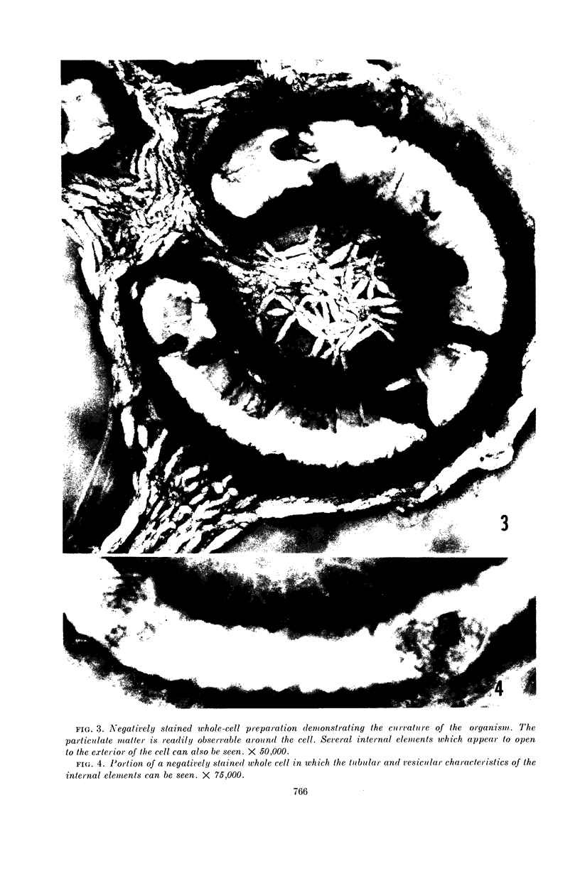
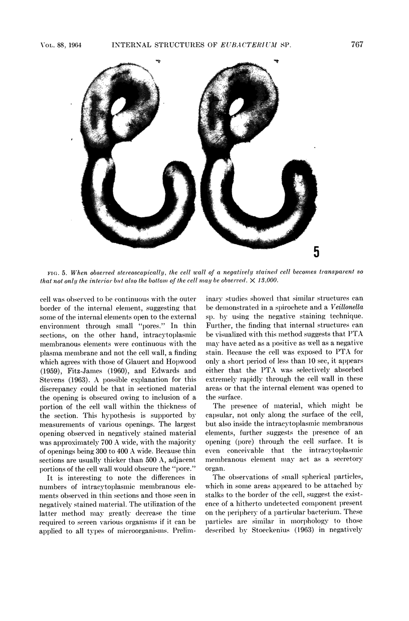
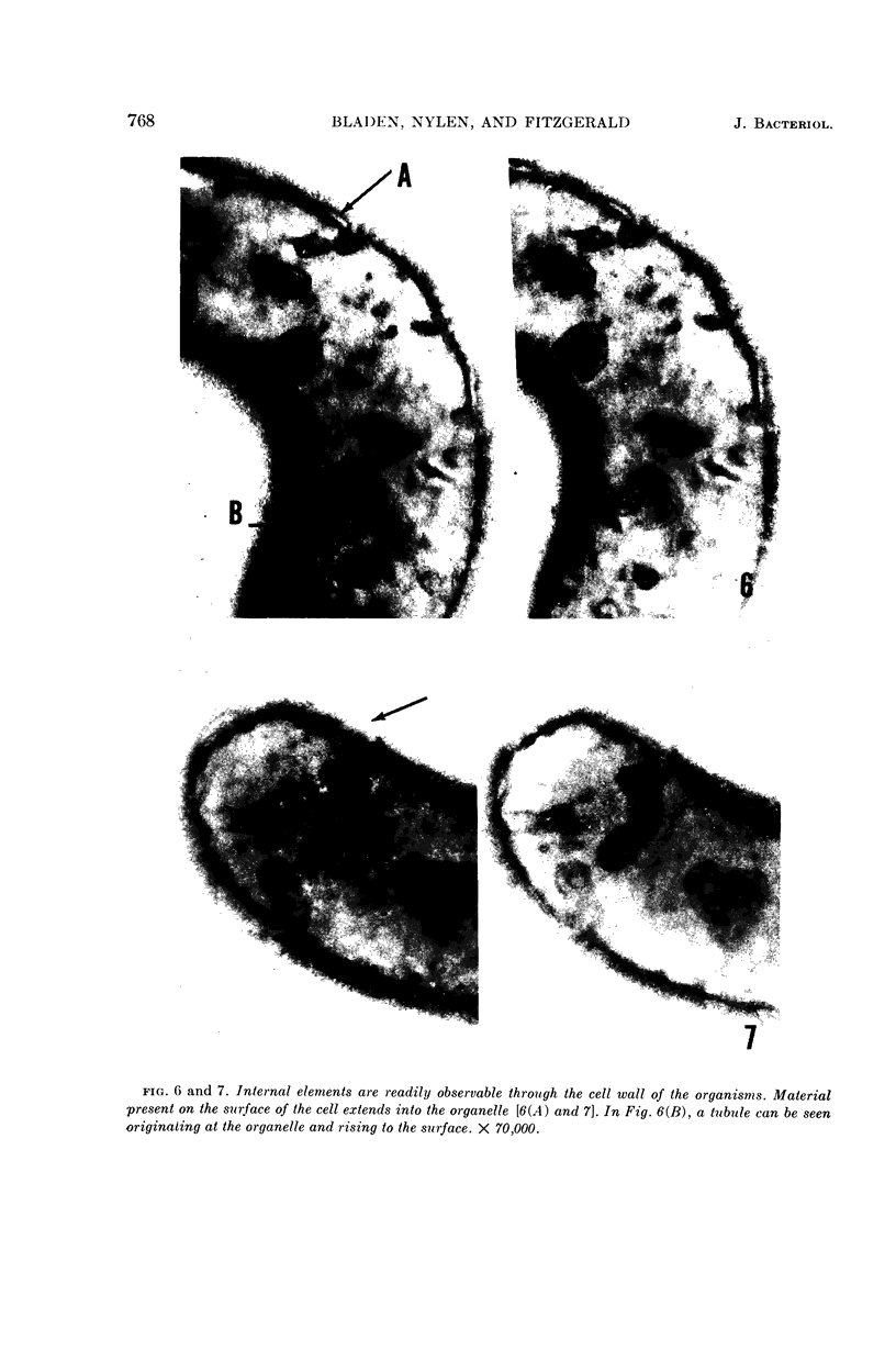
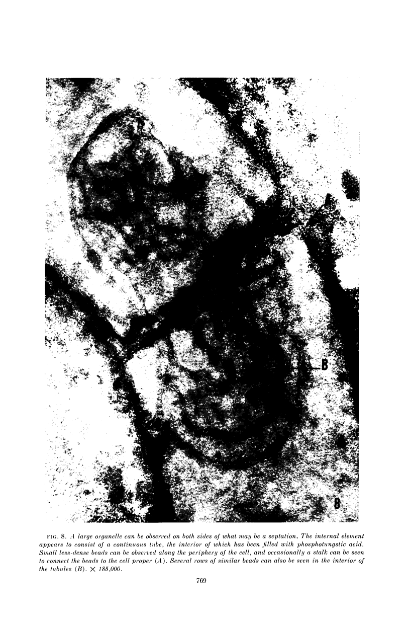
Bladen, Howard A. (National Institute of Dental Research, Bethesda, Md.), Marie U. Nylen, and Robert J. Fitzgerald. Internal structures of a Eubacterium sp. demonstrated by the negative staining technique. J. Bacteriol. 88:763–770. 1964.—Thin sections as well as negatively stained whole cells of a Eubacterium sp. isolated from the rat cecum were examined in a Siemens Elmiskop I electron microscope. The cell wall usually appeared in thin sections as a single dense layer about 130 A thick; however, occasionally it was demonstrated to be composed of three layers. The plasma membrane was approximately 130 A wide, and was composed of a denselight-dense arrangement. Intracytoplasmic membranous elements continuous with the plasma membrane were also observed in thin sections. When whole cells were negatively stained with phosphotungstic acid, the cell wall became transparent, and structures comparable in morphology to intracytoplasmic membranous elements were observed. These were demonstrated by stereomicroscopy to be in the interior environment of the cell, and appeared in many cases continuous with the cell wall. Frequently, they seemed to open to the exterior of the cell through what may be termed a pore. Small stalked structures similar to those described in mitochondria were observed along the periphery of the cell and occasionally along the walls of the internal elements.
Full text
PDF
Images in this article
Selected References
These references are in PubMed. This may not be the complete list of references from this article.
- BLADEN H. A., WATERS J. F. ELECTRON MICROSCOPIC STUDY OF SOME STRAINS OF BACTEROIDES. J Bacteriol. 1963 Dec;86:1339–1344. doi: 10.1128/jb.86.6.1339-1344.1963. [DOI] [PMC free article] [PubMed] [Google Scholar]
- CHAPMAN G. B., HANKS J. H., WALLACE J. H. An electron microscope study of the disposition and fine structure of Mycobacterium lepraemurium in mouse spleen. J Bacteriol. 1959 Feb;77(2):205–211. doi: 10.1128/jb.77.2.205-211.1959. [DOI] [PMC free article] [PubMed] [Google Scholar]
- CHAPMAN G. B., HILLIER J. Electron microscopy of ultra-thin sections of bacteria I. Cellular division in Bacillus cereus. J Bacteriol. 1953 Sep;66(3):362–373. doi: 10.1128/jb.66.3.362-373.1953. [DOI] [PMC free article] [PubMed] [Google Scholar]
- EDWARDS M. R., STEVENS R. W. FINE STRUCTURE OF LISTERIA MONOCYTOGENES. J Bacteriol. 1963 Sep;86:414–428. doi: 10.1128/jb.86.3.414-428.1963. [DOI] [PMC free article] [PubMed] [Google Scholar]
- FITZ-JAMES P. C. Participation of the cytoplasmic membrane in the growth and spore fromation of bacilli. J Biophys Biochem Cytol. 1960 Oct;8:507–528. doi: 10.1083/jcb.8.2.507. [DOI] [PMC free article] [PubMed] [Google Scholar]
- GLAUERT A. M., HOPWOOD D. A. A membranous component of the cytoplasm in Streptomyces coelicolor. J Biophys Biochem Cytol. 1959 Dec;6:515–516. doi: 10.1083/jcb.6.3.515. [DOI] [PMC free article] [PubMed] [Google Scholar]
- GLAUERT A. M., HOPWOOD D. A. The fine structure of Streptomyces coelicolor. I. The cytoplasmic membrane system. J Biophys Biochem Cytol. 1960 Jun;7:479–488. doi: 10.1083/jcb.7.3.479. [DOI] [PMC free article] [PubMed] [Google Scholar]
- GLAUERT A. M. The fine structure of bacteria. Br Med Bull. 1962 Sep;18:245–250. doi: 10.1093/oxfordjournals.bmb.a069988. [DOI] [PubMed] [Google Scholar]
- IMAEDA T., OGURA M. Formation of intracytoplasmic membrane system of mycobacteria related to cell division. J Bacteriol. 1963 Jan;85:150–163. doi: 10.1128/jb.85.1.150-163.1963. [DOI] [PMC free article] [PubMed] [Google Scholar]
- KELLENBERGER E., RYTER A., SECHAUD J. Electron microscope study of DNA-containing plasms. II. Vegetative and mature phage DNA as compared with normal bacterial nucleoids in different physiological states. J Biophys Biochem Cytol. 1958 Nov 25;4(6):671–678. doi: 10.1083/jcb.4.6.671. [DOI] [PMC free article] [PubMed] [Google Scholar]
- MUDD S., WINTERSCHEID L. C., DeLAMATER E. D., HENDERSON H. J. Evidence suggesting that the granules of mycobacteria are mitochondria. J Bacteriol. 1951 Oct;62(4):459–475. doi: 10.1128/jb.62.4.459-475.1951. [DOI] [PMC free article] [PubMed] [Google Scholar]
- SHINOHARA C., FUKUSHI K., SUZUKI J. Mitochondria-like structures in ultrathin sections of Mycobacterium avium. J Bacteriol. 1957 Sep;74(3):413–415. doi: 10.1128/jb.74.3.413-415.1957. [DOI] [PMC free article] [PubMed] [Google Scholar]
- STOECKENIUS W. Some observations on negatively stained mitochondria. J Cell Biol. 1963 May;17:443–454. doi: 10.1083/jcb.17.2.443. [DOI] [PMC free article] [PubMed] [Google Scholar]
- VAN ITERSON W. Some features of a remarkable organelle in Bacillus subtilis. J Biophys Biochem Cytol. 1961 Jan;9:183–192. doi: 10.1083/jcb.9.1.183. [DOI] [PMC free article] [PubMed] [Google Scholar]