Abstract
The Esx secretion pathway is conserved across Gram-positive bacteria. Esx-1, the best-characterized system, is required for virulence of Mycobacterium tuberculosis, although its precise function during infection remains unclear. Esx-3, a paralogous system present in all mycobacterial species, is required for growth in vitro. Here, we demonstrate that mycobacteria lacking Esx-3 are defective in acquiring iron. To compete for the limited iron available in the host and the environment, these organisms use mycobactin, high-affinity iron-binding molecules. In the absence of Esx-3, mycobacteria synthesize mycobactin but are unable to use the bound iron and are impaired severely for growth during macrophage infection. Mycobacteria thus require a specialized secretion system for acquiring iron from siderophores.
Keywords: secretion, siderophore
The thick, waxy mycobacterial cell wall functions as an exceptionally efficient barrier against a range of environmental and host insults. The relatively impermeable structure also poses a challenge, because it impedes the exchange of metabolites between the bacterium and its host during chronic intracellular infection. Mycobacteria have evolved both general and specialized systems to transport molecules across their cell walls (1). One such class of transporters is the Esx (type VII) secretion pathway (2).
The genome of Mycobacterium tuberculosis, the causative agent of tuberculosis, contains five paralogous esx loci (3–5). Esx-1 is the best described. Substrates of this secretion system include the esx-1-encoded EsxB (Esat-6), EsxA (Cfp-10) and Rv3881c (6, 7), and EspA and EspB, encoded by an unlinked but evolutionarily related genetic locus (8, 9). Although Esx-1 and its substrates are required for virulence of pathogenic mycobacteria (10–12) and conjugation in M. smegmatis (13, 14), their precise contributions to these processes have not been determined (1, 2).
Even less is known about the other Esx systems. Preliminary characterization of Esx-5 has revealed that the system exports various proteins containing Pro-Pro-Glu (PPE) and polymorphic GC-rich sequences (PGRS) and modulates macrophage responses (15, 16). Neither protein export nor biological function has been reported for the products of esx-2, esx-3, and esx-4.
In a genome-wide scan of M. tuberculosis after growth on laboratory medium, we were able to recover transposon insertions in all of the esx loci except esx-3 (17), suggesting that Esx-3 may be essential for mycobacterial growth in vitro. Previous reports suggest that expression of this locus is regulated by the metal-regulated repressors IdeR and Zur (18, 19). Besides divalent cation availability, in vitro growth and stress conditions such as acid, hydrogen peroxide, nitric oxide, SDS, stationary phase, and biofilms appear to alter esx-3 expression (20–24).
We show here that Esx-3 is required for growth of M. bovis BCG and M. smegmatis under iron deprivation. Further, we demonstrate that Esx-3 is a secretion system and that export of esx-3-encoded substrates is iron-dependent. Genetic epistasis experiments reveal a link between iron acquisition via the mycobactin siderophore pathway and Esx-3 protein export. Although mycobacteria that lack Esx-3 detect and initiate genetic programs in response to iron deprivation, they are unable to procure the metal from mycobactin or multiply normally in macrophages. Esx-3 is thus a specialized secretion system that is required for mycobactin-mediated iron acquisition and for survival during infection.
Results
Esx-3 Is Required for Growth under Iron-Limited Conditions.
To investigate the apparent growth requirement for Esx-3 (25), we used gene set enrichment analysis (GSEA) (26) to test for coordinated transcriptional regulation of M. tuberculosis esx genes under 75 experimental conditions (27) that perturb growth in vitro. Significant up-regulation of esx-3 gene expression occurs in response to five of the 75 treatments, four of which are iron chelators (Table 1). Furthermore, of all genomic windows for which we used GSEA to test transcriptional responses to chelation, esx-3 is one of the most highly up-regulated, ranking between two loci that encode enzymes that synthesize the siderophore mycobactin (Table 2) (28, 29). These findings suggest that Esx-3 functions in response to iron deprivation in M. tuberculosis, and they correspond well with published data that show an increase in IdeR-regulated esx-3 transcription in response to iron starvation (19).
Table 1.
Conditions affecting esx-3 transcription
| Treatment* | Transcription change direction | Nominal P value | False discovery rate q value† |
|---|---|---|---|
| 111895 | Up | 0 | 0.00 |
| Dipyridyl | Up | 0 | 0.00 |
| Deferoxamine | Up | 0 | 0.04 |
| Clofazimine | Up | 0 | 0.04 |
| Ascididemin | Up | 0 | 0.03 |
Gene set enrichment analysis was performed as described in ref. 26. Expression changes with a nominal P value <0.05 and a false discovery rate q value <0.05 were considered significant.
*Treatments from ref. 27: 111895, dipyridyl, deferoxamine, and ascididemin are known chelators. Clofazimine is an antituberculosis drug with an unknown mechanism of action.
†False discovery rate q value is the fraction of observations expected to be false positives.
Table 2.
Genomic windows up-regulated in low iron*
| Gene range† | Nominal P value | False discovery rate q value‡ | Annotation§ |
|---|---|---|---|
| rv1338-rv1362c | 0.00 | 0.03 | irtAB, fadD33, rv1347c |
| rv0278c-rv0302 | 0.00 | 0.02 | esx-3 |
| rv2380c-rv2403c | 0.00 | 0.05 | Mycobactin synthesis |
| rv2360c-rv2384 | 0.00 | 0.05 | Mycobactin synthesis |
| rv1180-rv1204c | 0.00 | 0.05 | Polyketide synthesis |
*The 199 gene sets from overlapping 25-gene windows covering the Mycobacterium tuberculosis genome were tested by gene set enrichment analysis for transcriptional changes in response to chelator treatments from ref. 27.
†Identification of the first and last gene in the window are given. The gene sets consisted of these flanking genes plus annotated coding loci between them that have unique probes present on the arrays used in ref. 27.
‡False discovery rate q value is the fraction of observations expected to be false positives.
§Summary of the gene content of each window.
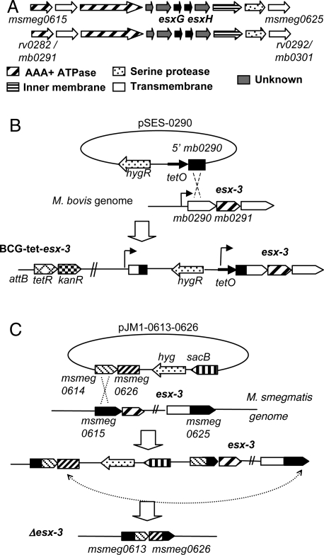
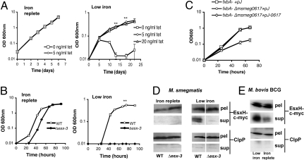
To test whether Esx-3 is important for survival in iron-limiting conditions, we first sought to construct mutants defective in Esx-3 production. As predicted by the transposon insertion data (17), we were unable to recover M. tuberculosis or M. bovis BCG esx-3 deletion mutants through homologous recombination, suggesting that this gene is indeed essential for growth (Table S1). Therefore, we generated a conditional M. bovis BCG esx-3 mutant strain by replacing the native promoter upstream of the first gene in the locus, mb0290, with a tetracycline-regulated promoter (BCG-tet-esx-3; Fig. 1B). These mutants are able to grow without inducer (Fig. 2A), because the promoter is not repressed completely in the absence of tetracycline (Fig. S1A). We also found that, unlike pathogenic mycobacteria, the environmental organism M. smegmatis is able to tolerate deletion of the entire esx-3 locus (Fig. 1C) and does not have a growth defect in complete medium. Development of these two experimental systems that bypass the growth requirement related to Esx-3 allowed us to probe Esx-3 function. Both mycobacterial strains experience severe growth retardation in the absence of exogenous iron (Fig. 2 A and B).
Fig. 1.
Construction of the mycobacterial esx-3 mutants. Schematic representation of the esx-3 loci from Mycobacterium smegmatis (upper) and in Mycobacterium tuberculosis/Mycobacterium bovis bacillus Calmette–Guérin (BCG) (lower) (A) and strategies used to generate the M. bovis BCG esx-3 conditional mutant (BCG-tet-esx-3) (B) and the M. smegmatis Δesx-3 mutant (C).
Fig. 2.
Esx-3 is required for Mycobacterium bovis bacillus Calmette–Guérin (BCG) and Mycobacterium smegmatis growth and for secretion of esx-3-encoded proteins under iron-deprived conditions. Growth of BCG-tet-esx-3 in iron-replete 7H9 medium or low iron glycerol alanine salts Tween-80 medium in the presence or absence of the inducer anhydrotetracycline (n = 3; **, P = 0.0002) (A). Growth of wild-type (WT) and Δesx-3 M. smegmatis in low iron (100 μM 2,2′-dipyridyl) or iron-replete (12.5 μM FeCl3) Sauton's medium (n = 3; **, P = 0.0014) (B). Strains were subcultured in low iron Sauton's medium before inoculation. Growth of fxbA− and fxbA− Δmsmeg0617 M. smegmatis containing pJEB402 with or without the msmeg0617 gene in low iron Sauton's medium (C). Strains were not subcultured before inoculation. Anti-c-myc and anti-ClpP1P2 immunoblotting of cell lysates and culture supernatants of WT and Δesx-3 M. smegmatis containing pTetG-esxGH-c-myc (D) or WT M. bovis BCG containing pMV762-esxG-6Xhis-esxH-c-myc (E) in low iron or iron-replete Sauton's medium. All data are representative of at least three independent experiments. Error bars represent the standard deviations.
Esx-3 Is an Iron-Responsive Secretion System.
The organization of the mycobacterial esx-3 and esx-1 loci is similar: esx-3 encodes EsxG and EsxH, which are paralogous to the small, secreted proteins EsxB (Cfp-10) and EsxA (Esat-6) encoded by esx-1 (Fig. 1A) (5). Mutations in several esx-1 genes abolish export of EsxB and EsxA (10, 30–35). To determine if the export of EsxH is dependent on Esx-3, we compared amounts of the heterologously-expressed, epitope-tagged protein in the cell-associated and supernatant fractions of wild-type and Δesx-3 M. smegmatis. We find that EsxH-c-myc secretion is dependent on the presence of Esx-3 (Fig. 2D). Protein export, moreover, is enhanced in both M. smegmatis and M. bovis BCG under low iron conditions (Fig. 2 D and E). Thus, Esx-3 does indeed function as a secretion system that functionally responds to iron deprivation.
Esx-3 Interacts with the Mycobactin Pathway of Iron Acquisition.
Mycobacteria acquire iron from at least two classes of siderophore scavenging systems: exochelin and mycobactin (36). Although M. smegmatis produces both exochelin and mycobactin (37), pathogenic mycobacteria use only the pathway involving mycobactin and its structurally-related soluble form, carboxymycobactin (36). Expression of the mycobactin biosynthetic loci covaries with esx-3 in our GSEA study and under several published conditions (20).
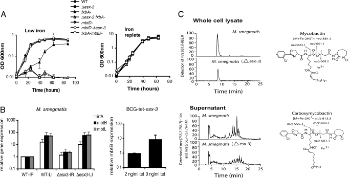
To test whether Esx-3 genetically interacts with either of the siderophore pathways, we constructed M. smegmatis mutants with insertions in mbtD (mbtD−), which encodes a polyketide synthase required for mycobactin synthesis, or in fxbA (fxbA−), which encodes a formyl transferase required for exochelin synthesis (Fig. S2), and grew them without subculturing (in contrast to Fig. 2B) to produce an intermediate level of attenuation. Deletion of esx-3 or inactivation of mbtD has little effect on M. smegmatis growth under these more permissive, low iron conditions (Fig. 3A). In contrast, inactivation of fxbA causes a marked growth defect, likely because exochelin is the primary iron acquisition system in M. smegmatis (37). Combining mutations in different iron acquisition pathways should produce a synthetic growth defect under conditions of iron starvation, whereas combining mutations in the same pathway should not. fxbA− mbtD−, which lacks siderophores, and fxbA− Δesx-3 both fail to grow in low iron medium (Fig. 3A). In contrast, the mbtD− Δesx-3 strain does not exhibit an additive increase in low iron growth inhibition (Fig. 3A), strongly suggesting that Esx-3 functions in iron acquisition via the mycobactin pathway.
Fig. 3.
Esx-3 interacts with the mycobactin pathway but is not required for sensing iron or for siderophore synthesis. (A) Growth of wild-type (WT), Δesx-3, exochelin (fxbA−), and mycobactin (mbtD−) insertional biosynthesis mutants and double mutants (fxbA− Δesx-3 and fxbA− mbtD−) in low iron or iron-replete Sauton's medium (n = 3; *, P < 0.001). Strains were not subcultured before inoculation. (B) Quantitative RT-PCR for mbtB, mbtL, and irtA expression in WT and Δesx-3 Mycobacterium smegmatis (n = 3) in low iron (LI) or iron-replete (IR) medium and for mbtB in bacillus Calmette–Guérin (BCG)-tet-esx-3 in low iron medium. Data representative of three independent experiments are shown. Error bars represent the standard deviations. (C) The LC-MS analysis of lipids extracted from the whole cell lysates of M. smegmatis with chlorofrom and methanol yielded mycobactin chromatograms in which the m/z (881.4), elution time, and fragments (right) correspond to the expected and actual measurements of an authentic standard. Conditioned supernatants were subjected to LC-MS detection in mass windows corresponding to the known masses of carboxymycobactins [m/z (756.7–757.7) + 14n, where n is 0, 1, 2, 3, 4] and [m/z (757.7–758.7) + 14n] (upper). The elution time and collision-induced dissociation MS fragmentation (right) of the putative carboxymycobactins are consistent with those of an authentic carboxymycobactin standard.
Requirement of Esx-3 for Iron-Limited Growth Can Be Complemented.
In BCG-tet-esx-3, the low iron growth defect is reversed by induction of Esx-3 (Fig. 2A). Because we were unable to complement fxbA− Δesx-3 or Δesx-3 M. smegmatis with a cosmid containing the M. tuberculosis esx-3 locus, we combined the fxbA− insertional mutation with a single gene deletion in the esx-3 gene msmeg0617. The M. tuberculosis esx-1 paralog rv3870/3871 encodes an AAA+ ATPase that is required for Esx-1-mediated secretion, virulence, and conjugation in multiple mycobacterial species (1, 2). We find that fxbA− Δmsmeg0617 has a pronounced low iron growth defect that permits robust complementation by episomal expression of msmeg0617 (Fig. 2C). Complementation of a single esx-3 gene deletion further supports a role for the secretion system in growth under iron-deprived conditions.
Impaired Iron Sensing or Reduced Mycobactin Abundance Do Not Account for the esx-3 Defect.
Several models could explain the genetic interaction between Esx-3 and the mycobactin pathway. Mycobacteria that lack Esx-3 might fail to increase mycobactin production in response to iron deprivation. A second possibility is that esx-3 mutants are able to synthesize mycobactin but that the siderophore might not be exported from the cell or be functional. Finally, Esx-3 might be required for utilization of iron-bound mycobactin.
Mycobacteria up-regulate the expression of siderophore biosynthesis genes when starved for iron (19). Could strains that lack Esx-3 fail to sense low iron conditions and therefore fail to induce gene expression? To test this possibility, we compared transcript abundance of the iron-responsive genes mbtB, mbtL, and irtA in wild-type and Δesx-3 M. smegmatis in the presence or absence of iron. mbtB and mbtL reside in distinct genomic loci and encode mycobactin biosynthetic enzymes (28, 29), whereas irtA encodes part of an importer/exporter system for mycobactin (38, 39). Both strains increase transcription of these genes in response to iron deprivation (Fig. 3B). In fact, mbtB transcript abundance in uninduced BCG-tet-esx-3, which has low esx-3 expression (Fig. S1B), is higher than that in the induced mutant (Fig. 3B). These data suggest that Esx-3 is not necessary for iron sensing in M. smegmatis or M. bovis BCG but that its depletion in M. bovis BCG, which lacks alternate iron acquisition systems such as the exochelin pathway, may result in greater perceived iron starvation.
Although esx-3 mutants do not appear to be defective for iron sensing, they might be unable to respond to iron deprivation by producing functional siderophores or, in the case of the soluble carboxymycobactin, by transporting them across the cell wall. To test whether Esx-3 is required for mycobactin synthesis, we analyzed the siderophores in the whole cell lysates or conditioned supernatants of wild-type and Δesx-3 M. smegmatis with LC-MS. Analysis of samples that were normalized for mass and found to have unaltered signals for unrelated lipids showed that the ion current corresponding to molecules matching the expected mass of the iron–mycobactin complex (Fig. 3C) and related molecular variants are decreased moderately in the Δesx-3 strain. Carboxymycobactin detected as sodium adducts or as iron complexes in supernatants is detected at similar levels in wild-type and Δesx-3 M. smegmatis (Fig. 3C), and individual molecular variants in the spectrum of carboxymycobactins were preserved (Fig. S3). Collision-induced dissociation MS of the major mycobactin and carboxymycobactin species shows no changes in structure compared with those of standard compounds and prior analyses (Fig. S4). Although loss of Esx-3 does not change the structures or alter the individual molecular species of mycobactin and carboxymycobactin, there is a small but consistent decrease in the total pools of the siderophores. The moderate changes in total mycobactin abundance are unlikely to account for the striking low iron growth phenotype in the absence of Esx-3, because the fxbA− mbtB− M. smegmatis strain, which does not produce mycobactin, has a quantitatively similar growth defect in low iron to that of the fxbA− Δesx-3 strain (Fig. 3A), which produces high amounts of mycobactin.
Esx-3 Is Required for Mycobactin Utilization.
In response to iron deprivation, mycobacteria that lack Esx-3 appear to produce mycobactin and carboxymycobactin and export carboxymycobactin appropriately. To test whether these mutants are impaired for utilization of the siderophore, we monitored growth of fxbA− strains deficient in Esx-3 or mycobactin production in the presence or absence of exogeneous, iron-bound mycobactin S or carboxymycobactin S. Whereas the low iron growth of the fxbA− mbtB− strain can be rescued almost completely by purified mycobactin or carboxymycobactin, neither siderophore rescues the fxbA− Δesx-3 strain to fxbA− growth levels (Fig. 4 A–C). Thus, M. smegmatis that lacks Esx-3 is unable to use iron-bound mycobactin.
Fig. 4.
Esx-3 is required for optimal utilization of iron-bound mycobactin. Growth curves for fxbA−, fxbA− mbtD−, and fxbA− Δesx-3 Mycobacterium smegmatis in low iron Sauton's medium (n = 3; **, P = 0.0005) (A), in the presence of 10 ng/mL ferri-mycobactin S (n = 3; **, P = 0.0017) (B), or 20 ng/mL ferri-carboxymycobactin S (n = 3; **, P = 0.0003) (C). All data are representative of at least three independent experiments. Error bars represent the standard deviations.
Esx-3 Is Required for Growth in Macrophages.
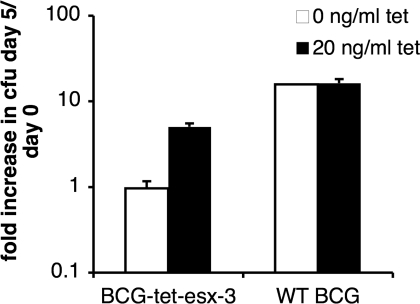
Iron is limiting in many environmental conditions, particularly in an infected host (40). Consistent with a role for Esx-3 in mycobactin-mediated iron acquisition, we find that BCG-tet-esx-3 grows significantly more slowly in J774 macrophages in the absence of inducer (Fig. 5). Preliminary studies also suggest that BCG-tet-esx-3 multiplies slower than wild-type M. bovis BCG in the spleen and lungs of SCID mice (Fig. S5 A and B). Hence, Esx-3 is likely important for M. bovis BCG growth both in vitro and in vivo.
Fig. 5.
Esx-3 is required for mycobacterial infection of macrophages. Growth of wild-type (WT) M. bovis BCG and BCG-tet-esx-3 in the presence or absence of anhydrotetracycline in J774 macrophages (n = 3).
Discussion
We demonstrate that Esx-3, a group of proteins of previously unknown function, is a specialized secretion system required for mycobactin-mediated iron acquisition. The wide variety of conditions that perturb esx-3 expression (18–24, 27) may, in part, reflect the ubiquitous need for iron in enzymes involved in multiple cellular processes (40). Alternatively, Esx-3 may transport additional substrates in addition to those involved in iron uptake.
The moderate reduction in total mycobactin in the absence of Esx-3 does not appear to be responsible for the growth defect seen under iron deprivation, given that the addition of purified mycobactin fails to rescue the fxbA− Δesx-3 strain (Fig. 4). Further experimentation is necessary to determine whether the decrease in mycobactin pools in the absence of Esx-3 reflects mechanism or whether it is a consequence of perturbation of mycobacterial iron acquisition, normally a carefully orchestrated process.
How might a specialized secretion system like Esx-3 contribute to siderophore utilization? In other bacteria, siderophore-mediated iron acquisition requires the participation of multiple extracellular components (40). Although Gram-negative organisms have numerous mechanisms for siderophore secretion and uptake across the outer membrane, analogous systems for navigating the substantial cell wall of Gram-positive bacteria are poorly defined (40). The only known uptake system for mycobactin is via IrtB (38, 39), an ABC transporter confined to the cytoplasmic membrane (39). Esx-3 could have direct or indirect roles in iron transport. For example, the secretion system might export or position mycobactin-binding proteins in the cell wall. In this model, mycobacteria that lack Esx-3 or its transported substrates are defective for the delivery of iron-bound mycobactin to IrtB or possibly to alternate uptake systems. Alternatively, Esx-3 might regulate the activity of mycobactin transporters or have more global effects on the cell wall. The complex structure of the Gram-positive cell wall is a significant barrier to import or export. The Esx transport systems may provide a mechanism for moving compounds within and across the thick cell wall barriers of Gram-positive bacteria.
Materials and Methods
Bacterial Strains, Media, and Reagents.
For iron starvation experiments, M. bovis BCG was cultured in iron-replete 7H9 medium (SI Materials and Methods) to late stationary phase, then diluted 1:100 or 1:250 in glycerol alanine salts Tween-80 medium lacking ferric ammonium citrate (41). Before infection experiments, bacteria were grown in 7H9 medium in the presence or absence of 5 ng/mL anhydrotetracycline. M. smegmatis was cultured in 7H9 medium, subcultured once in chelated Sauton's medium (SI Materials and Methods), and then diluted 1:1,000 in chelated Sauton's medium without antibiotics containing either 100 μM 2,2′-dipyridyl or 12.5 μM FeCl3. For epistasis experiments, bacteria were diluted directly 1:1,000 from 7H9 medium into chelated Sauton's medium. The mycobactin and carboxymycobactin complementation experiments were performed similarly, except that 2,2′-dipyridyl was not added. Purified ferri-mycobactin S and ferri-carboxymycobactin S were kind gifts of Colin Ratledge (University of Hull, Hull, United Kingdom).
Mycobacterial Mutant Construction.
Deletion and promoter replacements were constructed by transforming M. tuberculosis, M. bovis BCG, and M. smegmatis with suicide vectors containing antibiotic resistance markers and the counterselectable marker sacB. Details of the selection process are contained in SI Materials and Methods.
Gene Set Enrichment Analysis.
We used GSEA to test for coordinated transcriptional up- or down-regulation in 75 conditions in a published expression compendium (26, 27). Genes represented by identical or nearly identical microarray probes were removed from gene sets consisting of one esx locus each to avoid artifacts resulting from cross-hybridization. The GSEA enrichment was considered significant if it resulted in a nominal P value <0.05 and a false discovery rate <0.05. All five treatments meeting our criteria for significance resulted in increased transcription of esx-3. To assess the importance of the esx-3 locus among all possible loci in M. tuberculosis, we performed GSEA using 199 gene sets, each containing 25 genes in overlapping sliding windows covering the entire M. tuberculosis genome. We do not believe that small differences in the relative rankings are meaningful at this level of resolution.
Immunoblotting.
Wild-type and Δesx-3 M. smegmatis were transformed with pTetG-esxGH-c-myc, an episomal plasmid derived from pUV15tetOR (42), containing esxG and c-myc C-terminally tagged esxH from M. smegmatis under a tetracycline-inducible promoter. M. bms BCG were transformed with pMV762-esxG-6XHis-esxH-c-myc, an episomal plasmid that is derived from pMV762 (43) and contains poly-His N-terminally tagged esxG and c-myc C-terminally tagged esxH from M. bovis BCG. See SI Materials and Methods for details of protein isolation under low iron and iron-replete conditions and subsequent analysis.
RNA Extraction and Quantitative RT-PCR.
Standard procedures are described in SI Materials and Methods.
Mass Spectrometry.
M. smegmatis were diluted 1:500 from 7H9 medium into chelated Sauton's medium without Tween-80 containing 0.1 μM FeCl3. Cultures were grown to saturation, centrifuged, washed, and resuspended in the same medium. After 2 days of growth, bacteria were centrifuged to separate the whole cell and supernatant fractions, and the latter was filtered to remove bacteria and debris. Lipids were extracted from the culture supernatant by adding 6 N HCl and ethyl acetate and then centrifuging at 800 × g for 30 min at room temperature. The upper organic phase containing the lipids was collected, evaporated to dryness, and resuspended in 1:1 chloroform/methanol. Lipids were extracted from the cell wall fraction as described in ref. 44. Details of the mass spectrometry are contained in SI Materials and Methods.
Macrophage and Animal Infections.
Infection of mouse macrophage J774 cells by M. bovis BCG or BCG-tet-esx-3 at a multiplicity of infection of 1:1 and mixed, i.v. infection of CB-17 SCID mice (The Jackson Laboratory) by 105 cfu of BCG-tet-esx-3 and 106 cfu of wild-type M. bovis BCG (10:1 ratio) were assessed by standard procedures (SI Materials and Methods).
Statistical Analysis.
Differences between groups were evaluated using a paired Student's t test.
Supplementary Material
Acknowledgments.
We gratefully acknowledge Dr. Colin Ratledge for helpful advice and for providing ferri-mycobactin S and ferri-carboxymycobactin S. We also thank Dr. Alfred Goldberg for providing the anti-ClpP1P2 antibody. This work was supported by the Burroughs Wellcome Fund and the National Institutes of Health Grants R01 AI48704 and R0171155.
Note Added in Proof.
Recently, Serafini et al. (45) showed that Esx-3 is required for M. tuberculosis growth under iron-restricted conditions.
Footnotes
The authors declare no conflict of interest.
This article is a PNAS Direct Submission.
This article contains supporting information online at www.pnas.org/cgi/content/full/0900589106/DCSupplemental.
References
- 1.DiGiuseppe Champion PA, Cox JS. Protein secretion systems in Mycobacteria. Cell Microbiol. 2007;9:1376–1384. doi: 10.1111/j.1462-5822.2007.00943.x. [DOI] [PubMed] [Google Scholar]
- 2.Abdallah AM, et al. Type VII secretion—Mycobacteria show the way. Nat Rev Microbiol. 2007;5:883–891. doi: 10.1038/nrmicro1773. [DOI] [PubMed] [Google Scholar]
- 3.Cole ST, et al. Deciphering the biology of Mycobacterium tuberculosis from the complete genome sequence. Nature. 1998;393:537–544. doi: 10.1038/31159. [DOI] [PubMed] [Google Scholar]
- 4.Tekaia F, et al. Analysis of the proteome of Mycobacterium tuberculosis in silico. Tuber Lung Dis. 1999;79:329–342. doi: 10.1054/tuld.1999.0220. [DOI] [PubMed] [Google Scholar]
- 5.Gey Van Pittius NC, et al. The ESAT-6 gene cluster of Mycobacterium tuberculosis and other high G+C Gram-positive bacteria. Genome Biol. 2001;2:RESEARCH0044. doi: 10.1186/gb-2001-2-10-research0044. [DOI] [PMC free article] [PubMed] [Google Scholar]
- 6.McLaughlin B, et al. A mycobacterium ESX-1-secreted virulence factor with unique requirements for export. PLoS Pathog. 2007;3:e105. doi: 10.1371/journal.ppat.0030105. [DOI] [PMC free article] [PubMed] [Google Scholar]
- 7.Xu J, et al. A unique Mycobacterium ESX-1 protein co-secretes with CFP-10/ESAT-6 and is necessary for inhibiting phagosome maturation. Mol Microbiol. 2007;66:787–800. doi: 10.1111/j.1365-2958.2007.05959.x. [DOI] [PubMed] [Google Scholar]
- 8.Fortune SM, et al. Mutually dependent secretion of proteins required for mycobacterial virulence. Proc Natl Acad Sci USA. 2005;102:10676–10681. doi: 10.1073/pnas.0504922102. [DOI] [PMC free article] [PubMed] [Google Scholar]
- 9.MacGurn JA, Raghavan S, Stanley SA, Cox JS. A non-RD1 gene cluster is required for Snm secretion in Mycobacterium tuberculosis. Mol Microbiol. 2005;57:1653–1663. doi: 10.1111/j.1365-2958.2005.04800.x. [DOI] [PubMed] [Google Scholar]
- 10.Lewis KN, et al. Deletion of RD1 from Mycobacterium tuberculosis mimics bacille Calmette–Guérin attenuation. J Infect Dis. 2003;187:117–123. doi: 10.1086/345862. [DOI] [PMC free article] [PubMed] [Google Scholar]
- 11.Pym AS, Brodin P, Brosch R, Huerre M, Cole ST. Loss of RD1 contributed to the attenuation of the live tuberculosis vaccines Mycobacterium bovis BCG and Mycobacterium microti. Mol Microbiol. 2002;46:709–717. doi: 10.1046/j.1365-2958.2002.03237.x. [DOI] [PubMed] [Google Scholar]
- 12.Pym AS, et al. Recombinant BCG exporting ESAT-6 confers enhanced protection against tuberculosis. Nat Med. 2003;9:533–539. doi: 10.1038/nm859. [DOI] [PubMed] [Google Scholar]
- 13.Flint JL, Kowalski JC, Karnati PK, Derbyshire KM. The RD1 virulence locus of Mycobacterium tuberculosis regulates DNA transfer in Mycobacterium smegmatis. Proc Natl Acad Sci USA. 2004;101:12598–12603. doi: 10.1073/pnas.0404892101. [DOI] [PMC free article] [PubMed] [Google Scholar]
- 14.Coros A, Callahan B, Battaglioli E, Derbyshire KM. The specialized secretory apparatus ESX-1 is essential for DNA transfer in Mycobacterium smegmatis. Mol Microbiol. 2008;69:794–808. doi: 10.1111/j.1365-2958.2008.06299.x. [DOI] [PMC free article] [PubMed] [Google Scholar]
- 15.Abdallah AM, et al. The ESX-5 secretion system of Mycobacterium marinum modulates the macrophage response. J Immunol. 2008;181:7166–7175. doi: 10.4049/jimmunol.181.10.7166. [DOI] [PubMed] [Google Scholar]
- 16.Abdallah AM, et al. A specific secretion system mediates PPE41 transport in pathogenic mycobacteria. Mol Microbiol. 2006;62:667–679. doi: 10.1111/j.1365-2958.2006.05409.x. [DOI] [PubMed] [Google Scholar]
- 17.Sassetti CM, Boyd DH, Rubin EJ. Genes required for mycobacterial growth defined by high density mutagenesis. Mol Microbiol. 2003;48:77–84. doi: 10.1046/j.1365-2958.2003.03425.x. [DOI] [PubMed] [Google Scholar]
- 18.Maciąg A, et al. Global analysis of the Mycobacterium tuberculosis Zur (FurB) regulon. J Bacteriol. 2007;189:730–740. doi: 10.1128/JB.01190-06. [DOI] [PMC free article] [PubMed] [Google Scholar]
- 19.Rodriguez GM, Voskuil MI, Gold B, Schoolnik GK, Smith I. ideR, an essential gene in Mycobacterium tuberculosis: Role of IdeR in iron-dependent gene expression, iron metabolism, and oxidative stress response. Infect Immun. 2002;70:3371–3381. doi: 10.1128/IAI.70.7.3371-3381.2002. [DOI] [PMC free article] [PubMed] [Google Scholar]
- 20.Schnappinger D, et al. Transcriptional adaptation of Mycobacterium tuberculosis within macrophages: Insights into the phagosomal environment. J Exp Med. 2003;198:693–704. doi: 10.1084/jem.20030846. [DOI] [PMC free article] [PubMed] [Google Scholar]
- 21.Manganelli R, Voskuil MI, Schoolnik GK, Smith I. The Mycobacterium tuberculosis ECF sigma factor σE: Role in global gene expression and survival in macrophages. Mol Microbiol. 2001;41:423–437. doi: 10.1046/j.1365-2958.2001.02525.x. [DOI] [PubMed] [Google Scholar]
- 22.Ojha A, Hatfull GF. The role of iron in Mycobacterium smegmatis biofilm formation: The exochelin siderophore is essential in limiting iron conditions for biofilm formation but not for planktonic growth. Mol Microbiol. 2007;66:468–483. doi: 10.1111/j.1365-2958.2007.05935.x. [DOI] [PMC free article] [PubMed] [Google Scholar]
- 23.Betts JC, Lukey PT, Robb LC, McAdam RA, Duncan K. Evaluation of a nutrient starvation model of Mycobacterium tuberculosis persistence by gene and protein expression profiling. Mol Microbiol. 2002;43:717–731. doi: 10.1046/j.1365-2958.2002.02779.x. [DOI] [PubMed] [Google Scholar]
- 24.Maciąg A, Piazza A, Riccardi G, Milano A. Transcriptional analysis of ESAT-6 cluster 3 in Mycobacterium smegmatis. BMC Microbiol. 2009;9:48. doi: 10.1186/1471-2180-9-48. [DOI] [PMC free article] [PubMed] [Google Scholar]
- 25.Sassetti CM, Rubin EJ. Genetic requirements for mycobacterial survival during infection. Proc Natl Acad Sci USA. 2003;100:12989–12994. doi: 10.1073/pnas.2134250100. [DOI] [PMC free article] [PubMed] [Google Scholar]
- 26.Subramanian A, et al. Gene set enrichment analysis: A knowledge-based approach for interpreting genome-wide expression profiles. Proc Natl Acad Sci USA. 2005;102:15545–15550. doi: 10.1073/pnas.0506580102. [DOI] [PMC free article] [PubMed] [Google Scholar]
- 27.Boshoff HIM, et al. The transcriptional responses of Mycobacterium tuberculosis to inhibitors of metabolism: Novel insights into drug mechanisms of action. J Biol Chem. 2004;279:40174–40184. doi: 10.1074/jbc.M406796200. [DOI] [PubMed] [Google Scholar]
- 28.Krithika R, et al. A genetic locus required for iron acquisition in Mycobacterium tuberculosis. Proc Natl Acad Sci USA. 2006;103:2069–2074. doi: 10.1073/pnas.0507924103. [DOI] [PMC free article] [PubMed] [Google Scholar]
- 29.Quadri LEN, Sello J, Keating TA, Weinreb PH, Walsh CT. Identification of a Mycobacterium tuberculosis gene cluster encoding the biosynthetic enzymes for assembly of the virulence-conferring siderophore mycobactin. Chem Biol. 1998;5:631–645. doi: 10.1016/s1074-5521(98)90291-5. [DOI] [PubMed] [Google Scholar]
- 30.Brodin P, et al. Dissection of ESAT-6 system 1 of Mycobacterium tuberculosis and impact on immunogenicity and virulence. Infect Immun. 2006;74:88–98. doi: 10.1128/IAI.74.1.88-98.2006. [DOI] [PMC free article] [PubMed] [Google Scholar]
- 31.Burts ML, Williams WA, DeBord K, Missiakas DM. EsxA and EsxB are secreted by an ESAT-6-like system that is required for the pathogenesis of Staphylococcus aureus infections. Proc Natl Acad Sci USA. 2005;102:1169–1174. doi: 10.1073/pnas.0405620102. [DOI] [PMC free article] [PubMed] [Google Scholar]
- 32.Converse SE, Cox JS. A protein secretion pathway critical for Mycobacterium tuberculosis virulence is conserved and functional in Mycobacterium smegmatis. J Bacteriol. 2005;187:1238–1245. doi: 10.1128/JB.187.4.1238-1245.2005. [DOI] [PMC free article] [PubMed] [Google Scholar]
- 33.Gao LY, et al. A mycobacterial virulence gene cluster extending RD1 is required for cytolysis, bacterial spreading and ESAT-6 secretion. Mol Microbiol. 2004;53:1677–1693. doi: 10.1111/j.1365-2958.2004.04261.x. [DOI] [PubMed] [Google Scholar]
- 34.Guinn KM, et al. Individual RD1-region genes are required for export of ESAT-6/CFP-10 and for virulence of Mycobacterium tuberculosis. Mol Microbiol. 2004;51:359–370. doi: 10.1046/j.1365-2958.2003.03844.x. [DOI] [PMC free article] [PubMed] [Google Scholar]
- 35.Hsu T, et al. The primary mechanism of attenuation of bacillus Calmette–Guérin is a loss of secreted lytic function required for invasion of lung interstitial tissue. Proc Natl Acad Sci USA. 2003;100:12420–12425. doi: 10.1073/pnas.1635213100. [DOI] [PMC free article] [PubMed] [Google Scholar]
- 36.Ratledge C, Dover LG. Iron metabolism in pathogenic bacteria. Annu Rev Microbiol. 2000;54:881–941. doi: 10.1146/annurev.micro.54.1.881. [DOI] [PubMed] [Google Scholar]
- 37.Ratledge C, Ewing M. The occurrence of carboxymycobactin, the siderophore of pathogenic mycobacteria, as a second extracellular siderophore in Mycobacterium smegmatis. Microbiology. 1996;142:2207–2212. doi: 10.1099/13500872-142-8-2207. [DOI] [PubMed] [Google Scholar]
- 38.Rodriguez GM, Smith I. Identification of an ABC transporter required for iron acquisition and virulence in Mycobacterium tuberculosis. J Bacteriol. 2006;188:424–430. doi: 10.1128/JB.188.2.424-430.2006. [DOI] [PMC free article] [PubMed] [Google Scholar]
- 39.Farhana A, et al. Mechanistic insights into a novel exporter-importer system of Mycobacterium tuberculosis unravel its role in trafficking of iron. PLoS ONE. 2008;3:e2087. doi: 10.1371/journal.pone.0002087. [DOI] [PMC free article] [PubMed] [Google Scholar]
- 40.Miethke M, Marahiel MA. Siderophore-based iron acquisition and pathogen control. Microbiol Mol Biol Rev. 2007;71:413–451. doi: 10.1128/MMBR.00012-07. [DOI] [PMC free article] [PubMed] [Google Scholar]
- 41.De Voss JJ, et al. The salicylate-derived mycobactin siderophores of Mycobacterium tuberculosis are essential for growth in macrophages. Proc Natl Acad Sci USA. 2000;97:1252–1257. doi: 10.1073/pnas.97.3.1252. [DOI] [PMC free article] [PubMed] [Google Scholar]
- 42.Ehrt S, et al. Controlling gene expression in mycobacteria with anhydrotetracycline and Tet repressor. Nucleic Acids Res. 2005;33:e21. doi: 10.1093/nar/gni013. [DOI] [PMC free article] [PubMed] [Google Scholar]
- 43.Steyn AJC, Joseph J, Bloom BR. Interaction of the sensor module of Mycobacterium tuberculosis H37Rv KdpD with members of the Lpr family. Mol Microbiol. 2003;47:1075–1089. doi: 10.1046/j.1365-2958.2003.03356.x. [DOI] [PubMed] [Google Scholar]
- 44.Matsunaga I, et al. Mycobacterium tuberculosis pks12 produces a novel polyketide presented by CD1c to T cells. J Exp Med. 2004;200:1559–1569. doi: 10.1084/jem.20041429. [DOI] [PMC free article] [PubMed] [Google Scholar]
- 45.Serafini , et al. Characterization of a Mycobacterium tuberculosis Esx-3 conditional mutant: Essentially and rescue by iron and zinc. J Bacteriol. 2009;191:6340–6344. doi: 10.1128/JB.00756-09. [DOI] [PMC free article] [PubMed] [Google Scholar]
Associated Data
This section collects any data citations, data availability statements, or supplementary materials included in this article.


