Abstract
The aim of this study is to investigate the effects of theanine, a tea characteristic amino acid, on human lung cancer and leukemia cells. In the present study, we have demonstrated that theanine suppressed the in vitro and ex vivo growth of human non-small cell lung cancer A549 and leukemia K562 cell lines in dose- and time-dependant manners. In addition, theanine displayed the inhibitory effect on the migration of A549 cells. More importantly, theanine enhanced the anticancer activity of anticancer agents such as trichostatin A (the histone deacetylase inhibitor), berbamine and norcantharidin (the anticancer drugs in China) by strongly reducing the viability and/or migration rate in A549 cells. In addition, theanine significantly suppressed A549 cell invasion. Suppression of A549 cell migration may be one of the important mechanisms of action of theanine against the A549 cell invasion. Our present results suggest that theanine may have the wide therapeutic and/or adjuvant therapeutic application in the treatment of human lung cancer and leukemia.
Keywords: Theanine, Trichostatin A, Berbamine, Human lung cancer, Leukemia, Growth, Migration, Invasion
Introduction
Tea is the most popular beverage in the world, next to water. There have been more than 2,000 papers including our previous studies reporting the effects of tea and/or tea components against cancer and more than 700 papers showing the anticancer activities of tea polyphenol EGCG in teas. However, the effects of theanine (γ-glutamylethylamide), a characteristic amino acid in tea, against some human cancers have not yet been well studied although the anticancer activities of theanine against some cancers have been reported (Sugiyama and Sadzuka 1998; Sugiyama et al. 1999; Sadzuka et al. 2002). There have been reports showing that theanine is transported through the intestinal brush-border membrane (Kitaoka et al. 1996), incorporated into the serum, liver and brain (Terashima et al. 1999), and excreted into urine as itself or after being degraded into glutamic acid and ethylamine (Terashima et al. 1999). Our previous study indicated that theanine and the sera from theanine-fed rats inhibited the invasion of rat hepatoma cells in vitro and ex vivo (Zhang et al. 2001) and the hepatoma growth in vivo (Zhang et al. 2002). In order to further disclose the anticancer activity of theanine, we investigated the effects of theanine on the growth of human lung cancer cell line A549 and human leukemia cell line K562 as well as the migration and invasion of A549 cells. In the present study, we have demonstrated that theanine significantly suppressed the in vitro and ex vivo growth of both cancer cell lines and A549 cell migration and invasion in vitro. Theanine also enhanced the anticancer activity of anticancer agents by strongly reducing the viability and/or migration rate of A549 cells.
Materials and methods
Materials
Matrigel, fibronectin and Boyden chambers were purchased from BD Inc., and Costar, Corning, Inc., NY, respectively. Theanine, norcantharidin (NCTD), trichostatin A (TSA), berbamine (BER), RPMI 1640, penicillin, streptomycin, fetal bovine serum (FBS), 3-[4,5-dimethylthiazol-2-yl]-2,5-diphenyl-tetrazolium bromide (MTT), trypsin/EDTA, propidium iodide (PI), and all other chemicals employed in this study were purchased from Sigma Chemical Co. (St. Louis, MO).
Animal experimentation and preparation of sera from theanine-fed rabbits
These were done according to our published methods with slight modifications (Zhang et al. 2001). Briefly, New Zealand White rabbits (3.5–4 kg; from Luye Pharmaceutical Company, Yantai, China) were treated in accordance with guidelines established by the Animal Care and Use Committee at Yantai University. Theanine was orally intubated to the rabbits once daily at a dose of 100 mg/mL/kg body weight for 3 days. On the 3rd day, the blood was then collected at 0, 0.5, 1, and 1.5 h from the rabbits (fasted for 16 h) after oral intubation of theanine. The collected blood was left to clot for 2 h at room temperature and centrifuged twice at 3,000g at 4 °C for 20 min. The sera were sterilized by filtration and then heated at 56 °C for 30 min. The prepared sera were aliquoted, and stored at −80 °C until ex vivo growth assay.
Cell culture and in vitro and ex vivo growth assays
The A549 lung adenocarcinoma cell line and K562 erythroblastoid leukemia were obtained from the American Type Culture Collection. Both human cancer cell lines were incubated in RPMI 1640 medium containing 10% heat-inactivated FBS, glutamine (2 mM), penicillin (100 U/mL) and streptomycin (100 μg/mL) at 37 °C in a humidified incubator with 95% air/5% CO2 atmosphere. The in vitro and ex vivo assays were done according to our published methods (Zhang et al. 1999, 2001). The cells in control groups were treated with DMSO vehicle in vitro assay (0.1%, final concentration). The cells were cultured in RPMI 1640 medium supplemented with 10% FBS (in the case of in vitro assay) containing different concentrations of theanine or in combination of existing anticancer agents (TSA, NCTD, BER), or 10% rabbits sera (in the case of ex vivo assay) obtained at different time points after theanine was orally intubated the rabbits. Cell viability was measured 24, 48, and 72 h after the treatments using MTT assay kit. The MTT method is based on the method of Zhang et al. (1999). Experiments were performed in triplicate.
In vitro migration and invasion assays
Tumor cell migration and invasion were measured by examining cell migration and invasion through fibronectin- and Matrigel-coated polycarbonate filters, respectively, using modified transwell chambers. In brief, A549 cells (5 × 10⁴) were seeded into the upper chamber in 200 μL of serum-free medium containing theanine at the concentrations of 0–500 μM, respectively; the lower compartment was filled with 0.66 mL of RPMI 1640 media supplemented with 10% of FBS (as a chemoattractant). The cells in control group were treated with DMSO (0.1%, final concentration). After incubation for 6 h (in the case of migration assay) and 16 h (in the case of invasion assay) at 37 °C, the cells that migrated or invaded to the lower surface of the filter were fixed and stained using propidium iodide. The cells on the upper side of the filter were removed using a rubber scraper. The migrated or invaded cells on the underside of the filter were counted and recorded for images under a fluorescent microscope (Nikon, TE2000-U, Japan). Experiments were performed in triplicate.
Statistical analysis
The data were expressed as mean ± SD and analyzed by the SPSS 13.0 software to evaluate the statistical difference. One-way or two-way ANOVA followed by the appropriate post hoc test (Bonferroni) was used to establish whether significant differences existed among groups. For confirming the synergistic effect between theanine and TSA, BER, or NCTD, comparison was made by two-way ANOVA followed by Bonferroni post hoc test. Values among different treatment groups at different times were compared. Mean concentrations and cell viability, migration or invasion (%) are shown for each group; Asterisk p < 0.05. For all tests, p values less than 0.05 were considered statistically significant. All statistical tests were two-sided.
Results and discussion
In vitro and ex vivo effects of theanine on the growth in A549 and K562 cells and the anticancer activity of anticancer agents against A549 growth
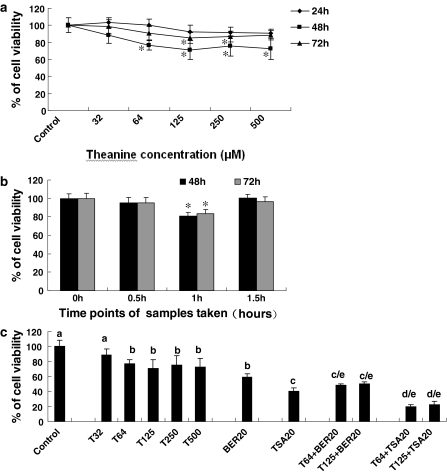
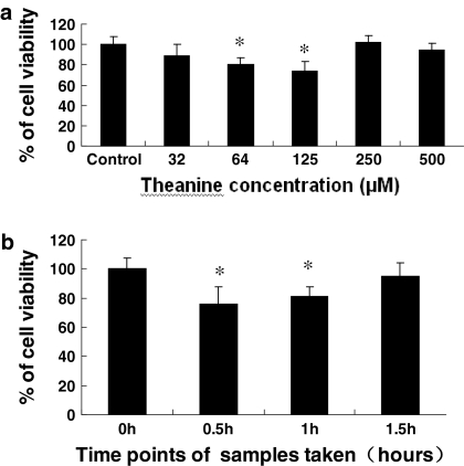
We first confirmed that theanine significantly suppressed the growth of human non-small cell lung cancer (NSCLC) A549 cell line after the cells were treated for 48 h with theanine at 32–500 μM. Theanine at the concentrations of 125 and 250 μM showed the significant inhibition of A549 cell growth although it did not display significant effect at the concentrations of 32, 64 and 500 μM after the cells were treated for 72 h (Fig. 1a). The dose–response curve indicates that theanine at the concentrations of 0–125 μM dose–dependently inhibited A549 cell growth but the growth inhibition decreased as the theanine concentrations increased from 125 to 250 and 500 μM. The short time (24 h) or long time (72 h) treatment of A549 cells with theanine did not produce the optimum inhibition. This suggests that the peak inhibition of A549 cell growth is the 48 h treatment with theanine at a concentration of 125 μM. The ex vivo assay showed that the rabbit sera obtained 1 h after oral intubation of theanine in the rabbits significantly suppressed the growth of A549 cells after the cells were treated with the sera for 48 h, while the 0.5 and 1.5 h rabbit sera did not show a significant inhibitory effect (Fig. 1b). Moreover, theanine at the concentrations of 64 and 125 μM enhanced the anticancer activity of the anticancer agents berbamine (BER) at 20 μM (an anticancer drug in China) and trichostatin A at 20 μg/L (a histone deacetylase inhibitor) by reducing the viability of A549 cells after the cells were treated for 48 h with the agents (Fig. 1c). In addition, theanine at concentrations of 64 and 125 μM significantly inhibited the growth of the human leukemia K562 cell line after the cells were treated for 48 h with theanine (Fig. 2a). The rabbit sera obtained 0.5 and 1 h after oral intubation of theanine in the rabbits significantly suppressed the growth of K562 cells after the cells have been treated with these sera for 48 h (Fig. 2b). These results confirm that theanine has a certain bioavailability by oral administration and the peak inhibition of cancer cell growth is at 0.5–1 h after oral intubation of theanine. This finding is consistent with the results of previous work in rats (Terashima et al. 1999) in which a peak concentration of theanine in serum was obtained 1 h after intragastrical administration of theanine. This finding is also consistent with our previous results in which the peak inhibition of proliferation in rat hepatoma AH109A cells by theanine-fed rat sera was at 1 h after oral intubation of theanine in rats (Zhang et al. 2001). In our previous study, we found out that theanine at concentrations of 25–1,600 μM did not suppress the proliferation of rat hepatoma cells in vitro (Zhang et al. 2001) but it significantly inhibited hepatoma growth in vivo (Zhang et al. 2002). The mechanisms of action of theanine against the growth of human cancer A549 and K562 cell lines require further study.
Fig. 1.
In vitro (a, c) and ex vivo (b) effects of theanine and sera from theanine-fed rabbits on the growth of human lung cancer A549 cells and the anticancer activity of anticancer agents against A549 growth. The cells were treated for 24, 48, and 72 h with theanine at the concentrations indicated (a) or the sera taken from rabbits (n = 6 for each group at different time points) at 0 h (as the control group), 0.5, 1 and 1.5 h after oral administration of theanine in rabbits (b), respectively. (c) In vitro effects of theanine on the anticancer activity of anticancer agents against A549 cell growth. The cells were treated for 48 h with theanine at 32–500 μM (T32, T64, T125, T250, T500) in the absence or presence of the anticancer agents berbamine (BER) at 20 μM (an anticancer drug in China) and trichostatin A (TSA) at 20 μg/L (a histone deacetylase inhibitor). The effects on A549 cell growth were examined by the MTT assay in vitro and ex vivo as described in the section of “Materials and methods”. The data are presented as the mean ± SD (Bar) for each group (n = 6). The figures (a, b and c) are the representative of three similar experiments performed. Comparison was made by two-way ANOVA followed by Bonferroni post hoc test to establish whether significant differences existed between the groups. * p < 0.05. Values with different letters (a–d) differ significantly (p < 0.05). c/e and d/e represent the significant synergistic effects (T64 + BER20, p < 0.001, two-way ANOVA; T64 + TSA20, p < 0.001, two-way ANOVA) compared with the treatment with its individual compound alone
Fig. 2.
In vitro (a) and ex vivo (b) effects of theanine and sera from theanine-treated rabbits on the growth of human leukemia K562 cells. The cells were treated for 48 h with theanine at the concentrations indicated (a) or the sera taken from rabbits (n = 6 for each group at different time points) at 0 h (as the control group), 0.5, 1 and 1.5 h after oral administration of theanine in rabbits (b), respectively. The effects on the K562 cell growth were examined by the MTT assay in vitro and ex vivo as described in the section of “Materials and methods”. The data are presented as the mean ± SD (Bar) for each group (n = 6). The figures are the representative of three similar experiments performed. Statistical analysis was carried out using the ANOVA and Bonferroni test. * p < 0.05
In vitro effects of theanine on the A549 cell migration and invasion and anticancer activity of anticancer agents against A549 cell migration
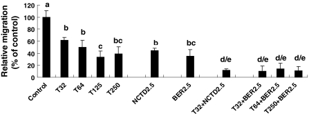
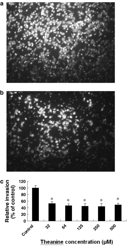
The presence of metastasis is the main cause of morbidity and mortality in millions of patients with cancer. During the complicated process of metastasis, the invasion of cancer cells is the most important and characteristic step. Clearly, an agent which could efficiently inhibit the growth and invasion of cancer cells would be a hopeful candidate to suppress cancer progression and metastasis and thus could reduce mortality. The migration of cancer cells is one of the important steps during invasion. Therefore, we examined the effects of theanine on the migration and invasion in A549 cells. The migration assay indicated that theanine at concentrations of 32–250 μM significantly suppressed the migration of A549 cells after the cells were treated for 6 h with theanine (Fig. 3). The dose–response curve shows that theanine at the concentrations of 0–125 μM dose–dependently inhibits A549 cell migration but the migration inhibition did not increase as the theanine concentration increased to 250 μM. This dose–effect on the migration (Fig. 3) shows a trend consistent with the trend of the dose–effect of theanine on the growth of A549 cells after treatment for 48 h (Fig. 1a). More importantly, theanine at the concentration of 32 μM enhanced the inhibitory effects of the anticancer agents NCTD and BER (anticancer drugs in China) against A549 cell migration after the cells were treated for 6 h with theanine in the presence of NCTD at 2.5 μM or BER at 2.5 μM (Fig. 3). In addition, the invasion assay further confirmed that theanine at concentrations of 32–500 μM significantly suppressed the invasion of A549 cells after the cells were treated for 16 h with theanine (Fig. 4a, b and c). The dose–response curve shows no evident dose–dependent effect on the invasion of A549 cells after treatment with theanine at concentrations of 0–500 μM. This dose–response is different from the effects of theanine on the growth (Fig. 1a) and migration (Fig. 3) of A549 cells. Theanine at concentrations of 32, 64 and 125 μM reduced cell migration by 39, 50 and 67%, respectively, but reduced cell invasion by 48, 54 and 55% (Fig. 4c), respectively. These data suggest that the migration inhibition by theanine may play a very important role in the invasion suppression although other mechanisms of action may be involved in the activity of theanine against A549 cell invasion. Theanine at the concentrations of 32–500 μM did not reduce the viability of A549 cells after treatment of the cells for 24 h with theanine (Fig. 1a). NCTD and BER at 2.5 μM also did not affect the viability of A549 cells after the treatment for 24 h (data not shown). These results suggest that the inhibition of A549 cell migration and invasion by the treatment with theanine alone or in combination with its synergistic anticancer agents NCTD and BER does not result from reduced cell viability of A549 cells. To understand the mechanisms of action of theanine against A549 cell invasion, we examined the effect of theanine on MMP-9 and MMP-2 activation in A549 cells by gelatin zymography analysis. The result indicated that theanine did not significantly affect the MMP-9/MMP-2 activation in A549 cells (data not shown). Nevertheless, our present results have demonstrated that reduction of A549 cell migration by theanine is one of the important mechanisms of action of theanine against A549 cell invasion. The mechanisms of action of theanine against the migration and invasion of A549 cells require further investigation.
Fig. 3.
Effects of theanine on the migration of human lung cancer A549 cells and the anticancer activity of anticancer agents NCTD and BER against A549 cell migration. The cells were treated for 6 h with theanine (T) at the concentrations of 32–250 μM (T32, T64, T125, T250) in the absence or presence of norcantharidin (NCTD) at 2.5 μM and berbamine (BER) at 2.5 μM. The cells in the control group were treated with DMSO vehicle (0.1%, final concentration). The effects on the migration of A549 cells were examined by the migration assay as described in the section of “Materials and methods”. The data are presented as the mean ± SD (Bar) for each group (n = 6). The figure is the representative of three similar experiments performed. Comparison was made by two-way ANOVA followed by Bonferroni post hoc test to establish whether significant differences existed between the groups. Values with different letters (a–d) differ significantly (p < 0.05). d/e Represents the significant synergistic effects (T32 + NCTD2.5, p < 0.001, two-way ANOVA; T32 + BER2.5, p < 0.001, two-way ANOVA) compared with the treatment with its individual compound alone
Fig. 4.
In vitro effects of theanine on the invasion of human lung cancer A549 cells. The effects on the invasion of A549 cells were examined by the invasion assay as described in the section of “Materials and methods”. (a) and (b) The photos show propidium iodide-stained A549 cells that invaded through Matrigel-coated transwell chamber. The cells were treated for 16 h with either the vehicle (a) (0.1% DMSO as the control) or theanine (b) at 125 μM. (c) Reduction of the relative invasion of A549 cells by theanine. The cells were treated for 16 h with theanine at the concentrations indicated in the figure. Relative invasion (%) ± SD (n = 6) is shown for the indicated theanine concentrations and the cells in the control group were treated with DMSO vehicle (0.1%, final concentration). The figure is the representative of three similar experiments performed. Statistical analysis was carried out using the ANOVA and Bonferroni test. * p < 0.05
It should be mentioned that theanine has displayed an anticancer activity comparable to EGCG in the suppression of some kinds of cancer cells such as A549 and hepatoma AH109A cells in vitro and/or in in vivo models (Kweon et al. 2006; Zhang et al. 2002). This is the first time to report that theanine significantly reduces cell viability of human non-small cell lung cancer and leukemia cells as well as cell migration and invasion of human lung cancer cells. More importantly, theanine enhanced the anticancer activity of anticancer agents such as trichostatin A (a histone deacetylase inhibitor), berbamine and norcantharidin (anticancer drugs in China) by strongly reducing viability and/or migration rate in A549 cells. These findings suggest that there will be many important mechanisms of action of theanine to be disclosed; theanine may have a great therapeutic potential and/or adjuvant therapeutic effects on human NSCLC and leukemia. There have been more than 700 papers reporting the activities of EGCG against cancer. Whereas, there have been only 23 papers reporting the theanine activity against cancer in PubMed as yet. Theanine, as a characteristic amino acid in tea, plays very important roles in contributing to the activity of tea against cancer. It is necessary to pay great attentions to the activities of theanine against cancer.
Abnormal growth and metastasis of cancer cells are regarded as the important biological characteristics of cancers. As an important anticancer component in tea, theanine has been reported to enhance antitumor activity by inhibiting the efflux of anticancer drugs pirarubicin and adriamycin from M5076 ovarian sarcoma cells (Sugiyama and Sadzuka 1998; Sugiyama et al. 1999; Sadzuka et al. 2002). Our previous results confirmed that theanine inhibited the invasion of rat hepatoma cells in vitro and ex vivo (Zhang et al. 2001) and hepatoma growth in vivo (Zhang et al. 2002) although theanine did not show an inhibitory effect on the proliferation of rat hepatoma AH109A cells in vitro. There have been studies reporting that theanine suppressed the growth in hepatoma, prostate cancer, and colon cancer cells (Friedman et al. 2007). Our results confirm for the first time the anticancer activity of theanine against growth of human lung cancer and leukemia cells as well as migration and invasion of human lung cancer cells. In the present study, we have shown that theanine significantly suppressed the growth of human lung cancer A549 and leukemia K562 cells in vitro and ex vivo. In addition, we have demonstrated that theanine also significantly inhibited the migration and invasion of A549 cells. More importantly, theanine enhanced the anticancer activity of the anticancer agents trichostatin A (a histone deacetylase inhibitor), NCTD and BER by strongly reducing cell viability and/or migration rate of A549 cells that are cells from non-small cell lung cancer (NSCLC) which constitutes ~80% of lung cancer cases and remains an aggressive lung cancer associated with a poor patient survival. Taken together, our present findings suggest that theanine may have a wide therapeutic and/or adjuvant therapeutic application in the treatment of human NSCLC and leukemia.
Acknowledgments
Grant support: This work is supported in part by grants from the Ministry of Education of the People’s Republic of China to G.Z., from the Ministry of Human Resources and Social Security of the People’s Republic of China to G.Z., the Projects of Yantai University to G.Z., and Projects from the Department of Science and Technology of Shandong Province to G.Z. (Y2008C71; 2009GG10002089).
Conflict of interest statement The authors declare no conflict of interest.
Abbreviations
- BER
Berbamine
- T
Trichostatin A
- Bay
Bay 11-7082
- S
Celecoxib
References
- Friedman M, Mackey BE, Kim HJ, Lee IS, Lee KR, Lee SU, Kozukue E, Kozukue N (2007) Structure–activity relationships of tea compounds against human cancer cells. J Agric Food Chem 55:243–253 [DOI] [PubMed]
- Kitaoka S, Hayashi H, Yokogoshi H, Suzuki Y (1996) Transmural potential changes associated with the in vitro absorption of theanine in the guinea pig intestine. Biosci Biotechnol Biochem 60:1768–1771 [DOI] [PubMed]
- Kweon MH, Adhami VM, Lee JS, Mukhtar H (2006) Constitutive overexpression of Nrf2-dependent heme oxygenase-1 in A549 cells contributes to resistance to apoptosis induced by epigallocatechin 3-gallate. J Biol Chem 281:33761–33772 [DOI] [PubMed]
- Sadzuka Y, Yamashita Y, Sugiyama T, Sonobe T (2002) Effect of dihydrokainate on the antitumor activity of doxorubicin. Cancer Lett 179:157–163 [DOI] [PubMed]
- Sugiyama T, Sadzuka Y (1998) Enhancing effects of green tea components on the antitumor activity of adriamycin against M5076 ovarian sarcoma. Cancer Lett 133:19–26 [DOI] [PubMed]
- Sugiyama T, Sadzuka Y, Nagasawa K, Ohnishi N, Yokoyama T, Sonobe T (1999) Membrane transport and antitumor activity of pirarubicin, and comparison with those of doxorubicin. Jpn J Cancer Res 90:775–780 [DOI] [PMC free article] [PubMed]
- Terashima T, Takido T, Yokogoshi H (1999) Time-dependent changes of amino acids in the serum, liver, brain and urine ofrats administered with theanine. Biosci Biotechnol Biochem 63:615–618 [DOI] [PubMed]
- Zhang G, Miura Y, Yagasaki K (1999) Effects of green, oolong and black teas and related components on the proliferation and invasion of hepatoma cells in culture. Cytotechnology 31:37–44 [DOI] [PMC free article] [PubMed]
- Zhang G, Miura Y, Yagasaki K (2001) Inhibitory effects of theanine and sera from theanine-fed rats on receptor-mediated cancer cell beneath mesothelial-cell monolayers. Cytotechnology 36:195–200 [DOI] [PMC free article] [PubMed]
- Zhang G, Miura Y, Yagasaki K (2002) Effects of dietary powdered green tea and theanine on tumor growth and hyperlipidemia in hepatoma-bearing rats. Biosci Biotechnol Biochem 66:711–716 [DOI] [PubMed]


