Abstract
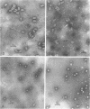
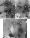
Rosenblum, E. D. (The University of Texas, Dallas), and Sue Tyrone. Serology, density, and morphology of staphylococcal phages. J. Bacteriol. 88:1737–1742. 1964.—A correlation between serology, buoyant density, and morphology has been demonstrated for six serological groups of staphylococcal phages. Four morphological types have been observed and represent the following serological groups: (i) group A, (ii) groups B, F, and L, (iii) group D, and (iv) group G. The correlations were useful in the detection of serological variation among several staphylococcal typing phages.
Full text
PDF
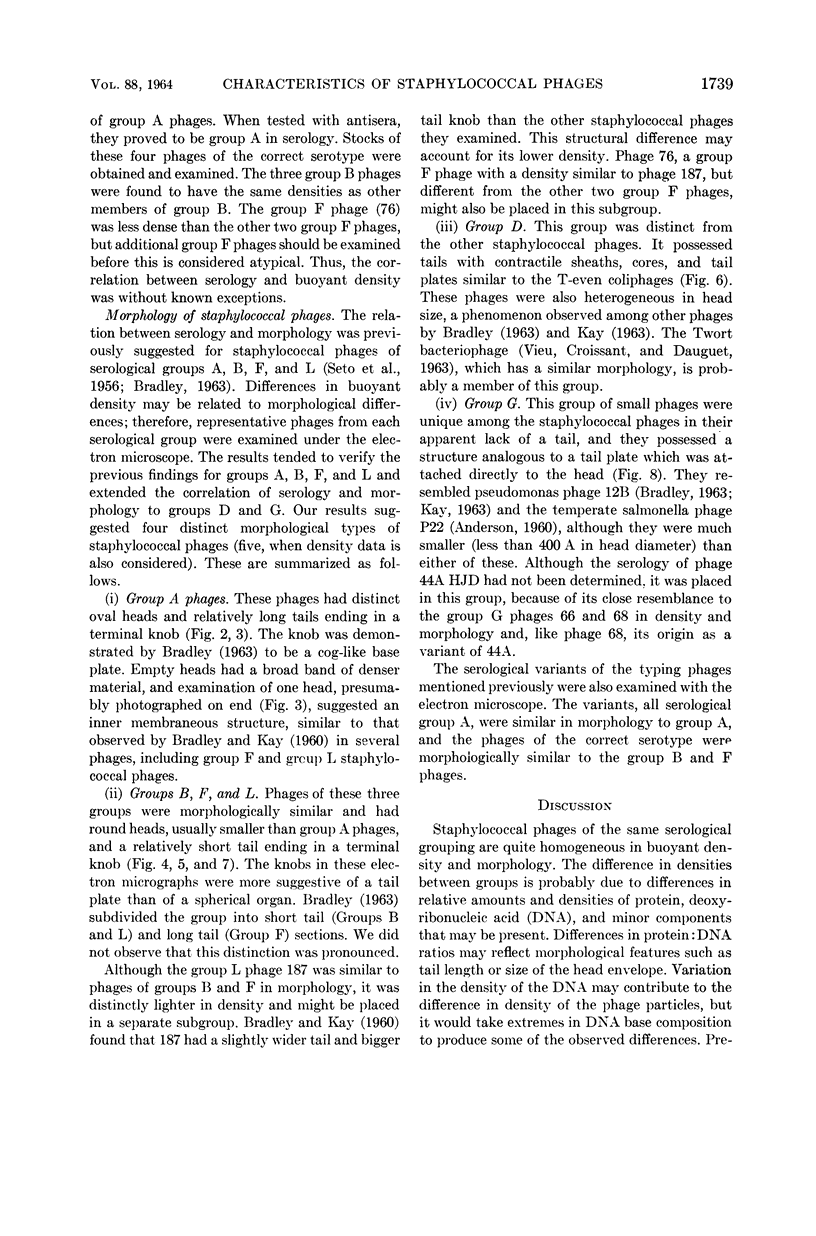
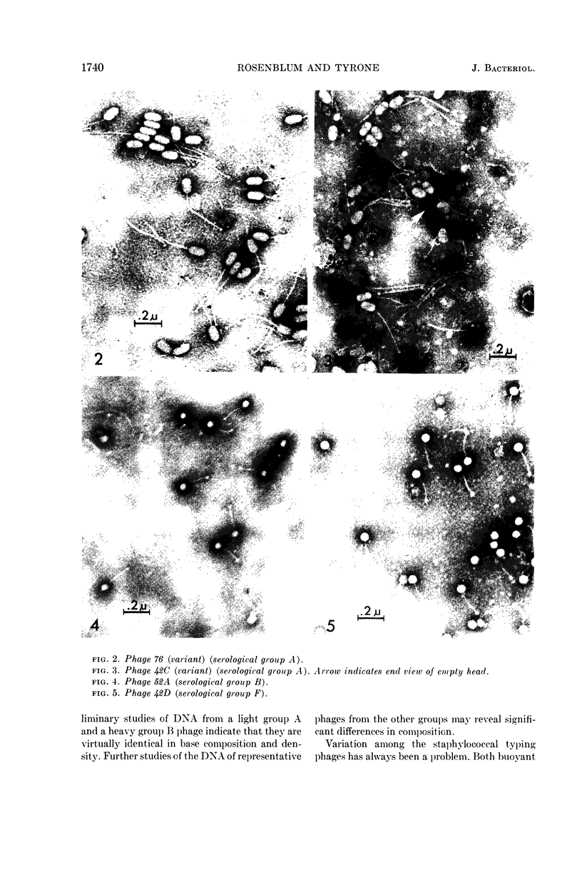
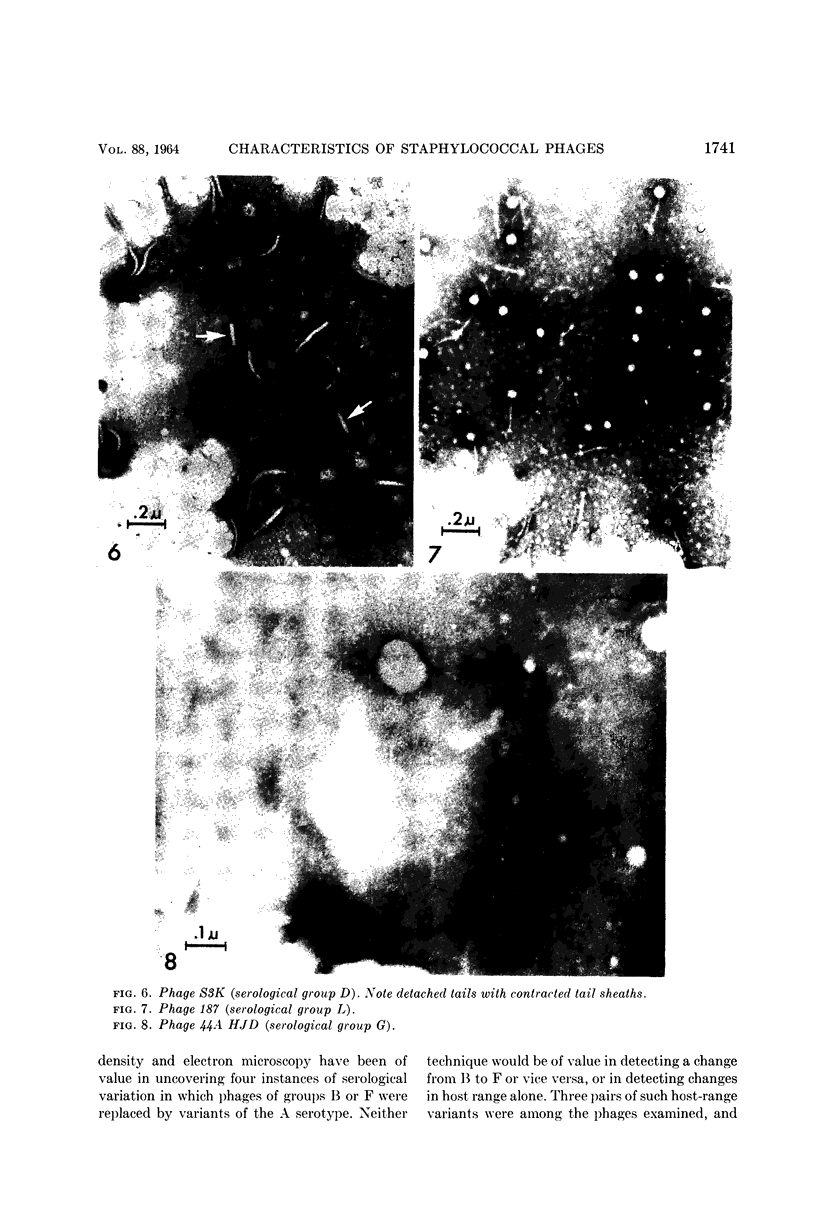
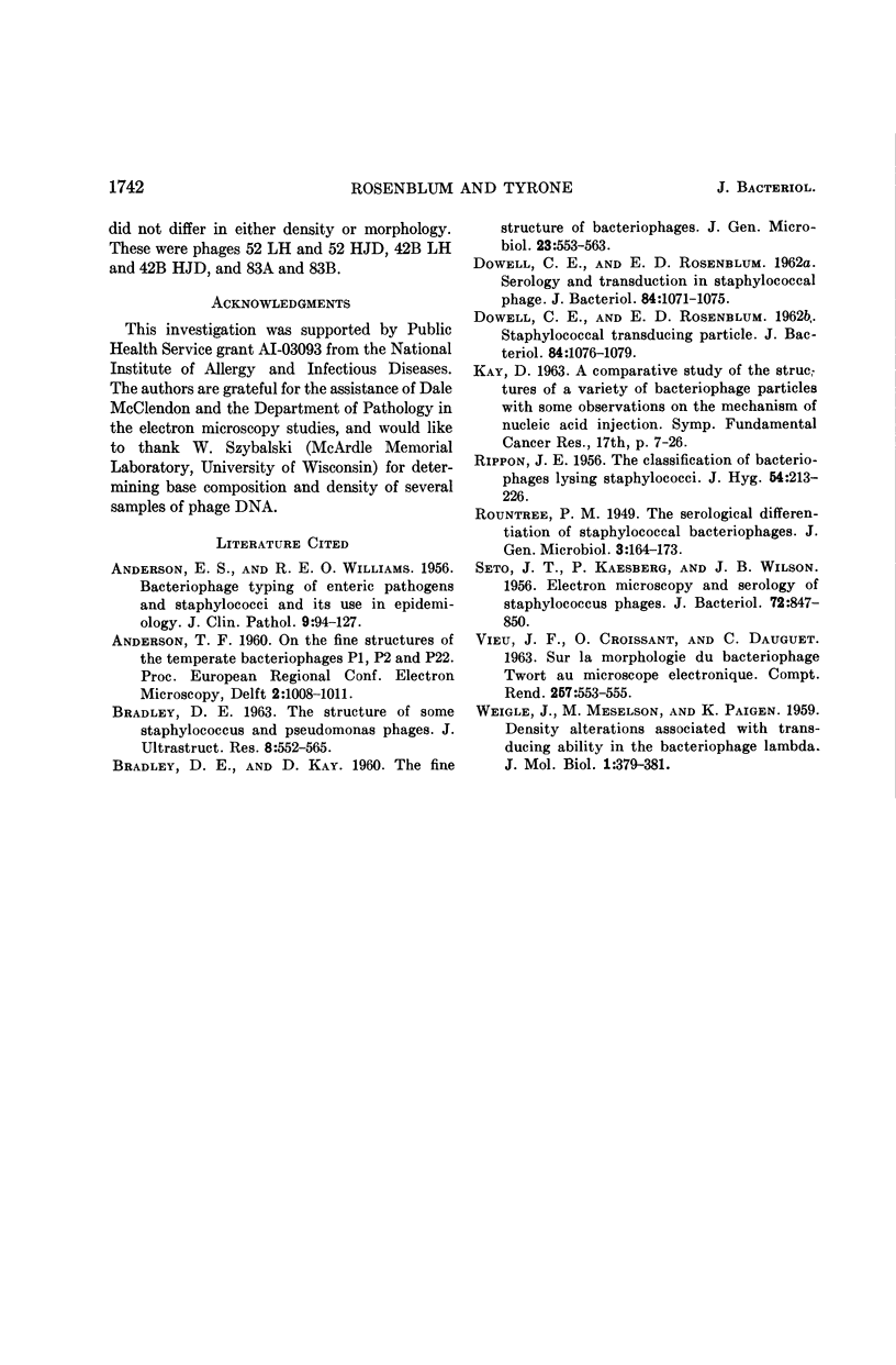
Images in this article
Selected References
These references are in PubMed. This may not be the complete list of references from this article.
- ANDERSON E. S., WILLIAMS R. E. Bacteriophage typing of enteric pathogens and staphylococci and its use in epidemiology. J Clin Pathol. 1956 May;9(2):94–127. doi: 10.1136/jcp.9.2.94. [DOI] [PMC free article] [PubMed] [Google Scholar]
- BRADLEY D. E. The structure of some Staphylococcus and Pseudomonas phages. J Ultrastruct Res. 1963 Jun;8:552–565. doi: 10.1016/s0022-5320(63)80055-6. [DOI] [PubMed] [Google Scholar]
- Dowell C. E., Rosenblum E. D. SEROLOGY AND TRANSDUCTION IN STAPHYLOCOCCAL PHAGE. J Bacteriol. 1962 Nov;84(5):1071–1075. doi: 10.1128/jb.84.5.1071-1075.1962. [DOI] [PMC free article] [PubMed] [Google Scholar]
- Dowell C. E., Rosenblum E. D. STAPHYLOCOCCAL TRANSDUCING PARTICLE. J Bacteriol. 1962 Nov;84(5):1076–1079. doi: 10.1128/jb.84.5.1076-1079.1962. [DOI] [PMC free article] [PubMed] [Google Scholar]
- KAESBERG P., SETO J. T., WILSON J. B. Electron microscopy and serology of staphylococcus phages. J Bacteriol. 1956 Dec;72(6):847–850. doi: 10.1128/jb.72.6.847-850.1956. [DOI] [PMC free article] [PubMed] [Google Scholar]
- RIPPON J. E. The classification of bacteriophages lysing staphylococci. J Hyg (Lond) 1956 Jun;54(2):213–226. doi: 10.1017/s0022172400044478. [DOI] [PMC free article] [PubMed] [Google Scholar]
- VIEU J. F., CROISSANT O., DAUGUET C. [On morphology of the Twort bacteriophage under the electron microscope]. C R Hebd Seances Acad Sci. 1963 Jul 8;257:553–555. [PubMed] [Google Scholar]


