Abstract
Control and preventive measures for gonococcal infections are based on precise epidemiological characteristics of N. gonorrhoeae isolates. In the present study the potential utility of opa-typing and ribotyping for molecular epidemiological study of consecutive gonococcal strains was determined. Sixty gonococcal isolates were subjected to ribotyping with two restriction enzymes, AvaII and HincII, and opa-typing with TaqI and HpaII for epidemiological characterization of gonococcal population. Ribotyping with AvaII yielded 6 ribotype patterns while twelve RFLP patterns were observed with HincII. Opa-typing of the 60 isolates revealed a total 54 opa-types, which 48 were unique and 6 formed clusters. Fifty-two opa-types were observed with TaqI-digested PCR product while opa-typing with HpaII demonstrated 54 opa-types. The opa-types from isolates that were epidemiologically unrelated were distinct, whereas those from the sexual contacts were identical. The results showed that opa-typing is highly useful for characterizing gonococcal strains from sexual contacts and has more discriminatory than ribotyping that could differentiate between gonococci of the same ribotype. The technique even with a single restriction enzyme has a high level of discrimination (99.9%) between epidemiologically unrelated isolates. In conclusion, the molecular methods such as opa-typing and ribotyping can be used for epidemiological characterization of gonococcal strains.
1. Introduction
Neisseria gonorrhoeae is the causative agent of gonorrhea, which continues to be one the most common sexually transmitted infections worldwide [1, 2]. Control and preventive measures for infection are based on epidemiological analyses as well as antibiotic susceptibility of the N. gonorrhoeae isolates. The process of typing of Neisseria gonorrhoeae isolates can be used to identify the population of strains that circulate in different communities, temporal, and geographic changes among the strains, transmission patterns, and antibiotic resistance outbreaks [3, 4].
Various typing methods have been developed to characterize N. gonorrhoeae isolates. The widely used phenotypic methods are based on auxotyping, serotyping, and antimicrobial susceptibility testing. However, these methods have some limitations such as reproducibility and discriminatory power [5–9]. Therefore, different molecular genetic methods have been developed for the characterization of gonococcal strains include ribotyping [10, 11], pulsed-field gel electrophoresis (PFGE) [12], arbitrarily primed PCR [13], Amplified Fragment length polymorphism (AFLP) [14], opa-typing [15, 16], and sequence typing based on one or more genes [4, 13, 16, 17].
Ribotyping refers to the use of Southern blot analysis for detecting polymorphisms that are associated with the ribosomal DNA regions. The method has appeared to be good reproducibility, stability, and typeability for characterization of gonococcal isolates. However, it provides less discrimination between isolates of N. gonorrhoeae [10, 11].
Opa-typing is a PCR-based RFLP technique, which relies on amplifying the 11 opa genes and subsequent digestion with restriction enzymes, to generate restriction fragment length polymorphism. The method has shown to be highly discriminatory enough to be applied to large collections of gonococcal isolates [15]. The opa-typing is also useful to confirm self-reported sexual contacts and to identify sex partners and short chains of transmission [15, 16, 18].
There are hardly any data about molecular epidemiological analysis of N. gonorrhoeae isolates by opa-typing and ribotyping in India. Epidemiological analysis of N. gonorrhoeae isolates by genotypic methods has not been carried out in India so far.
In this study, we determined the potential utility of opa-typing and ribotyping for differentiation of gonococcal strains.
2. Material and Methods
2.1. Gonococcal Isolates and Culture Conditions
Sixty consecutive N. gonorrhoeae strains isolated from among 62 males with urethritis, 22 females with endocervicitis, and 10 sexual contacts of these patients attending the STD clinic of Lok Nayak Hospital, New Delhi from January, 2006 to June, 2007 were studied. All gonococcal isolates were subjected to opa-typing and ribotyping for epidemiological characterization.
The samples were inoculated directly onto the selective Modified Thayer-Martin medium (MTM) medium and incubated at 35–36.5°C containing 5%CO2 for 48 hours. The colonies suspected to be N. gonorrhoeae were identified by gram stain, oxidase, superoxol tests, and rapid carbohydrate utilization test (RCUT). Gonococcal isolates were stored at −70°C [19].
2.2. Isolation of Genomic DNA
Genomic DNA was isolated from confluent (18–24 hours) growth of gonococcal cells by using a Wizard genomic DNA purification kit (Promega Corp., Madison, USA) according to the manufacturer's instructions. Briefly, the bacterial cell was suspended in 500 μL of Tris-EDTA buffer (pH 7.8). The suspensions were pelleted and resuspended in 500 μL of Nuclei lysis solution (Promega) and incubated for 5 minutes at 80°C. However, 4 μL of RNase (10 mg/mL) (Sigma Chemical Co., USA) was added to cell lysate and incubated for 1 hour at 37°C. The lysate was adjusted to 0.7 M NaCl and 1% cetyltrimethylammonium bromide (CTAB), mixed thoroughly, and incubated for 10 minutes at 65°C in order to precipitate cell wall debris, denatured proteins, and polysaccharides. The supernatant was extracted twice with phenol/chloroform/isoamyl alcohol (25 : 24 : 1) and twice with chloroform/isoamyl alcohol (24 : 1). Chromosomal DNA was precipitated with isopropanol, washed with 70% ethanol, resuspended in 50 μL of Tris-EDTA buffer (pH 7.8), and stored at −20°C.
2.3. Ribotyping
Chromosomal DNA was digested with AvaII and HincII, and the restricted fragments were separated by electrophoresis on 0.8% agarose gels in Tris-borate (TB) buffer at 40 V for 16 hours. DNA fragments were transferred to positively charged nylon membrane (Roche Diagnostic GmbH, Mannheim, Germany) from agarose gels by the method of Southern. The blot was hybridized with the addition of digoxigenin-dUTP labeled cDNA probe at 68°C for 18–24 hours [20].
However, 16S and 23S rRNA from E. coli (Roche Diagnostic GmbH, Mannheim, Germany) was reverse transcribed into cDNA with SuperScript II reverse transcriptase (Invitrogen, Calif, USA). The cDNA was then labeled by random priming with digoxigenin-dUTP according to the manufacturer's directions (Roche Diagnostic GmbH, Mannheim, Germany).
The sizes of the hybridized fragments from at least four blots were averaged after measuring distances of band migration in comparison with the Digoxigenin labeled DNA molecular weight marker III (Roche Diagnostic GmbH, Mannheim, Germany).
2.4. Opa-Typing
Opa-typing was performed as described previously [16]. Briefly, opa genes were amplified from gonococcal chromosomal DNA by PCR. Amplification was carried out in a 50 μL volume containing 0.5 μg of each opa forward and reverse primer (opa-01, 5′-ATGTCAGGCGGATTTAGCC-3′, and opa-04, 5′-AATGAGGCTTCGTGGGTTTTG-3′), 100 ng of chromosomal DNA, 0.2 mM of each dNTP, 2.5 U of Taq DNA polymerase, and the buffer provided by the manufacturer with 75 mM Tris HCl (pH 9.0), 2 mM MgCl2, 50 mM KCl, 20 mM (NH4)2SO4 (Biotools, B&M Labs, S.A., Madrid, Spain). Cycle conditions were set at 94°C for 5 minutes (hot-start); 30 cycles of 94°C for 1 minute, 64°C for 1 minute, and 72°C for 1 minute; a final extension reaction at 72°C for 10 minutes. The PCR products (≈670 bp) were precipitated with ethanol, washed with 70% ethanol, and resuspended in 50 μL of TE buffer. The opa gene fragments (1.5 μg) were digested overnight at 65°C with 10 U of TaqI in a volume of 30 μL using the buffer recommended by manufacturer. An equivalent aliquot was digested overnight at 37°Cwith 10 U of Hpa II. The resulting restriction fragments was fractionated on a precast 12% polyacrylamide gel and visualized by ethidium bromide staining.
2.5. Analysis of the Typing Methods
The reproducibility of ribotyping and opa-typing was examined by analysis of the same bacterial strain on three separate occasions. The stability of the techniques was examined by daily subcultures of N. gonorrhoeae isolates. The discriminatory index for two methods was calculated as described previously [21]. The relatedness values were calculated as the proportion of unweighted mismatches of fragments between the different patterns. Computed similarity cluster analysis of the patterns was done by the unwieghted pair-group average algorithm, using the Dice coefficient to analyze the similarities of the banding patterns. The phylogenetic tree (dendogram) was constructed by DNASTAR software package.
2.6. Statistical Analysis
Statistical methods like Chi-square test and Fisher's exact test were performed to determine the significant association of various demographic data using the SPSS version 13.0 for Windows.
3. Results
A total of 60 gonococcal strains were isolated from 52 (83.87%) out of 62 men with urethritis, 4 (18.18%) out of 22 women with endocervicitis, and 4 (40%) out of 10 sexual contacts of these cases. All gonococcal isolates were subjected to opa-typing and ribotyping for epidemiological characterization.
3.1. Ribotyping
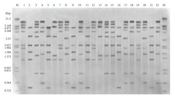
Chromosomal DNAs from 60 isolates were digested with HincII and AvaII. Hybridization of AvaII fragments with labeled rRNA probes yielded 6 ribotype patterns (Table 1). Twenty-three of the 60 isolates demonstrated pattern 1; 16 isolates had patterns 2; 12 isolates demonstrated pattern 3; 4 isolates had pattern 4; 3 isolates pattern 5; 2 isolates pattern 6. Analysis with HincII produced highly resolved fragments suitable for analysis, and fragment sizes ranged from 0.46 to 18.36 kb (Figure 1). Twelve RFLP patterns were observed with HincII, from which 16 of the 60 isolates demonstrated pattern 1; 10 isolates had pattern 2; 8 isolates had pattern 3; patterns 4–6 comprised 4 isolates each; patterns 7–9 contained 3 isolates each; patterns 10 and 11 had 2 isolates each. One isolate demonstrated pattern 12. All isolates recovered from index case and their sexual contacts were found to be identical by ribotyping.
Table 1.
Discriminatory indices of typing methods for gonococcal isolates.
| Typing method | number of type patterns | Discriminatory index |
|---|---|---|
| Ribotyping | ||
| With AvaII | 6 | 0.75 |
| With HincII | 12 | 0.87 |
|
| ||
| Opa-typing | ||
| With Taq I | 52 | 0.995 |
| With Hpa II | 54 | 0.997 |
Figure 1.
Examples of HincII ribotype patterns of consecutive gonococcal isolates: lane M: digoxigenin labeled DNA molecular weight marker III (Roche Diagnostics Corp., Indianapolis, USA); lanes: 1, 7, 12, and 19: ribotype pattern RH6; lanes: 2, 5, 9, 11, 16, and 21: ribotype pattern RH1; lanes: 3, 14, and 5: ribotype pattern RH9; lanes: 4, 10, 18, and 22: ribotype pattern RH3; lanes: 6 and 13: ribotype pattern RH11; lanes: 8, 17, and 20: ribotype pattern RH7.
The characteristic isolate banding patterns with HincII remained unchanged when DNA was prepared from randomly selected strains grown on separate occasions, and the ribotypes did not change after three individual strains were subcultured 1, 10, and 20 times.
3.2. Opa-Typing
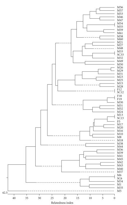
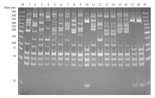
For opa-typing, the 11 opa genes were amplified and the PCR products were digested with restriction enzymes TaqI and HpaII. Dendogram obtained from opa-typing with HpaII is shown in Figure 2. Cluster analysis of HpaII opa-type patterns revealed a total 54 opa-types, from which 48 (88.9%) were unique and 6 (11.1%) formed clusters (Table 1 and Figure 2). Of the 48 isolates with unique opa-types, none were known to be from sexual contacts. Fifty-two opa-types were observed with TaqI-digested PCR product. Forty-five had unique opa-type while 7 formed clusters. Opa-typing with TaqI was slightly less discriminatory than typing with HpaII (Table 1). The opa-types from isolates that were epidemiologically unrelated (unconnected) were distinct, whereas those from the sexual contacts were identical. Isolates M54 and M55 could not be distinguished by both enzymes. Examples of the HpaII opa-type patterns are given in Figure 3.
Figure 2.
Dendogram of the cluster analysis of the 60 consecutive gonococcal isolates obtained after opa-typing with HpaII. The relatedness values were calculated as the proportion of unweighted mismatches of fragments between the different patterns. The dendogram was generated by DNASTAR software package.
Figure 3.
Examples of different patterns of HpaII opa-types of consecutive gonococcal isolates. Lane M: molecular weight marker (New England Biolabs, USA); lanes 1–18: N. gonorrhoeae isolates; sexual contacts: lanes 13, 14 (HpaII opa-type 13), lanes 15, 16 (HpaII opa-type 14), lanes 17, 18 (HpaII opa-type 15).
PCR amplifications of the opa genes were performed from three isolates of N. gonorrhoeae grown on separate occasions, from which the opa-types were identical. The stability of the opa-typing during subculture in vitro was examined by obtaining opa-types from DNA prepared from daily subcultures of three isolates of N. gonorrhoeae. No differences in the opa-types of the isolates were seen after 1, 10, and 20 subcultures.
4. Discusion
The ability to identify accurately the strains of infectious agents that cause disease is central to epidemiological surveillance and public health decisions. The widely available typing systems have the potential to provide information useful to the control and prevention of N. gonorrhoeae infections and to guide health interventions. All typing systems for epidemiological studies should have established reproducibility, and be highly discriminating in order to distinguish epidemiologically unrelated infections. The shortcomings of phenotypic typing methods have led to development of molecular genetic methods, which minimize problems with reproducibility or discriminatory power [5–9, 12, 15, 16].
There is hardly any data about epidemiological studies of N. gonorrhoeae isolates based on molecular typing in India. Therefore, our aim was to assess molecular typing of N. gonorrhoeae isolates using ribotyping and opa-typing.
Ribotyping has the advantage over restriction endonuclease analysis in that the RFLP patterns contain fewer bands for comparison because only fragments with rRNA sequences are visualized. More pronounced heterogeneity of rRNA hybridization patterns could be generated by restriction endonuclease that have different restriction sites within the rRNA genes. In our study, RFLP patterns generated with HincII (12 ribotype patterns) showed greater heterogeneity than those generated by AvaII (6 ribotype patterns). On the basis of combined HincII and AvaII-generated RFLP patterns, 60 isolates of N. gonorrhoeae were classified into 16 ribotype patterns, and no isolate was nontypeable. The results of this study showed that ribotyping was much simpler to interpret than the complex restriction endonuclease patterns of total chromosomal DNA. The technique was also found to be both stable and reproducible. The results suggested that there may be sufficient heterogeneity of rRNA patterns within N. gonorrhoeae isolates and the method may to be useful for differentiation of strains in epidemiological studies.
A similar study has shown that RFLP patterns generated by HincII could be used to differentiate 43 isolates of N. gonorrhoeae into nine patterns [22]. A previous study of ribotype patterns generated by combination of SmaI and AvaII digests of N. gonorrhoeae differentiated 23 proline-citrulline-uracil requiring (PCU−) auxotypes into four groups [10]. A Canadian study indicated that the ribotyping by SmaI could be classified 27 ornithine-uracil-hypoxanthine (OUH−) or citrulline-uracil-hypoxanthine (CUH−) or ornithine-hypoxanthine (OH−)/IA-2 auxotypes of N. gonorrhoeae into five ribotype patterns [11]. The present study showed that common DNA bands were present in the RFLP patterns for all the N. gonorrhoeae isolates in accordance with previous studies [10, 11, 22].
Opa-typing is a technique based on diversity (polymorphism) of opa genes of N. gonorrhoeae. A single gonococcus possesses 11 different opa genes, which encodes the outer membrane proteins. The extensive variation and rapid evolution of the opa gene repertoire make it likely that gonococci with identical opa-types are very closely related genetically and epidemiologically. Major potential use of opa-typing is the identification of groups of individuals in sexual networks by the isolation of gonococci with the same opa-type from gonococcal populations recovered in a restricted geographic area within a limited time period [15].
In this study, opa-typing of the 60 isolates revealed a total of 54 opa-types, from which 48 (88.9%) were represented by single isolates. The opa-types from isolates that were epidemiologically unrelated were distinct, while those from the sexual contacts were identical. There were only two consecutive isolates being indistinguishable by opa-typing. As the gonococci within these were indistinguishable by ribotyping, and were from patients living in the same part of Delhi attending the STD clinic within a short time period, it is possible that they were from individuals who were connected by a short chain of infection transmission or were form common core group (sexual contacts). This discrepancy was also found in a previous study [15].
Opa-typing identified all of the isolates from sexual contacts. This result indicates that opa-typing is highly useful for characterizing gonococcal strains from sexual contacts in full agreement with previous studies [15, 16, 18].
We found opa-typing to be more discriminatory than ribotyping and could differentiate between gonococci of the same ribotype. The technique even with a single restriction enzyme (HpaII) has a high level of discrimination (99.7%) between epidemiologically unrelated isolates in concordance with results in previous studies [15, 16, 18, 23].
The results of both methods indicate that gonococcal infection among the patients studied was not derived from common core groups. In conclusion, the molecular methods such as opa-typing and ribotyping can be used for epidemiological characterization of gonococcal strains.
References
- 1.Centers for Disease Control. Special focus: Trends in Reportable Sexually Transmitted Diseases in the United States. 2004.
- 2.Gerbase AC, Rowley JT, Mertens TE. Global epidemiology of sexually transmitted diseases. Lancet. 1998;351(supplement 3):2–4. doi: 10.1016/s0140-6736(98)90001-0. [DOI] [PubMed] [Google Scholar]
- 3.Unemo M, Olcén P, Jonasson J, Fredlund H. Molecular typing of Neisseria gonorrhoeae isolates by pyrosequencing of highly polymorphic segments of the porB gene. Journal of Clinical Microbiology. 2004;42(7):2926–2934. doi: 10.1128/JCM.42.7.2926-2934.2004. [DOI] [PMC free article] [PubMed] [Google Scholar]
- 4.Bash MC, Zhu P, Gulati S, McKnew D, Rice PA, Lynn F. Por variable-region typing by DNA probe hybridization is broadly applicable to epidemiologic studies of Neisseria gonorrhoeae . Journal of Clinical Microbiology. 2005;43(4):1522–1530. doi: 10.1128/JCM.43.4.1522-1530.2005. [DOI] [PMC free article] [PubMed] [Google Scholar]
- 5.Palomares JC, Lozano MC, Perea EJ. Antibiotic resistance, plasmid profile, auxotypes and serovars of Neisseria gonorrhoeae strains isolated in Sevilla (Spain) Genitourinary Medicine. 1990;66(2):87–90. doi: 10.1136/sti.66.2.87. [DOI] [PMC free article] [PubMed] [Google Scholar]
- 6.Lind I, Arborio M, Bentzon MW, et al. The epidemiology of Neisseria gonorrhoeae isolates in Dakar, Senegal 1982–1986: antimicrobial resistance, auxotypes and plasmid profiles. Genitourinary Medicine. 1991;67(2):107–113. doi: 10.1136/sti.67.2.107. [DOI] [PMC free article] [PubMed] [Google Scholar]
- 7.Brett MSY, Davies HGD, Blockley JR, Heffernan HM. Antibiotic susceptibilities, serotypes and auxotypes of Neisseria gonorrhoeae isolated in New Zealand. Genitourinary Medicine. 1992;68(5):321–324. doi: 10.1136/sti.68.5.321. [DOI] [PMC free article] [PubMed] [Google Scholar]
- 8.Ng L-K, Carballo M, Dillon J-AR. Differentiation of Neisseria gonorrhoeae isolates requiring proline, citrulline, and uracil by plasmid content, serotyping, and pulsed-field gel electrophoresis. Journal of Clinical Microbiology. 1995;33(4):1039–1041. doi: 10.1128/jcm.33.4.1039-1041.1995. [DOI] [PMC free article] [PubMed] [Google Scholar]
- 9.Unemo M, Olcén P, Albert J, Fredlund H. Comparison of serologic and genetic porB-based typing of Neisseria gonorrhoeae: consequences for future characterization. Journal of Clinical Microbiology. 2003;41(9):4141–4147. doi: 10.1128/JCM.41.9.4141-4147.2003. [DOI] [PMC free article] [PubMed] [Google Scholar]
- 10.Ng L-K, Dillon J-AR. Typing by serovar, antibiogram, plasmid content, riboprobing, and isoenzyme typing to determine whether Neisseria gonorrhoeae isolates requiring proline, citrulline, and uracil for growth are clonal. Journal of Clinical Microbiology. 1993;31(6):1555–1561. doi: 10.1128/jcm.31.6.1555-1561.1993. [DOI] [PMC free article] [PubMed] [Google Scholar]
- 11.Li H, Dillon J-AR. Utility of ribotyping, restriction endonuclease analysis and pulsed-field gel electrophoresis to discriminate between isolates of Neisseria gonorrhoeae of serovar IA-2 which require arginine, hypoxanthine or uracil for growth. Journal of Medical Microbiology. 1995;43(3):208–215. doi: 10.1099/00222615-43-3-208. [DOI] [PubMed] [Google Scholar]
- 12.Xia M, Whittington WL, Holmes KK, Plummer FA, Roberts MC. Pulsed-field gel electrophoresis for genomic analysis of Neisseria gonorrhoeae . Journal of Infectious Diseases. 1995;171(2):455–458. doi: 10.1093/infdis/171.2.455. [DOI] [PubMed] [Google Scholar]
- 13.Hobbs MM, Alcorn TM, Davis RH, et al. Molecular typing of Neisseria gonorrhoeae causing repeated infections: evolution of porin during passage within a community. Journal of Infectious Diseases. 1999;179(2):371–381. doi: 10.1086/314608. [DOI] [PubMed] [Google Scholar]
- 14.Spaargaren J, Stoof J, Fennema H, Coutinho R, Savelkoul P. Amplified fragment length polymorphism fingerprinting for identification of a core group of Neisseria gonorrhoeae transmitters in the population attending a clinic for treatment of sexually transmitted diseases in Amsterdam, The Netherlands. Journal of Clinical Microbiology. 2001;39(6):2335–2337. doi: 10.1128/JCM.39.6.2335-2337.2001. [DOI] [PMC free article] [PubMed] [Google Scholar]
- 15.O'Rourke M, Ison CA, Renton AM, Spratt BG. Opa-typing: a high-resolution tool for studying the epidemiology of gonorrhoea. Molecular Microbiology. 1995;17(5):865–875. doi: 10.1111/j.1365-2958.1995.mmi_17050865.x. [DOI] [PubMed] [Google Scholar]
- 16.Viscidi RP, Demma JC, Gu J, Zenilman J. Comparison of sequencing of the por gene and typing of the opa gene for discrimination of Neisseria gonorrhoeae strains from sexual contacts. Journal of Clinical Microbiology. 2000;38(12):4430–4438. doi: 10.1128/jcm.38.12.4430-4438.2000. [DOI] [PMC free article] [PubMed] [Google Scholar]
- 17.Lundbäck D, Fredlund H, Berglund T, Wretlind B, Unemo M. Molecular epidemiology of Neisseria gonorrhoeae- identification of the first presumed Swedish transmission chain of an azithromycin-resistant strain. APMIS. 2006;114(1):67–71. doi: 10.1111/j.1600-0463.2006.apm_332.x. [DOI] [PubMed] [Google Scholar]
- 18.Howie F, Young H, McMillan A. The diversity of the opa gene in gonococcal isolates from men who have sex with men. Sexually Transmitted Infections. 2004;80(4):286–288. doi: 10.1136/sti.2003.006775. [DOI] [PMC free article] [PubMed] [Google Scholar]
- 19.World Health Organization. Laboratory Diagnosis of Gonorrhoea. Geneva, Switzerland: WHO; 1999. (South East Asia Series no. 33). [Google Scholar]
- 20.Sambrook J, Russell DW. Molecular Cloning: A Laboratory Manual. 3rd edition. chapter 6. Vol. 1. Cold Spring Harbor, NY, USA: Cold Spring Harbor Laboratory Press; 2001. [Google Scholar]
- 21.Hunter PR, Gaston MA. Numerical index of the discriminatory ability of typing systems: an application of Simpson's index of diversity. Journal of Clinical Microbiology. 1988;26(11):2465–2466. doi: 10.1128/jcm.26.11.2465-2466.1988. [DOI] [PMC free article] [PubMed] [Google Scholar]
- 22.Poh CL, Khng HP, Lim CK, Loh GK. Molecular typing of Neisseria gonorrhoeae by restriction fragment length polymorphisms. Genitourinary Medicine. 1992;68(2):106–110. doi: 10.1136/sti.68.2.106. [DOI] [PMC free article] [PubMed] [Google Scholar]
- 23.Palmer HM, Leeming JP, Turner A. Investigation of an outbreak of ciprofloxacin-resistant Neisseria gonorrhoeae using a simplified opa-typing method. Epidemiology and Infection. 2001;126(2):219–224. doi: 10.1017/s0950268801005209. [DOI] [PMC free article] [PubMed] [Google Scholar]


