

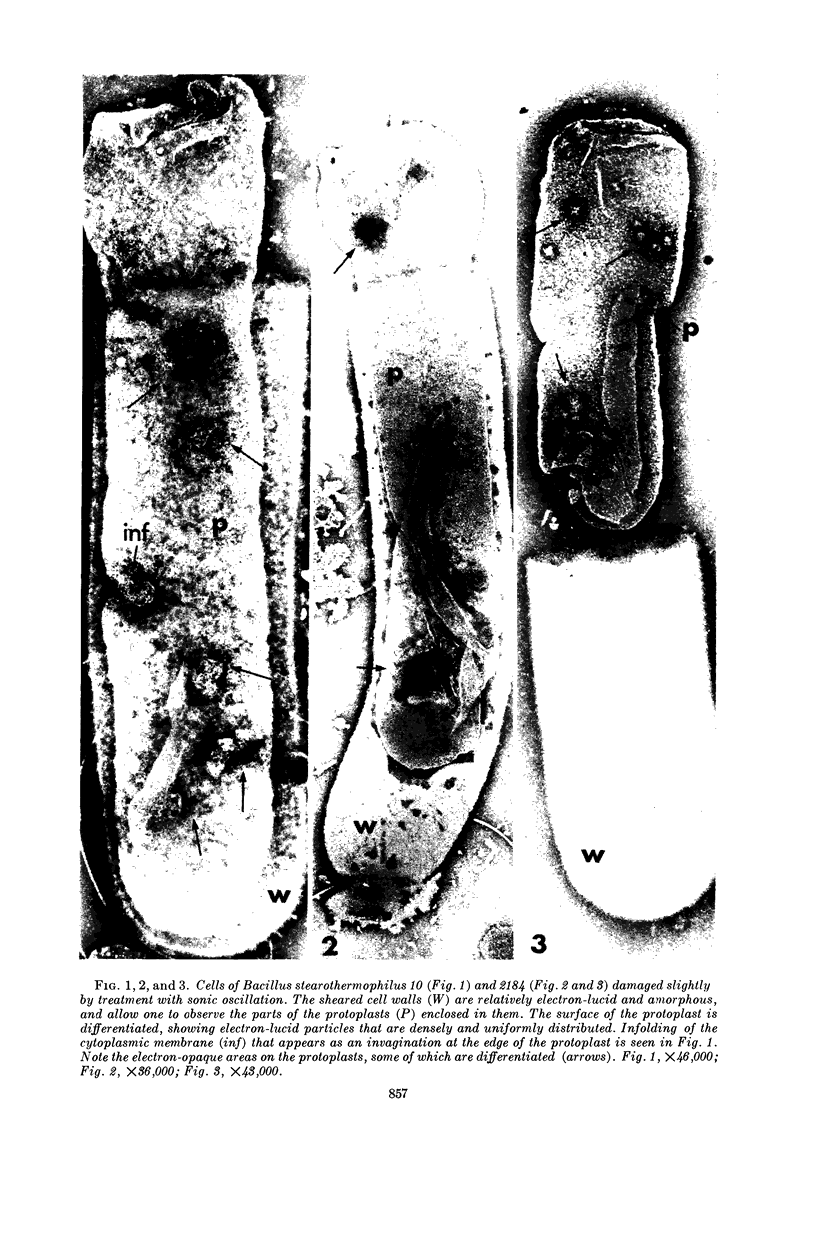
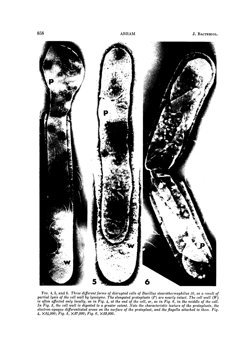
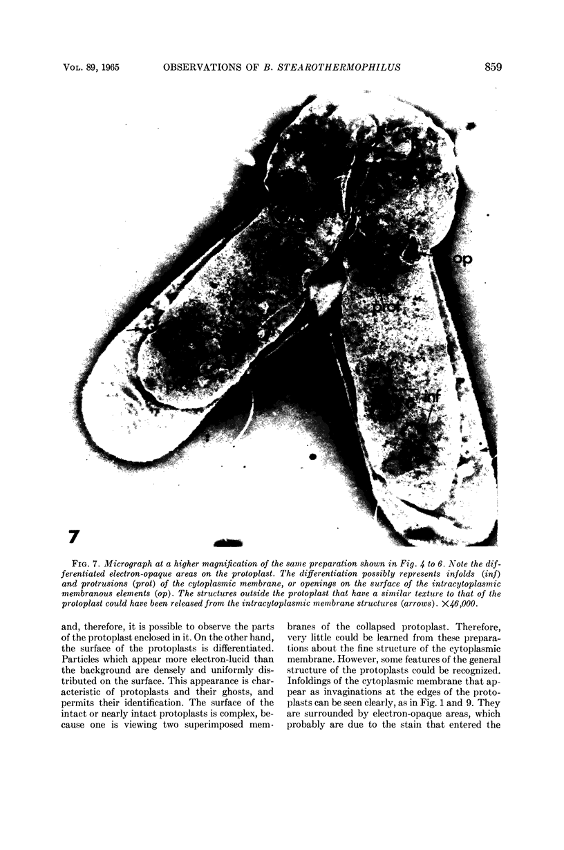
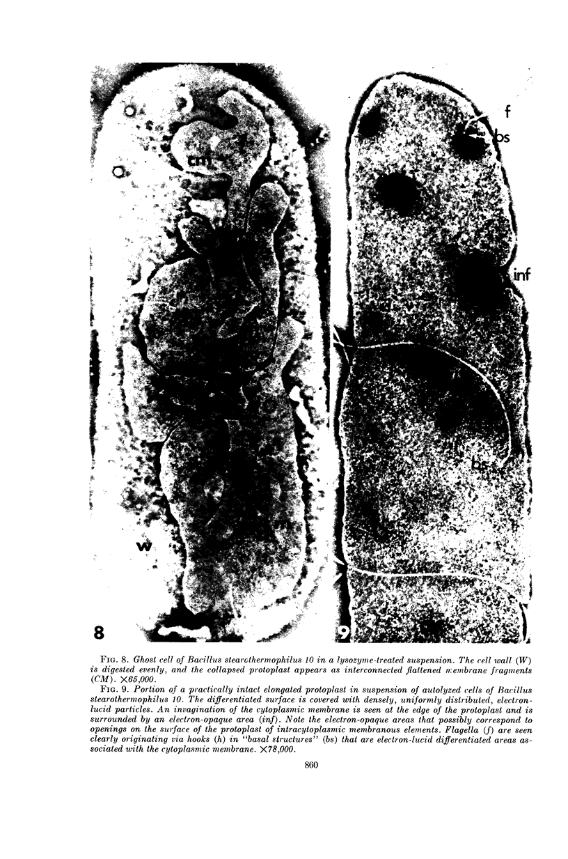
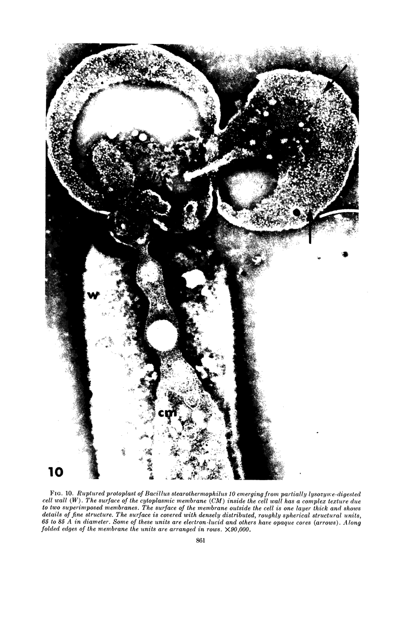
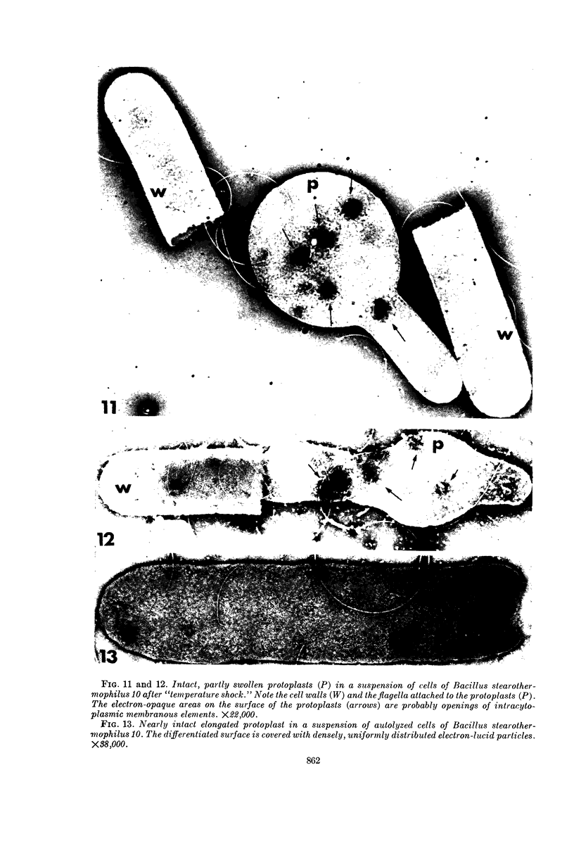
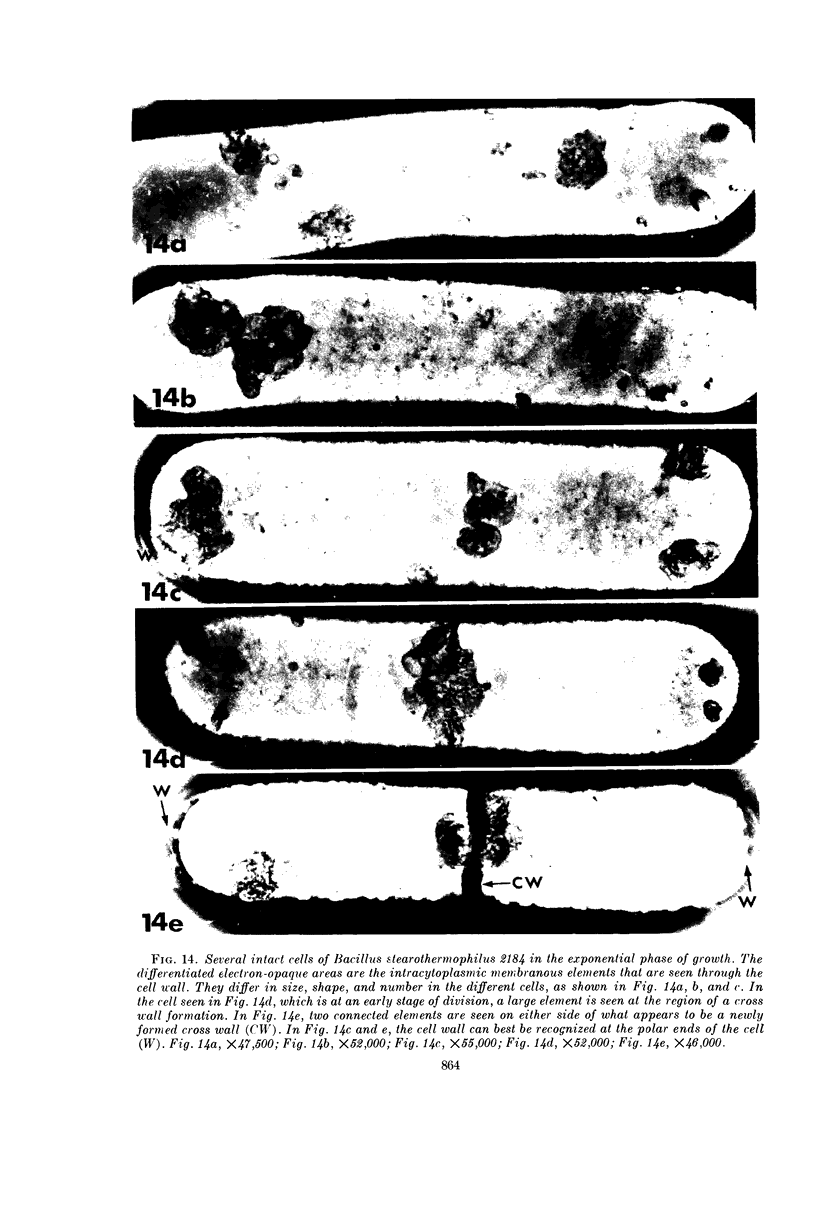
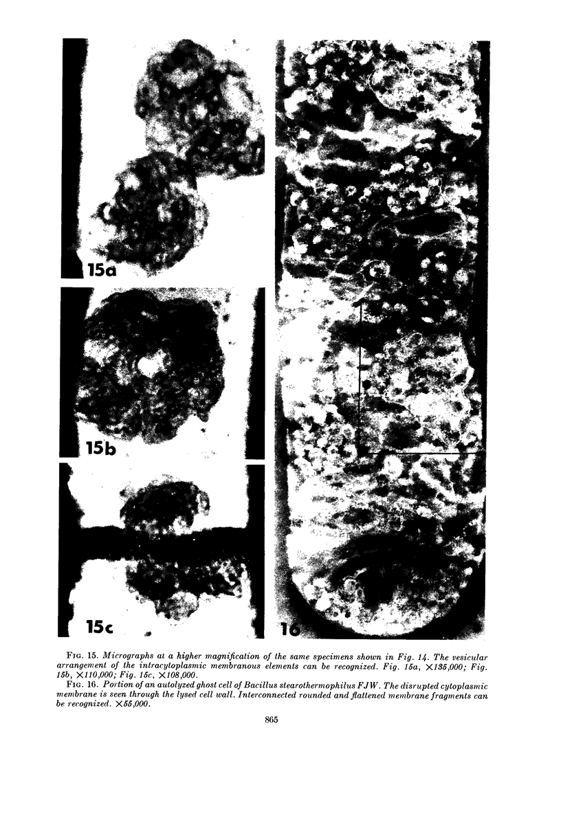
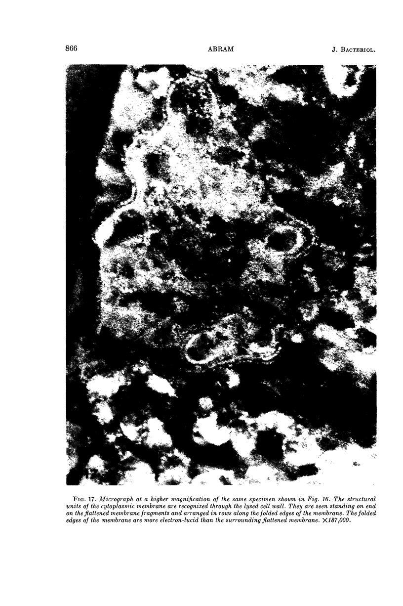
Abstract
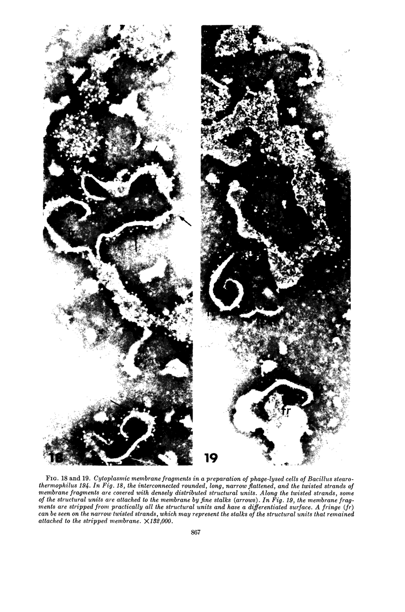
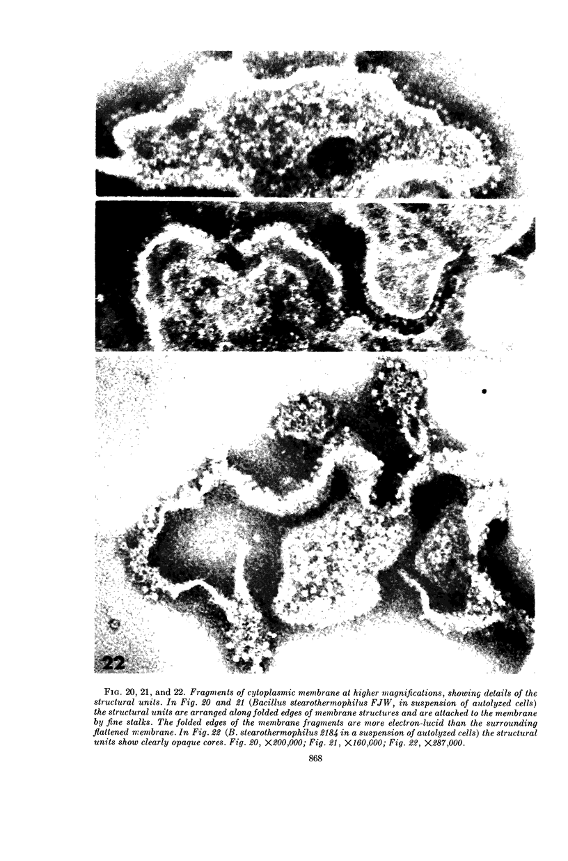
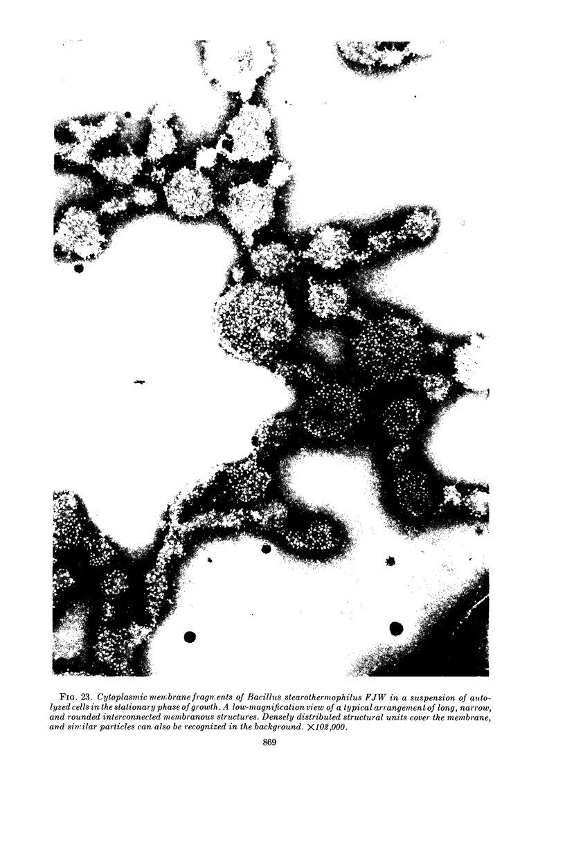
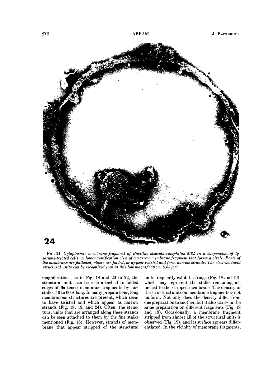
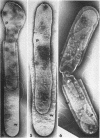
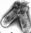
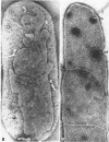
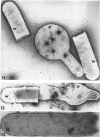
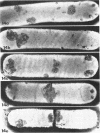
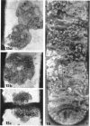
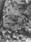
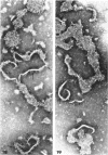
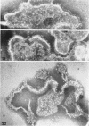
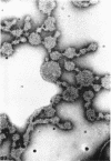
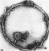
Abram, Dinah (Purdue University, Lafayette, Ind.). Electron microscope observations on intact cells, protoplasts, and the cytoplasmic membrane of Bacillus stearothermophilus. J. Bacteriol. 89:855–873. 1965.—Negatively stained preparations of protoplasts and fragments of cytoplasmic membranes from cells of Bacillus stearothermophilus ruptured by treatment with sonic oscillation, partial lysis with lysozyme, autolysis, or phage infection were examined electron microscopically. Specimens of intact cells also were examined by the same technique. The following structural details were revealed. Intact or nearly intact, partially swollen, elongated protoplasts and their ghosts have a characteristic differentiated surface texture and can easily be distinguished from the cell wall. Infoldings of the cytoplasmic membrane can be observed in these protoplasts, to which flagella are attached; the latter originate via hooks from “basal structures” that are in close association with the cytoplasmic membrane or part of it. Abundant intracytoplasmic membranous elements, which appear to be tubular or vesicular, can be seen in whole cells of three of the strains studied. The fine structure of the cytoplasmic membrane and probably that of its intracytoplasmic infoldings was observed on flattened and folded membrane fragments, one layer thick. Structural units, roughly spherical, 65 to 85 A in diameter, were present on one side of the cytoplasmic membrane, facing the cytoplasm. They were attached loosely to the membrane by fine stalks, 40 to 60 A long, and were easily detached, probably leaving the stalks behind them on the membrane. While the greater stability of membranes from thermophiles made this study of the fine structure possible, the structural units described were demonstrated also on cytoplasmic membranes from mesophiles.
Full text
PDF
Images in this article

Selected References
These references are in PubMed. This may not be the complete list of references from this article.
- BLADEN H. A., NYLEN M. U., FITZGERALD R. J. INTERNAL STRUCTURES OF A EUBACTERIUM SP. DEMONSTRATED BY THE NEGATIVE STAINING TECHNIQUE. J Bacteriol. 1964 Sep;88:763–770. doi: 10.1128/jb.88.3.763-770.1964. [DOI] [PMC free article] [PubMed] [Google Scholar]
- CHAPMAN G. B., HILLIER J. Electron microscopy of ultra-thin sections of bacteria I. Cellular division in Bacillus cereus. J Bacteriol. 1953 Sep;66(3):362–373. doi: 10.1128/jb.66.3.362-373.1953. [DOI] [PMC free article] [PubMed] [Google Scholar]
- EDWARDS M. R., STEVENS R. W. FINE STRUCTURE OF LISTERIA MONOCYTOGENES. J Bacteriol. 1963 Sep;86:414–428. doi: 10.1128/jb.86.3.414-428.1963. [DOI] [PMC free article] [PubMed] [Google Scholar]
- FERNANDEZ MORAN H., ODA T., BLAIR P. V., GREEN D. E. A MACROMOLECULAR REPEATING UNIT OF MITOCHONDRIAL STRUCTURE AND FUNCTION. CORRELATED ELECTRON MICROSCOPIC AND BIOCHEMICAL STUDIES OF ISOLATED MITOCHONDRIA AND SUBMITOCHONDRIAL PARTICLES OF BEEF HEART MUSCLE. J Cell Biol. 1964 Jul;22:63–100. doi: 10.1083/jcb.22.1.63. [DOI] [PMC free article] [PubMed] [Google Scholar]
- FERNANDEZ-MORAN H. Cell-membrane ultrastructure. Low-temperature electron microsopy and x-ray diffraction studies of lipoprotein components in lamellar systems. Circulation. 1962 Nov;26:1039–1065. doi: 10.1161/01.cir.26.5.1039. [DOI] [PubMed] [Google Scholar]
- FITZ-JAMES P. C. Participation of the cytoplasmic membrane in the growth and spore fromation of bacilli. J Biophys Biochem Cytol. 1960 Oct;8:507–528. doi: 10.1083/jcb.8.2.507. [DOI] [PMC free article] [PubMed] [Google Scholar]
- FITZ-JAMES P. FATE OF THE MESOSOMES OF BACILLUS MEGATERIUM DURING PROTOPLASTING. J Bacteriol. 1964 Jun;87:1483–1491. doi: 10.1128/jb.87.6.1483-1491.1964. [DOI] [PMC free article] [PubMed] [Google Scholar]
- GIBOR A., GRANICK S. PLASTIDS AND MITOCHONDRIA: INHERITABLE SYSTEMS. Science. 1964 Aug 14;145(3633):890–897. doi: 10.1126/science.145.3635.890. [DOI] [PubMed] [Google Scholar]
- GLAUERT A. M., BRIEGER E. M., ALLEN J. M. The fine structure of vegetative cells of Bacillus subtilis. Exp Cell Res. 1961 Jan;22:73–85. doi: 10.1016/0014-4827(61)90087-8. [DOI] [PubMed] [Google Scholar]
- GLAUERT A. M., HOPWOOD D. A. A membranous component of the cytoplasm in Streptomyces coelicolor. J Biophys Biochem Cytol. 1959 Dec;6:515–516. doi: 10.1083/jcb.6.3.515. [DOI] [PMC free article] [PubMed] [Google Scholar]
- GLAUERT A. M., HOPWOOD D. A. The fine structure of Streptomyces coelicolor. I. The cytoplasmic membrane system. J Biophys Biochem Cytol. 1960 Jun;7:479–488. doi: 10.1083/jcb.7.3.479. [DOI] [PMC free article] [PubMed] [Google Scholar]
- Green D. E., Blair P. V., Oda T. Isolation and Characterization of the Unit of Electron Transfer in Heart Mitochondria. Science. 1963 Apr 26;140(3565):382–382. doi: 10.1126/science.140.3565.382. [DOI] [PubMed] [Google Scholar]
- HUGHES D. E. The bacterial cytoplasmic membrane. J Gen Microbiol. 1962 Sep;29:39–46. doi: 10.1099/00221287-29-1-39. [DOI] [PubMed] [Google Scholar]
- HUNT A. L., RODGERS A., HUGHES D. E. Sub-cellular particles and the nicotinic acid hydroxylase system in extracts of. Biochim Biophys Acta. 1959 Aug;34:354–372. doi: 10.1016/0006-3002(59)90288-4. [DOI] [PubMed] [Google Scholar]
- IMAEDA T., OGURA M. Formation of intracytoplasmic membrane system of mycobacteria related to cell division. J Bacteriol. 1963 Jan;85:150–163. doi: 10.1128/jb.85.1.150-163.1963. [DOI] [PMC free article] [PubMed] [Google Scholar]
- LINNANE A. W., VITOLS E., NOWLAND P. G. Studies on the origin of yeast mitochondria. J Cell Biol. 1962 May;13:345–350. doi: 10.1083/jcb.13.2.345. [DOI] [PMC free article] [PubMed] [Google Scholar]
- Parsons D. F. Mitochondrial Structure: Two Types of Subunits on Negatively Stained Mitochondrial Membranes. Science. 1963 May 31;140(3570):985–987. doi: 10.1126/science.140.3570.985. [DOI] [PubMed] [Google Scholar]
- ROBERTSON J. D. The ultrastructure of cell membranes and their derivatives. Biochem Soc Symp. 1959;16:3–43. [PubMed] [Google Scholar]
- ROBINOW C. F. On the plasma membrane of some bacteria and fungi. Circulation. 1962 Nov;26:1092–1104. doi: 10.1161/01.cir.26.5.1092. [DOI] [PubMed] [Google Scholar]
- RYTER A., LANDMAN O. E. ELECTRON MICROSCOPE STUDY OF THE RELATIONSHIP BETWEEN MESOSOME LOSS AND THE STABLE L STATE (OR PROTOPLAST STATE) IN BACILLUS SUBTILIS. J Bacteriol. 1964 Aug;88:457–467. doi: 10.1128/jb.88.2.457-467.1964. [DOI] [PMC free article] [PubMed] [Google Scholar]
- SJOESTRAND F. S. A NEW REPEAT STRUCTURAL ELEMENT OF MITOCHONDRIAL AND CERTAIN CYTOPLASMIC MEMBRANES. Nature. 1963 Sep 28;199:1262–1264. doi: 10.1038/1991262a0. [DOI] [PubMed] [Google Scholar]
- STOECKENIUS W. Some observations on negatively stained mitochondria. J Cell Biol. 1963 May;17:443–454. doi: 10.1083/jcb.17.2.443. [DOI] [PMC free article] [PubMed] [Google Scholar]
- STUART D. C., Jr Fine structure of the nucleoid and internal membrane systems of Streptomyces. J Bacteriol. 1959 Aug;78:272–281. doi: 10.1128/jb.78.2.272-281.1959. [DOI] [PMC free article] [PubMed] [Google Scholar]
- VAN ITERSON W. Some features of a remarkable organelle in Bacillus subtilis. J Biophys Biochem Cytol. 1961 Jan;9:183–192. doi: 10.1083/jcb.9.1.183. [DOI] [PMC free article] [PubMed] [Google Scholar]