SUMMARY
The motor protein Kif3a and primary cilia regulate important developmental processes, but their roles in skeletogenesis remain ill defined. Here we created mice deficient in Kif3a in cartilage and focused on the cranial base and synchondroses. Kif3a deficiency caused cranial base growth retardation and dys-morphogenesis evident in neonatal animals by anatomical and μCT inspection. Kif3a deficiency also changed synchondrosis growth plate organization and function and the severity of these changes increased over time. By P7, mutant growth plates lacked typical zones of chondrocyte proliferation and hypertrophy and were instead composed of chondrocytes with an unusual phenotype characterized by strong collagen II expression, but barely detectable expression of Indian hedgehog (Ihh), collagen X, VEGF, MMP-13 and Osterix. Concurrently, unexpected developmental events occurred in perichondrial tissues that included excessive intramembranous ossification and formation of ectopic cartilage masses. Looking for possible culprits for the latter, we analyzed hedgehog signaling by monitoring expression of hedgehog effectors Patched-1 and Gli-1 and hedgehog binding surface component syndecan-3. Compared to controls, signaling was quite feeble within mutant growth plates already by P0, but was actually higher and widespread all along mutant perichondrial tissues. Lastly, we studied postnatal mice deficient in Ihh in cartilage; their cranial base defects only minimally resembled those in Kif3a-deficient mice. In sum, Kif3a and primary cilia make unique contributions to cranial base and synchondrosis development and their deficiency causes growth plate dysfunction, abnormal topography of hedgehog signaling, and un-physiologic responses and processes in perichondrial tissues including ectopic cartilage formation and excess intramembranous ossification.
Keywords: Kif3a, primary cilia, cranial base synchondroses, growth plate, hedgehog signaling, intramembranous ossification, ectopic cartilage
INTRODUCTION
Kif3a is an essential component of the intraflagellar transport (IFT) motor system of primary cilia (Rosenbaum and Witman, 2002). These organelles are microtubule-containing non-motile structures protruding from the surface of most vertebrate cells that were first described decades ago (Scherft and Daems, 1967; Sorokin, 1968). Recent studies have shown that primary cilia are important regulators of cell and tissue function and developmental processes. Mutations in primary cilia’s structural and functional components can cause serious human conditions, including polycyctic kidney disease and retinal degeneration (Ansley et al., 2003; Lin et al., 2003; Pazour et al., 2000). Similarly, altered primary cilia function can have damaging repercussions on limb patterning, neural tube formation and other major developmental processes (Huangfu et al., 2003; Murcia et al., 2000; Zhang et al., 2003). The importance and centrality of primary cilia in both health and disease reflect the fact that these organelles and their IFT system have turned out to mediate and transduce intercellular chemical, physical and biological signals (Davenport and Yoder, 2005), including those elicited by hedgehog proteins (Corbit et al., 2005; Haycraft et al., 2005; May et al., 2005). These proteins are well known regulators of multiple developmental processes (Bitgood and McMahon, 1995; McMahon et al., 2003). They are lipid-containing factors secreted by donor cells that diffuse through the surroundings and exert action on short- and long-range target cells (Gritli-Linde et al., 2001; Yin et al., 2002). The proteins act by binding to the cell surface receptor Patched and signals are transmitted by the receptor Smoothened via zinc-finger transcription factors Gli1, Gli2 and Gli3. Current thinking reviewed recently (Huangfu and Anderson, 2006; Singla and Reiter, 2006) is that following hedgehog interaction with Patched, Smoothened is activated and translocated to primary cilia via the ITF system, resulting in production of Gli activator forms that would translocate to the nucleus and activate hedgehog gene target expression. In the absence of hedgehog proteins, Smoothened would remain sequestered and inactive, and Gli proteins would be processed into repressor forms and move to the nucleus to inhibit hedgehog target gene expression.
A well documented target of hedgehog signaling is the developing axial and limb skeleton (Bitgood and McMahon, 1995; Koyama et al., 1996; Lanske et al., 1996). As a recent study from our groups shows, hedgehog signaling is also very important for early formation, growth and function of the cranial base and its synchondroses (Young et al., 2006). The synchondroses are cartilaginous palindromic structures each consisting of two mirror-image growth plates and termed ethmoidal, intrasphenoidal, spheno-occipital and intraoccipital according to their anatomical location. They are important for cranial base development and overall growth patterns (Ingervall and Thilander, 1972; Roberts and Blackwood, 1983), and defects in synchondrosis functioning are likely to contribute to craniofacial deformities such as those seen in Crouzon and Apert syndromes (Chen et al., 1999; Kreiborg et al., 1993). We found that synchondrosis development is severely affected in mice lacking Indian hedgehog. The Ihh−/− cartilaginous cranial bases were short and deficient in extracellular proteoglycan matrix. The mutant synchondroses displayed abnormal chondrocyte proliferation, hypertrophy and topographical arrangement with respect to perichondrium; as a consequence, endochondral ossification was deranged. In several but not all respects, these abnormalities are reminiscent of those seen in developing Ihh−/− long bones (St-Jacques et al., 1999), pointing to the pivotal nature and general requirement of hedgehog action in skeletogenesis. Given their roles in hedgehog signaling, primary cilia should be as essential for skeletal development and growth, and previous genetic studies certainly support that premise (Murcia et al., 2000; Zhang et al., 2003). Recent reports have described skeletal abnormalities in limbs, calvaria and cranial base in conditional mouse mutants lacking Kif3a or cilia-associated mechanosensor protein polycyctin-1 genes and mice with a missense mutation in the Pc1 gene (Olsen et al., 2005; Xiao et al., 2006), but the specific roles and functions of primary cilia in skeletogenesis and cartilage and growth plate function are still unclear. To obtain such insights and information, we created mice deficient in Kif3a in cartilage and focused on the cranial base and synchondroses, aiming to establish also whether and to what extent defects due to ciliary malfunction compare with those due to hedgehog malfunction. We find that the Kif3a-deficient cranial bases are abnormal and display both predicted and unexpected defects, the latter including a severe failure of hypertrophic cartilage formation, disregulation of hedgehog signaling topography and emergence of ectopic cartilage masses and excess intramembranous bone.
MATERIALS AND METHODS
Conditional Kif3a and Ihh ablation
Animals used in this study were maintained in accordance with the NIH Guide for the Care and Use of Laboratory Animals, and protocols were reviewed and approved by the IACUC at each of our respective Institutions. Homozygous mice deficient in Kif3a in cartilage were produced by sequential mating of Kif3afl/fl mice (Marszalek et al., 1999) with Col2a1-Cre transgenic mice (Ovdhinnikov et al., 2000). The genotype of transgenic mice was determined by PCR analyses of genomic DNA isolated from tail or liver. The Col2a1-Cre mice were identified by using Primer1: 5′ TGC TCT GTC CGT TTG CCG 3′ and Primer2: 5′ ACT GTG TCC AGA CCA GGC 3′, generating one band of about 720 bp. The loxP alleles or Kif3a were identified using three primers: Primer1: 5′ TCT GTG AGT TTG TGA CCA GCC 3′; Primer2: 5′ AGG GCA GAC GGA AGG GTG G 3′; and Primer 3: 5′ TGG CAG GTC AAT GGA CGC AG 3′. Homozygous Kif3a-deficient mice were born with Mendelian frequency and were studied at postnatal stages P0, P7 and P15.
Homozygous mice deficient in Ihh in postnatal cartilage were produced by sequential mating Ihhfl/fl mice (Maeda et al., 2006) with Col2a1-CreER transgenic mice (Nakamura et al., 2006). Genotyping of mice was performed by PCR using the following specific primers: fl-Ihh forward 5′-AGC ACC TTT TTT CTC GAC TGC CTG- 3′, fl-Ihh reverse 5′-TGT TAG GCC GAG AGG GAT TTC GTG-3′; Cre 275 5′-CGC GGT CTG GCA GTA AAA ACT ATC-3′, Cre 603 5′-CCC ACC GTC AGT ACG TGA GAT ATC- 3′. After an initial denaturation for 8 min at 94°C, amplification cycles consisted of denaturation at 94°C for 30 sec, annealing at 68°C for 30 sec, and 45 sec extension at 72°C for 35 cycles, followed by a final extension for 10 min at 72°C. The expected amplicons for the wild type Ihh allele were 320 bp, for the floxed Ihh allele 400 bp, and for the Cre allele 328 bp. To verify Cre activity and specificity postnatally, Col2a1-CreER mice were mated with Rosa R26R reporter mice, and resulting double transgenic mice were injected with 4-hydroxy-tamoxifen (Sigma) at P0 and evaluated for LacZ activity at P1, P4, P7 and P14 by histochemistry (Nakamura et al., 2006).
Micro computed tomography, histology and immunohistochemistry
Skulls were fixed in buffered 4% paraformaldehyde overnight at 4°C, rinsed and subjected to micro computed tomography (μCT) using a μCT40 SCANCO Medical system. Samples were scanned using a 36 mm holder at 45 kvolts of energy, 12 μm scanning thickness and medium resolution. Two dimensional slice images were selected and used to generate three dimensional reconstructions with the following parameters: filter width sigma = 0.8, support level = 1.0, and threshold =173. The same values were used to analyze wild type and mutant samples at each specified time point. Three-dimensional images were rotated at specific angles to generate lateral and bird-eye views of the cranial base.
For histology and immunohistochemistry, fixed skulls were rinsed in DEPC-treated water and decalcified in 0.1M Tris, pH 7.5 buffer containing 0.1% DEPC, 10% EDTA-4 Na, and 7.5% polyvinyl pyrolidione (PVP). For routine histological analysis, 5 mm sections were stained with hematoxylin and eosin or fast green using standard procedures. For immunofluorescence staining, 10 μm sections were incubated with 1:200 dilution of anti-acetylated α-tubulin monoclonal antibody (Sigma, Clone 6-11B-1) followed by biotinylated secondary antibodies and Cy3 conjugated streptavidin (Vector laboratories) to yield signal. Sections were counterstained with YOPRO••-3 iodide (612/631) or 4′,6-diamidino-2-phenylindole dihydrochloride (DAPI) (Invitrogen) to reveal nuclei.
Gene expression analysis
Serial paraffin tissue sections were pretreated with 10 μg/ml proteinase K (Sigma) for 10 min at room temperature, post-fixed in 4% paraformaldehyde, washed with PBS containing 2 mg/ml glycine, and treated with 0.25% acetic anhydride in triethanolamine buffer (Koyama et al., 1996). Sections were hybridized with antisense or sense 35S-labeled probes (approximately 1×10⁶ DPM/section) at 50°C for 16 hrs. Mouse cDNA clones included: histone H4C (nt. 549–799; AY158963); PTHrP (nt. 66–1386; NM_008970); osteopontin (nt. 1–267; AF515708); Osterix (nt. 40–1727; NM_130458); VEGF (nt. 115–539; gi/249858); Ihh (nt. 897–1954; MN_010544); Patched-1 (nt. 81–841; NM_008957); Smoothened (nt. 450–1000; BC048091); collagen X (nt. 1302–1816; NM009925); and collagen II (nt. 1095–1344; X57982). After hybridization, slides were washed with 2X SSC containing 50% formamide at 50°C, treated with 20 μg/ml RNase A for 30 min at 37°C, and washed three times with 0.1X SSC at 50°C for 10 min/wash. Sections were dehydrated with 70, 90, and 100% ethanol for 5 min/step, coated with Kodak NTB-3 emulsion diluted 1:1 with water, and exposed for 10 to 14 days. Slides were developed with Kodak D-19 at 20°C and stained with hematoxylin. Dark and bright field images were captured using a digital camera, and dark field images were pseudo-colored using Adobe Photoshop software.
RESULTS
Cranial base and synchondroses are abnormal in conditional Kif3a-deficient mice
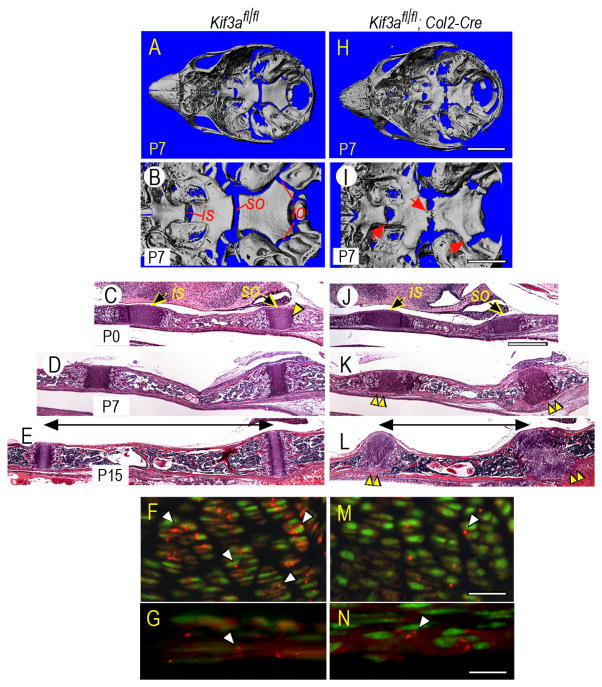
Gross anatomical inspection showed that the craniofacial skeleton was indeed defective in Kif3afl/fl;Col2a1-Cre mice (heretofore termed Kif3a-deficient mice) compared to Kif3afl/wt;Col2a1-Cre and Kif3afl/fl mice (heretofore termed control mice) particularly at postnatal stages. Thus, we processed the skulls of P0, P7 and P15 mice for μCT and histology, focusing on the cranial base. Control cranial bases displayed a typical elongated morphology along the antero-posterior axis (Fig. 1A) and their intrasphenoidal (is), spheno-occipital (so) and intra-occipital (io) synchondroses were well defined and distinct (Fig. 1B). Histologically, the synchondroses were fully cartilaginous reflecting their active growth status and flanked intervening regions of endochondral bone (Fig. 1C,D,E). In contrast, P7 Kif3a-deficient cranial bases were wider than controls (Fig. 1H), and their synchondroses were deformed (Fig. 1I) and even exhibited segments of mineralized tissue bridging the neighboring bones (Fig. 1I, arrows), a potentially catastrophic defect that would hamper growth. Their overall histological organization and shape were already defective at P0 (Fig. 1J) and much more so at P7 and P15 (Fig. 1K,L), and length of the intervening bone region was markedly decreased (Fig. 1E,L, horizontal bars) reflecting cranial base’s shortening along its antero-posterior axis. Immunostaining with antibodies to acetylated α-tubulin confirmed that primary cilia were obvious in control chondrocytes (Fig. 1F, arrowheads) but rare in Kif3a-deficient chondrocytes (Fig. 1M, arrowhead), attesting to effectiveness of Col2a1-Cre action. In addition, primary cilia were equally evident in perichondrial tissue of both control and Kif3a-deficient cranial bases (Fig. 1G,N, arrowheads), attesting to specificity of Col2a1-Cre action clearly limited to cartilage.
Fig. 1.
Kif-3a-deficient cranial bases and synchondroses are abnormal. (A–B,H–I) Skulls from P7 control (Kif3afl/fl) and Kif3a-deficient (Kif3afl/fl;Col2-Cre) mice were analyzed by μCT and are shown by bird-eye view at low and high magnification. Location of intrasphenoidal (is), spheno-occipital (so) and intra-occipital (io) synchondroses is indicated in controls; arrows point to defects in mutant specimens. (C–E,J–L) Parasagittal H&E-stained sections of control and Kif3a-deficient cranial bases displaying is and so synchondroses and intervening endochondral bone. Note that mutant synchondroses’ histology and organization are markedly abnormal compared to controls, and distance between the synchondroses is reduced as well (indicated by horizontal line). (F–G,M–N) Immunolocalization of acetylated α-tubulin in primary cilia (arrowheads) in P0 control (F–G) and Kif3a-deficient (M–N) synchondrosis growth plate and associated perichondrium. Scale bars: 5 mm for A,H; 2 mm for B,I; 300 μm for C–E and J–L; 100 μm for F,M; and 35 μm for G,N.
Gene expression analysis reveals dysfunction of mutant growth plates
Upon closer microscopic inspection, control synchondroses such as the spheno-occipital synchondrosis (Fig. 2A) displayed characteristic growth plate zones of small round resting chondrocytes (Fig. 2B, rz), flat shaped proliferating chondrocytes (Fig. 2B, pz), and oval pre-hypertrophic chondrocytes and large mature hypertrophic chondrocytes (Fig. 2C, phz and hz) followed by primary bone spongiosa intermixing with marrow (Fig. 2D, arrowheads). In sharp contrast, Kif3a-deficient growth plates lacked well-defined zones (Fig. 2I). In particular, small-sized resting and flat-shaped chondrocytes were conspicuously absent; instead, a mixed population of slightly enlarged and oval-to-round chondrocytes was present (Fig. 2J,K). In addition, the hypertrophic zone was very small (Fig. 2K), and there was an abrupt transition from cartilage to marrow and a concurrent reduction in primary spongiosa and endochondral bone (Fig. 2L) (Xiao et al., 2006). In line with absence of flat-shaped cells, mutant growth plates contained few proliferating chondrocytes as indicated by gene expression of mitotic marker histone 4C, (Fig. 2M,N,O), while the cells were abundant and organized in two distinct proliferative zones in control growth plates (Fig. 2E,F,G, arrowheads). Indeed, gene expression of PTHrP, a major regulator of growth plate chondrocyte proliferation (Lanske et al., 1996; Vortkamp et al., 1996), was strong in controls (Fig. 2H), but was low in mutant growth plates (Fig. 2P).
Fig. 2.
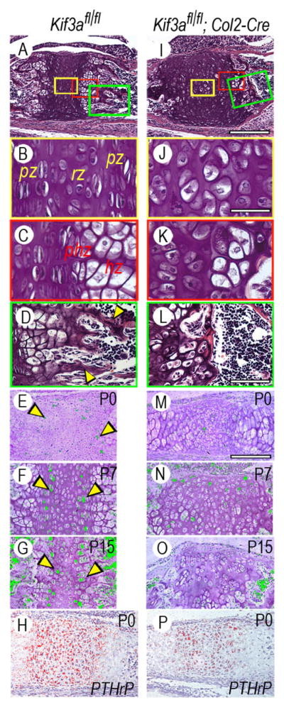
Synchondrosis growth plate organization and chondrocyte proliferation are deranged in Kif3a-deficient cranial bases. (A–D) Parasagittal sections of P7 control spheno-occipital synchondrosis. Note the presence of resting (rz), proliferative (pr), pre-hypertrophic (phz) and hypertrophic (hz) growth plate zones and primary bone spongiosa (arrowheads in D). Boxed colored areas in A are shown at higher magnification in B–D. (E–G) Presence and location of proliferating chondrocytes in control P0, P7 and P15 so synchondroses as revealed by histone 4C gene expression by in situ hybridization. Hybridization signal was given an artificial color for illustration purpose. Note the presence of two well-defined proliferative zones indicated by arrowheads flanking a central resting zone. (H) PTHrP gene expression in control P0 synchondrosis that characterizes resting and proliferative zones. (I–L) Parasagittal sections of P7 Kif3a-deficient so synchondrosis showing that the growth plate zone structure is totally abnormal (J–K) and that there is a near absence of primary spongiosa (L). (M–O) Near absence of histone 4C-expressing proliferating chondrocytes in Kif3a-deficient synchondroses. (P) PTHrP gene expression in Kif3a-deficient synchondrosis Scale bars: 300 μm for A,I; 40 μm for B,C,J,K; 80 μm for D,L; and 150 μm for E-H and M-P.
Growth plate disorganization and deficiency in primary spongiosa and endochondral bone suggested that the chondrocyte maturation process had been deranged in Kif3a-deficient synchondroses. In situ hybridization showed that compared to controls, the mutant chondrocytes did express low levels of Ihh and collagen X that are characteristic products of pre-hypertrophic and early hypertrophic chondrocytes (Fig. 3B,C,G,H), and equally low levels of MMP-13 and VEGF that are important for cartilage-to-endochondral bone transition (Fig. 3D,E,I,J) (Engsig et al., 2000; Zelzer et al., 2002). Deficiency in primary spongiosa (Fig. 4F) was reflected in the fact that expression of such bone markers and regulators as osteopontin, collagen I, Runx2 and Osterix was quite low in mutant (Fig. 4G,H,I,J) versus control specimens (Fig. 4B,C,D,E). Despite all these serious changes, mutant chondrocytes still exhibited strong collagen II expression as controls did (Fig. 3A,F), signifying that their basic differentiated phenotype was largely preserved.
Fig. 3.
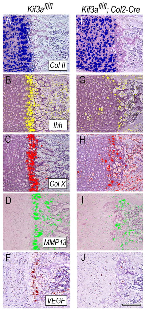
Gene expression of chondrocyte maturation-associated genes is depressed in Kif3a-deficient synchondroses. Serial sections from the medial portion of P7 control (A–E) and Kif3a-deficient (F–J) so synchondroses were processed for in situ hybridization analysis of indicated genes, using radiolabeled riboprobes. Hybridization signal was given artificial colors and images were superimposed to hematoxylin histologic images of corresponding field. Scale bar is 100 μm for A–J.
Fig. 4.
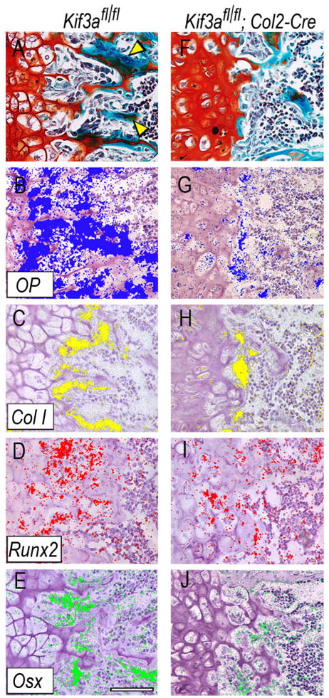
Gene expression of cartilage-to-bone-associated genes and formation of primary spongiosa are inhibited in Kif3a-deficient synchondroses. (A,F) Parasagittal serial sections of P7 control (A) and mutant so synchondrosis (F) were stained with fast green/Safranin O to reveal bone tissue. Note the marked reduction of primary bone spongiosa in mutant tissue (F) that is instead quite clear in controls (A, arrowheads). (B–E) and (G–J) Expression of indicated genes as revealed by in situ hybridization with serial sections of control (B–E) and mutant (G–J) tissue. Scale bar is 80 μm for A–J.
Excessive intramembranous ossification and ectopic cartilage formation
As they develop and mature in the growth plate, pre-hypertrophic and hypertrophic chondrocytes induce formation of intramembranous bone in adjacent perichondrial tissues, a process due to Ihh (Koyama et al., 1996; Nakamura et al., 1997; St-Jacques et al., 1999). Control synchondroses such as the spheno-occipital synchondrosis, conformed to this general rule. It displayed fast green-positive intramembranous bone flanking the Ihh-rich pre- and hypertrophic zones (Fig. 5A, arrowhead) and expressing collagen I, Runx2 and Osterix (Fig. 5B,C,D, arrowheads); importantly, there was no appreciable bone flanking the proliferative and resting zones as to be expected (Fig. 5A,B,C,D, arrows). In contrast, intramembranous bone had formed all along the growth plate in companion Kif3a-deficient synchondrosis, thus spanning the entire flank of the synchondrosis and bridging the neighboring occipital and sphenoidal bones (Fig. 5E,F,G,H). This excess intramembranous bone undoubtedly corresponds to the regions of mineralized tissue initially detected by μCT (Fig. 1I, arrows) and appreciable also by general histology (Fig. 1K,L, double arrowheads).
Fig. 5.
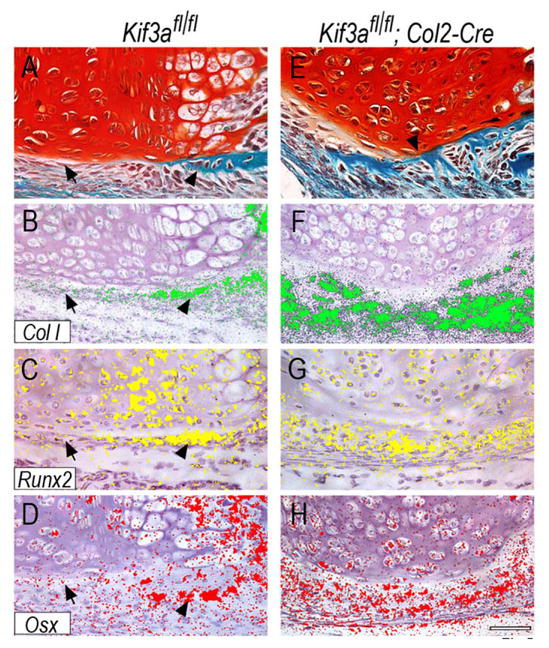
Intramembranous ossification is excessive near Kif3a-deficient synchondroses. Parasagittal serial sections of P7 control (A–D) and mutant so synchondroses were processed for staining with fast green/Safranin O (A,E) or in situ hybridization analysis of indicated genes. Note the presence of intramembranous bone collar adjacent to the pre-hypertrophic and hypertrophic zones in control (A–D, arrowhead) and its absence near the proliferative and resting zones (A–D, arrow) as to be expected. Note instead that intramembranous bone had formed all along the flank of the Kif3a-deficient synchondrosis (E–H). Scale bar is 75 μm for A–H.
Based on these findings, we asked whether there might be additional aberrations occurring in perichondrial tissues flanking Kif3a-deficient synchondroses. Further scrutiny did reveal that, surprisingly, ectopic cartilage masses had formed at several sites (Fig. 6D,E,F, arrows). The tissue was recognizable by its characteristic histology as well as collagen II gene expression and was located almost invariably in close proximity to the mutant synchondrosis growth plates (Fig. 6D,E,F, arrows), thus displaying a topographical arrangement also seen in pathological exostoses that form in close proximity of growth plates (Hecht et al., 1995). Ectopic cartilage masses were never seen in tissues flanking control synchondroses and their chondro-perichondrial boundary was extremely well defined and un-violated (Fig. 6A,B,C).
Fig. 6.
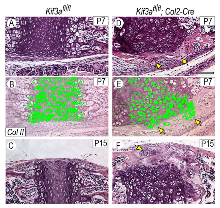
Presence of ectopic cartilage masses near Kif3a-deficient synchondroses. Sections of P7 and P15 control (A–C) and mutant (D–F) synchondroses were stained with H&E or processed for in situ hybridization analysis of collagen II expression. Ectopic cartilaginous masses forming in mutant specimens (arrows in D–E) are recognizable by their typical histology and expression of collagen II. Such phenomenon is never observed in control specimens where the chondro-perichondrial boundary is clear and un-violated (A–C). Scale bar is 75 μm for A–F.
Aberrant topography of hedgehog signaling
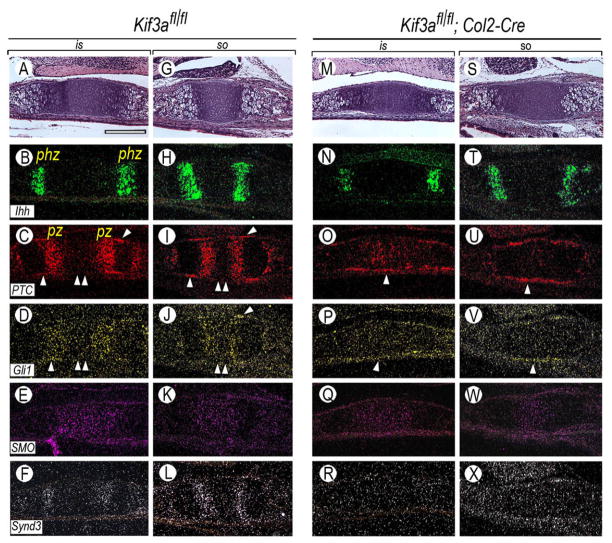
What could have triggered excessive intramembranous ossification and ectopic cartilage formation near mutant growth plates? An obvious possibility was that hedgehog signaling, and in particular the topography of signaling, had changed in mutant specimens, triggering broader responses in neighboring perichondrial tissues. To test this possibility, we determined the expression patterns of Ihh and its target genes Patched-1 and Gli1 in control and Kif3a-deficient synchondroses. We chose to focus on P0 hoping to detect as early changes in signaling patterns as possible. In controls, the three genes displayed predictable and distinct expression patterns, with abundant Ihh transcripts in pre-hypertrophic zones (phz) (Fig. 7A,B,G,H) and Patched-1 and Gli1 transcripts in preceding proliferative zones (pz) (Fig. 7C,D,I,J), reflecting the well established role of Ihh in chondrocyte proliferation (Shimo et al., 2004; St-Jacques et al., 1999). There was also clear Patched-1 and Gli1 expression in perichondrial tissue flanking the pre-hypertrophic and hypertrophic zones where the intramembranous bone collar normally forms (Fig. 7C,D,I,J, single arrowhead), but perichondrium flanking the resting and proliferative zones was negative (Fig. 7C,D,I,J, double arrowhead). Smoothened transcripts were present throughout the growth plates (Fig. 7E,K). In mutant synchondroses, Ihh expression was still strong at this stage (Fig. 7M,N,S,T), but expression of both Patched-1 and Gli1 was grossly abnormal. Patched-1 and Gli-1 transcripts were very low and scattered within the growth plates (Fig. 7O,P,U,V) but were evident along the entire length of perichondrial tissues (Fig. 7O,P,U,V, arrowhead), thus closely mirroring the location of excessive intramembranous bone formation. Smoothened expression appeared largely unaffected (Fig. 7Q,W).
Fig. 7.
Topography of hedgehog signaling is altered in Kif3a-deficient synchondroses. Serial section of P0 control (A–L) and mutant (M–X) is and so synchondroses were processed for expression analysis of indicated genes. Note in controls that Patched-1 and Gli-1 are expressed in proliferative zone (pz) and perichondrium flanking pre-hypertrophic and hypertrophic zones (C–D, I–J, single arrowhead), but not in perichondrium flanking the resting and proliferative zones (C–D, I–J, double arrowheads). In mutants, however, Patched-1 and Gli-1 are minimally expressed within the growth plates, but are expressed all along the perichondrial tissues (O–P, U–V, single arrowhead). Note also that syndecan-3 expression is mainly restricted to the proliferative zone in controls (F,L), but is extremely low in mutants (R,X). Expression of Ihh and Smoothened was similar in control (B,H, E,K) and mutant (N,T, Q,W) tissues at this stage. Scale bar is 150 μm for A–X.
To reinforce the idea that hedgehog signaling topography may be altered in mutant synchondroses, we asked whether expression of molecules needed to direct and restrict hedgehog signaling was altered as well. We focused on heparan sulfate proteoglycans known for their pivotal roles in hedgehog signaling logistics (Bellaiche et al., 1998; The et al., 1999) and on syndecan-3 in particular that we found to participate in such roles in long bone growth plates (Shimazu et al., 1996; Shimo et al., 2004). In controls, Syndecan-3 was expressed in the proliferative zones of synchondrosis growth plates (Fig. 7F,L) along with Patched-1 and Gli-1, in line with the idea that this syndecan may direct and limit Ihh action on proliferation (Pacifici et al., 2005). However, there was barely detectable syndecan-3 expression in Kif3a-deficient synchondroses (Fig. 7R,X).
Skeletal aberrations are caused by postnatal Ihh deficiency
The severe cranial base and synchondrosis abnormalities triggered by Kif3a deficiency in cartilage raised the question of whether they had a unique character or would resemble those elicited by deficiency in related genes, such as Ihh. Accordingly, we created conditional postnatal mice lacking Ihh in cartilage by treating neonatal Ihhfl/fl;Col2a-CreER mice with tamoxifen. Mice were sacrificed at P7 and P15 (heretofore termed Ihh-deficient mice); neonatal Ihhfl/fl littermates were treated with tamoxifen in parallel and served as controls. Ihh-deficient skulls exhibited antero-posterior shortening and abnormal synchondroses (Fig. 8E) relative to controls (Fig. 8E) reminiscent of the defects seen in Kif3a-deficient skulls above. At variance with the latter, however, the Ihh-deficient growth plates were entirely occupied by collagen X-expressing hypertrophic chondrocytes (Fig. 8F) and there was no detectable intramembranous ossification in flanking perichondrial tissues (Fig. 8G, arrowhead) relative to control (Fig. 8B,C). Defects were exacerbated by P15 when much of Ihh-deficient synchondroses had been replaced by endochondral bone and marrow and remnants of growth plate tissue were confined to a lateral and inconspicuous position (Fig. 8H), phenomena not seen in P15 Kif3a-deficient specimens.
Fig. 8.
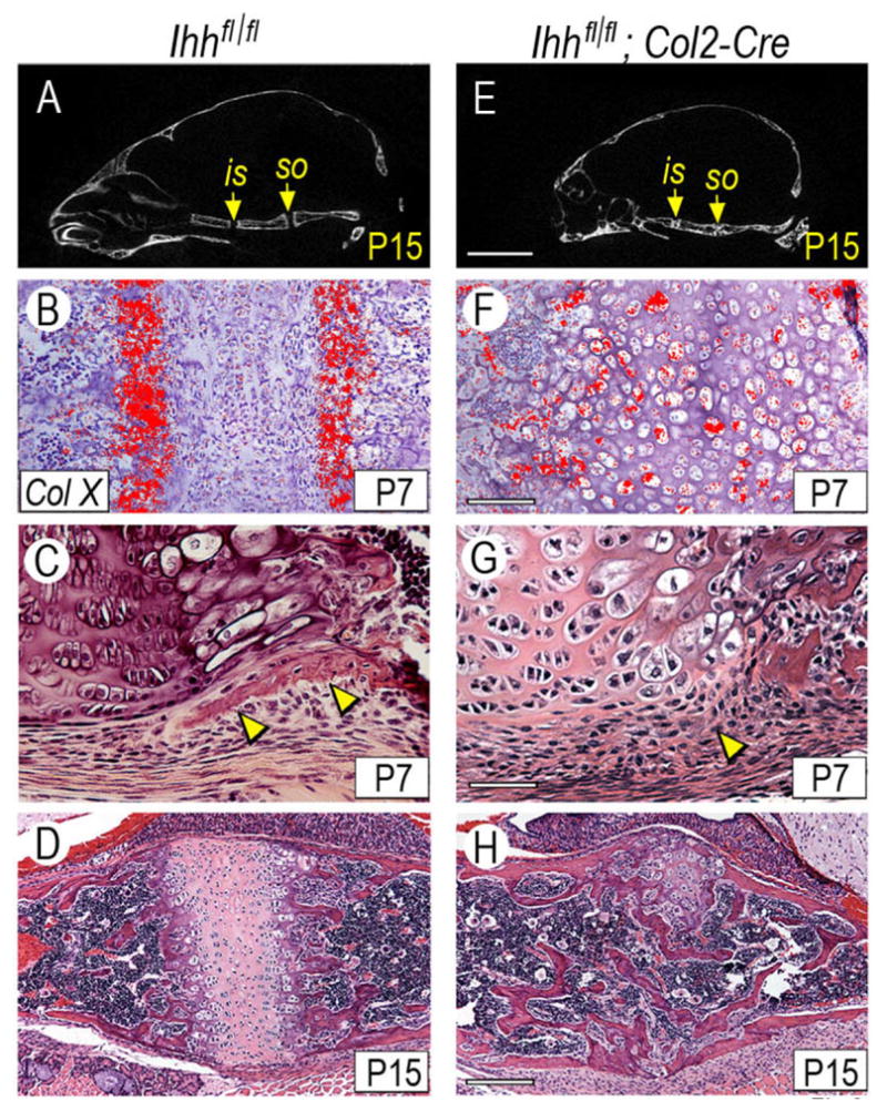
Conditional postnatal Ihh deficiency causes cranial base abnormalities. (A,E) Skulls from P15 control and Ihh-deficient mice were subjected to μCT analysis and one orthogonal plane through the cranial base is shown here. Note the presence of well defined is and so synchondroses in controls (A, arrows) and the ill-defined synchondroses and reduced antero-posterior length in mutants (E). (B–D, F–H) Sections from P7 and P15 control and mutant so synchondroses processed for collagen X gene expression (B,F) or histological analysis (C–D,G–H). Note that collagen X transcripts are restricted to hypertrophic zones in control (B) but are widespread throughout the mutant synchondrosis (F). Note also the presence of a well-formed intramembranous bone collar flanking the pre-hypertrophic and hypertrophic zones in control (C, arrowheads) that is undetectable in mutant (G, arrowhead). Note also that much of mutant synchondrosis is replaced by endochondral bone by P15 (H). Scale bar is 2 mm for A,E; 150 μm for B,F; 75 μm for C,G; and 250 μm for D,H.
DISCUSSION
In this study, we provide evidence that Kif3a and primary cilia are pivotal for cranial base and synchondrosis development and function. Kif3a deficiency alters the antero-posterior and lateral growth patterns of the cranial base, resulting in skull dys-morphogenesis reminiscent of defects seen in some Apert and Crouzon patients (Kreiborg et al., 1993). Kif3a deficiency affects as severely the behavior and functional organization of synchondrosis growth plates, resulting in a quasi-stasis of the chondrocyte maturation process and retardation of endochondral bone formation. Concurrently, there were unexpected and unpredicted alterations in developmental events occurring in synchondrosis perichondrial tissues that include excessive intramembranous ossification, most likely impacting very negatively on cranial base growth. Only some of these defects resemble those caused by conditional deficiency of Ihh, indicating that Kif3a and primary cilia make unique contributions to normal progression of cranial base and synchondrosis development, organization and function.
Growth plates are complex multi-facet structures in which chondrocyte shape, orientation and polarity, chondrocyte proliferation and phenotype, and chondrocyte maturation and hypertrophy must all be regulated in a coordinated manner. A well established mechanism delineated by work on developing long bones is the PTHrP-Ihh axis in which PTHrP produced by periarticular cells under Ihh influences regulates the rates of chondrocyte proliferation (Lanske et al., 1996). In line with this model, Ihh gene ablation in mouse embryos causes down-regulation of PTHrP expression and reduced proliferation (St-Jacques et al., 1999). This mechanism can certainly account for the decrease in chondrocyte proliferation in Kif3a-deficient growth plates that express low if any PTHrP. This would imply that synchondroses conform to, and obey, the PTHrP-Ihh axis (Young et al., 2006) and that a normal complement of Kif3a and primary cilia is needed to maintain it. Reduced proliferation was accompanied by absence of flat-shaped chondrocytes normally constituting the proliferative zone, absence of distinct resting, pre-hypertrophic and hypertrophic zones, and absence of a columnar cell organization. Such nearly chaotic organization of Kif3a-deficient growth plates thus suggests that the mutant chondrocytes may lack key topographical, navigational or mechanical mechanisms and devices that enable them to perceive location or cues within the growth plate and adopt appropriate zone-specific polarity, shape and phenotype. One such mechanism could be the planar cell polarity pathway (PCP), a form of non-canonical Wnt pathway mediated by primary cilia that is essential for cell orientation and mitotic spindle alignment in organs such as inner ear, kidney tubules and cranial cartilage (Dabdoub and Kelley, 2005; Fisher et al., 2006; Park et al., 2006). Importance and centrality of the PCP pathway in several systems make it likely that it operates in growth plates as well (Olsen et al., 2005) where it could orient chondrocyte mitotic spindle and division plane that are usually perpendicular to growth plate’s longitudinal axis (Dodds, 1930) or could promote the related process of columnar organization. Notably, since both PCP pathway and hedgehog proteins act via primary cilia, chondrocytes could control their polarity and orientation in relation to, and correlation with, their responses to Ihh which is distributed in the form of gradients within the growth plate (Gritli-Linde et al., 2001; Yin et al., 2002).
In addition to being disorganized, the Kif3a-deficient growth plates are occupied by enlarged oval-round chondrocytes that express collagen II but little if any maturation genes including collagen X, and are followed by a poor primary bone spongiosa. These phenotypic defects are rather unusual and clearly different from those seen in prenatal Ihh-null synchondroses (Young et al., 2006) or postnatal Ihh-deficient synchondroses described here in which there is exuberant and widespread chondrocyte hypertrophy followed by endochondral bone. One possibility is that the Kif3a-deficient chondrocytes are unable to respond to factors that promote their maturation and hypertrophy, including β-catenin-dependent Wnt proteins (Enomoto-Iwamoto et al., 2002; Tamamura et al., 2005) whose signaling and action may involve primary cilia (Germino, 2005). Alternatively, the topographical disorganization discussed above could have led to general intermingling and intermixing of chondrocytes with different phenotypic properties and maturation status within the mutant growth plates, creating conditions in which the cells interfere with each other’s phenotype. Previously, we showed that when immature chondrocytes are co-cultured with hypertrophic chondrocytes, the hypertrophic phenotype is suppressed (D’ Angelo and Pacifici, 1997).
One of the most intriguing and unexpected defects seen in Kif3a-deficient synchondroses is formation of excessive intramembranous bone and ectopic cartilage masses both readily appreciable by P7. These defects are preceded by alteration in hedgehog signaling topography delineated by Patched-1 and Gli-1 expression along the entire length of perichondrial tissues. Given that hedgehog proteins can stimulate osteogenesis and chondrogenesis (Enomoto-Iwamoto et al., 2000; Nakamura et al., 1997), excess hedgehog signaling in perichondrial tissues could have directly caused excessive intramembranous bone and cartilage formation. What remains unclear, however, is why there is excess signaling all along perichondrium to begin with, particularly starting at P0 when Ihh expression is still confined to fairly-restricted growth plate zones (see Fig. 7). One possibility is that the excess signaling is an autonomous local response of perichondrium to phenotypic defects in the adjacent growth plates. Alternatively, it could be due to the mutant chondrocytes themselves. For example, the Kif3a-deficient chondrocytes may lack means to control location and distribution of Ihh, allowing it to diffuse broadly and reach the entire length of perichondrium. This hypothesis is indirectly sustained by our observation that the Kif3a-deficient growth plates express little if any syndecan-3, likely to be a component of mechanisms limiting and restricting Ihh diffusion and action in the growth plate (Pacifici et al., 2005; Shimo et al., 2004).
At this regard, it is interesting to point out a recent study with mice lacking one allele of Ext2, a Golgi-associated glycosyltransferase needed for heparan sulfate synthesis (Stickens et al., 2005). The Ext2 heterozygous mice were found to be deficient in heparan sulfate and exhibited ectopic cartilaginous masses near their growth plates, thus mimicking exostoses present in Hereditary Multiple Exostosis patients who often have EXT-2 or EXT-1 mutations and reduced heparan sulfate levels (Hecht et al., 1995). The exact link between heparan sulfate deficiency and exostosis formation is still poorly understood. In the Ext2 study above, it was proposed that heparan sulfate deficiency reduces FGF signaling and tilts the balance in favor of BMP signaling, resulting in chondrocyte misbehavior, weakening of the bone collar, and formation of exostosis masses (Stickens et al., 2005). Our data showing reduced syndecan-3 expression in growth plate and excess hedgehog signaling in flanking perichondrial tissues point to hedgehog proteins and defective Kif3a-related mechanisms as culprits in exostosis formation as well. We do not know, however, how Kif3a deficiency and lack of primary cilia lead to a reduction in syndecan-3 expression. In previous studies, we did find that syndecan-3 over-expression in vivo and in vitro causes excess and widespread Ihh expression, suggesting that the two genes are coordinated and that such coordination was disrupted by Kif3a deficiency (Pacifici et al., 2005; Shimo et al., 2004). It will interesting to clarify whether Kif3a deficiency also affects expression of syndecan-4 and syndecan-2 that are normally expressed in proliferative zone and perichondrial tissues, respectively (David et al., 1993; Pacifici et al., 2005) and/or expression of other heparan sulfate proteoglycans including glypicans and perlecan. Given that hedgehogs, BMPs, FGFs and Wnts are all heparan sulfate-binding factors (Bernfield et al., 1999), the data may provide insights into how the various proteoglycans with their distinct structures and expression patterns may help to mediate and coordinate the functions of the many signaling factors and pathways that converge to orchestrate progression of skeletal development and how defects in these circuits could cause pathologies including HME.
Acknowledgments
This work was supported by NIH grants AR047543, AR046000 and AR050560
References
- Ansley SJ, Badano JL, Blacque OE, Hill J, Hoskins BE, Leitch CC, Kim JC, Ross AJ, Eichers ER, Teslovich TM, et al. Basal body dysfunction is a likely cause of pleiotropic Bardet-Biedl syndrome. Nature. 2003;425:628–633. doi: 10.1038/nature02030. [DOI] [PubMed] [Google Scholar]
- Bellaiche Y, The I, Perrimon N. Tout-velu is a Drosophila homologue of the putative tumor suppressor EXT-1 and is needed for Hh diffusion. Nature. 1998;394:85–88. doi: 10.1038/27932. [DOI] [PubMed] [Google Scholar]
- Bernfield M, Gotte M, Park PW, Reizes O, Fitzgerald ML, Lincecum J, Zako M. Functions of cell surface heparan sulfate proteoglycans. Annu Rev Biochem. 1999;68:729–777. doi: 10.1146/annurev.biochem.68.1.729. [DOI] [PubMed] [Google Scholar]
- Bitgood MJ, McMahon AP. Hedgehog and Bmp genes are coexpressed at many diverse sites of cell-cell interaction in the mouse embryo. Dev Biol. 1995;172:126–138. doi: 10.1006/dbio.1995.0010. [DOI] [PubMed] [Google Scholar]
- Chen L, Adar R, Yang X, Monsonego EO, Cuiling L, Hauschka PV, Yayon A, Deng CX. Gly369Cys mutation in mouse FGFR3 causes achrondroplasia by affecting both chondrogenesis and osteogenesis. J Clin Invest. 1999;104:1517–1525. doi: 10.1172/JCI6690. [DOI] [PMC free article] [PubMed] [Google Scholar]
- Corbit KC, Aanstad P, Singla V, Norman AR, Stainier DYR, Reiter JF. Vertebrate Smoothened functions at the primary cilium. Nature. 2005;437:1018–1021. doi: 10.1038/nature04117. [DOI] [PubMed] [Google Scholar]
- D’ Angelo M, Pacifici M. Articular chondrocytes produce factors that inhibit maturation of sternal chondrocytes in serum-free agarose cultures: a TGFβ independent process. J Bone Min Res. 1997;12:1368–1377. doi: 10.1359/jbmr.1997.12.9.1368. [DOI] [PubMed] [Google Scholar]
- Dabdoub A, Kelley MW. Planar cell polarity and a potential role for a Wnt morphogen gradient in stereociliary bundle orientation in the mammalian inner ear. J Neurobiol. 2005;64:446–457. doi: 10.1002/neu.20171. [DOI] [PubMed] [Google Scholar]
- Davenport JR, Yoder BK. An incredible decade for the primary cilium: a look at a once-forgotten organelle. Am J Physiol Renal Physiol. 2005;289:F1159–F1169. doi: 10.1152/ajprenal.00118.2005. [DOI] [PubMed] [Google Scholar]
- David G, Bai XM, vad der Schueren B, Marynen P, Cassiman JJ, van der Berghe H. Saptial and temporal changes in the expression of fibroglycan (syndecan-2) during mouse embryonic development. Development. 1993;119:841–854. doi: 10.1242/dev.119.3.841. [DOI] [PubMed] [Google Scholar]
- Dodds GS. Row formation and other types of arrangment of cartilage cells in endochondral ossification. Anat Rec. 1930;46:385–399. [Google Scholar]
- Engsig MT, Chen QJ, Vu TH, Pedersen AC, Therkidsen B, Lund LR, Henriksen K, Lenhard T, Foged NT, Werb Z, et al. Matrix metalloproteinase 9 and vascular endothelial growth factor are essential for osteoclast recruitment into developing long bones. J Cell Biol. 2000;151:879–889. doi: 10.1083/jcb.151.4.879. [DOI] [PMC free article] [PubMed] [Google Scholar]
- Enomoto-Iwamoto M, Kitagaki J, Koyama E, Tamamura Y, Wu C, Kanatani N, Koike T, Okada H, Komori T, Yoneda T, et al. The Wnt antagonist Frzb-1 regulates chondrocyte maturation and long bone development during limb skeletogenesis. Dev Biol. 2002;251:142–156. doi: 10.1006/dbio.2002.0802. [DOI] [PubMed] [Google Scholar]
- Enomoto-Iwamoto M, Nakamura T, Aikawa T, Higuchi Y, Yuasa T, Yamaguchi A, Nohno T, Noji S, Matsuya T, Kurisu K, et al. Hedgehog proteins stimulate chondrogenic cell differentiation and cartilage formation. J Bone Min Res. 2000;15:1659–1668. doi: 10.1359/jbmr.2000.15.9.1659. [DOI] [PubMed] [Google Scholar]
- Fisher EEl, Doyen A, Nato F, Nicolas JF, Torres V, Yaniv M, Pontoglio M. Defective planar cell polarity in polycystic kidney disease. Nature Genet. 2006;38:21–23. doi: 10.1038/ng1701. [DOI] [PubMed] [Google Scholar]
- Germino GG. Linking cilia to Wnts. Nature Genet. 2005;37:455–457. doi: 10.1038/ng0505-455. [DOI] [PubMed] [Google Scholar]
- Gritli-Linde A, Lewis P, McMahon AP, Linde A. The whereabouts of a morphogen: direct evidence for short- and graded long-range activity of hedgehog signaling peptides. Dev Biol. 2001;236:364–386. doi: 10.1006/dbio.2001.0336. [DOI] [PubMed] [Google Scholar]
- Haycraft CJ, Banizs B, Aydin-Son Y, Zhang Q, Michaud EJ, Yoder BK. Gli2 and Gli3 localize to cilia and require intraflagellar transport protein Polaris for processing and function. PLoS Genetics. 2005;1:480–488. doi: 10.1371/journal.pgen.0010053. [DOI] [PMC free article] [PubMed] [Google Scholar]
- Hecht JT, Hogue D, Strong LC, Hansen MF, Blanton SH, Wagner H. Hereditary multiple exostosis and chondrosarcoma: linkage to chromosome II and loss of heterozygosiity for EXT-linked markers on chromosome II and 8. Am J Hum Genet. 1995;56:1125–1131. [PMC free article] [PubMed] [Google Scholar]
- Huangfu D, Anderson KV. Signaling from Smo to Ci/Gli: conservation and divergence of hedgehog pathways from Drosophila to vertebrates. Development. 2006;133:3–14. doi: 10.1242/dev.02169. [DOI] [PubMed] [Google Scholar]
- Huangfu D, Liu A, Rakeman AS, Murcia NS, Niswander L, Anderson KV. Hedgehog signaling in the mouse requires intraflagellar transport proteins. Nature. 2003;426:83–87. doi: 10.1038/nature02061. [DOI] [PubMed] [Google Scholar]
- Ingervall B, Thilander B. The human spheno-occipital synchondrosis. I. The time of closure appraised macroscopically. Acta odontol Scand. 1972:30. doi: 10.3109/00016357209004602. [DOI] [PubMed] [Google Scholar]
- Koyama E, Leatherman JL, Noji S, Pacifici M. Early chick limb cartilaginous elements possess polarizing activity and express Hedgehog-related morphogenetic factors. Dev Dynam. 1996;207:344–354. doi: 10.1002/(SICI)1097-0177(199611)207:3<344::AID-AJA11>3.0.CO;2-4. [DOI] [PubMed] [Google Scholar]
- Kreiborg S, Marsh JL, Cohen MM, Liversage M, Pedersen H, Skovby F, Borgesen SE, Vannier MW. Comparative three-dimensional analysis of CT-scans of the calvaria and cranila base in Apert and Crouzon syndromes. J Craniomaxillofac Surg. 1993;21:181–188. doi: 10.1016/s1010-5182(05)80478-0. [DOI] [PubMed] [Google Scholar]
- Lanske B, Karaplis AC, Lee K, Lutz A, Vortkamp A, Pirro A, Karperien M, Defize LHK, Ho C, Mulligan R, et al. PTH/PTHrp receptor in early development and Indian hedgehog-regulated bone growth. Science. 1996;273:663–666. doi: 10.1126/science.273.5275.663. [DOI] [PubMed] [Google Scholar]
- Lin FTH, Cordes K, Siclair AM, Goldstein LS, Somlo S, Igarashi P. Kidney-specific inactivation of the KIF3A subunit of kinesin-II inhibits renal ciliogenesis and produces polycystic kidney disease. Proc Natl Acad Sci USA. 2003;100:5286–5291. doi: 10.1073/pnas.0836980100. [DOI] [PMC free article] [PubMed] [Google Scholar]
- Maeda Y, Suva LJ, Lanske B. Indian hedgehog (Ihh) is required for endochondral bone formation after birth. J Bone Min Res. 2006;21:S40. [Google Scholar]
- Marszalek JR, Ruiz-Lozano P, Roberts E, Chen KR, Goldstein LS. Situs inversus and embryonic ciliary morphogenesis defects in mouse mutants lacking the KIF3A subunit of kinesin-II. Proc Natl Acad Sci USA. 1999;96:5043–5048. doi: 10.1073/pnas.96.9.5043. [DOI] [PMC free article] [PubMed] [Google Scholar]
- May SR, Ashique AM, Karlen M, Wang B, Shen Y, Zarbalis K, Reiter JF, Ericson J, Peterson AS. Loss of retrograde motor for IFT disrupts localization of Smo to cilia and prevents the expression of both activator and repressor functions of Gli. Dev Biol. 2005;287:378–389. doi: 10.1016/j.ydbio.2005.08.050. [DOI] [PubMed] [Google Scholar]
- McMahon AP, Ingham PW, Tabin CJ. Developmental roles and clinical significance of hedgehog signaling. Curr Top Dev Biol. 2003;53:1–114. doi: 10.1016/s0070-2153(03)53002-2. [DOI] [PubMed] [Google Scholar]
- Murcia NS, Richards WG, Yoder BK, Mucenski ML, Dunlap JR, Woychik RP. The Oak Ridge Polycyctic Kidney (orpk) disease gene is required for left-right axis determination. Development. 2000;127:2347–2355. doi: 10.1242/dev.127.11.2347. [DOI] [PubMed] [Google Scholar]
- Nakamura E, Nguyen MT, Mackem S. Kinetics of tamoxifen-regulated Cre activity in mice using a cartilage-specific CreERT to assay temporal activity windows along the proximodistal limb skeleton. Dev Dyn. 2006;235:2603–2612. doi: 10.1002/dvdy.20892. [DOI] [PubMed] [Google Scholar]
- Nakamura T, Aikawa T, Enomoto-Iwamoto M, Iwamoto M, Higuchi Y, Pacifici M, Kinto N, Yamaguchi A, Noji S, Kurisu K, et al. Induction of osteogenic differentiation by hedgehog proteins. Biochem Biophys Res Commun. 1997;237:465–469. doi: 10.1006/bbrc.1997.7156. [DOI] [PubMed] [Google Scholar]
- Olsen BR, Kolpakova E, McBratney-Owen B, Li X, Zhou J, Fukai N. Genetic and epigenetic determinants of skeletal morphogenesis - Role of cellular polarity and ciliary function in skeletal development and growth. Oral Biosci Med. 2005;213:57–65. [Google Scholar]
- Ovdhinnikov DA, Deng JM, Ogunrinu G, Behringer RR. Col2a1-directed expression of Cre recombinase in differentiating chondrocytes in transgenic mice. Genesis. 2000;26:145–146. [PubMed] [Google Scholar]
- Pacifici M, Shimo T, Gentili C, Kirsch T, Freeman TA, Enomoto-Iwamoto M, Iwamoto M, Koyama E. Syndecan-3: a cell surface heparan sulfate proteoglycan important for chondrocyte proliferation and function during limb skeletogenesis. J Bone Min Metab. 2005;23:191–199. doi: 10.1007/s00774-004-0584-1. [DOI] [PubMed] [Google Scholar]
- Park TJ, Haigo SL, Wallingford JB. Ciliogenesis defects in embryos lacking inturned or fuzzy function are associated with failure of planar cell polarity and hedgehog signaling. Nature Genet. 2006;38:303–311. doi: 10.1038/ng1753. [DOI] [PubMed] [Google Scholar]
- Pazour GJ, Dickert BL, Vucica Y, Seeley ES, Rosenbaum JL, Witman GB, Cole DG. Chlamydomonas IFT88 and its mouse homologue polycystic kidney disease gene tg737, are required for assembly of cilia and flagella. J Cell Biol. 2000;151:709–718. doi: 10.1083/jcb.151.3.709. [DOI] [PMC free article] [PubMed] [Google Scholar]
- Roberts GJ, Blackwood HJ. Growth of the cartilages of the mid-line cranial base; a radiographic and histological study. J Anat. 1983;136:307–320. [PMC free article] [PubMed] [Google Scholar]
- Rosenbaum JL, Witman GB. Intraflagellar transport. Nature Reviews Mol Cell Biol. 2002;3:813–825. doi: 10.1038/nrm952. [DOI] [PubMed] [Google Scholar]
- Scherft JP, Daems WT. Single cilia in chondrocytes. J Ultrastruc Res. 1967;19:546–555. doi: 10.1016/s0022-5320(67)80080-7. [DOI] [PubMed] [Google Scholar]
- Shimazu A, Nah HD, Kirsch T, Koyama E, Leatherman JL, Golden EB, Kosher RA, Pacifici M. Syndecan-3 and the control of chondrocyte proliferation during endochondral ossification. Exp Cell Res. 1996;229:126–136. doi: 10.1006/excr.1996.0350. [DOI] [PubMed] [Google Scholar]
- Shimo T, Gentili C, Iwamoto M, Wu C, Koyama E, Pacifici M. Indian hedgehog and syndecan-3 coregulate chondrocyte proliferation and function during chick limb skeletogenesis. Dev Dyn. 2004;229:607–617. doi: 10.1002/dvdy.20009. [DOI] [PubMed] [Google Scholar]
- Singla V, Reiter JF. The primary cilium as the cell’s antenna: signaling at a sensory organelle. Science. 2006;313:629–633. doi: 10.1126/science.1124534. [DOI] [PubMed] [Google Scholar]
- Sorokin SP. Reconstructions of centriole formation and ciliogenesis in mammalian lungs. J Cell Sci. 1968;207:207–230. doi: 10.1242/jcs.3.2.207. [DOI] [PubMed] [Google Scholar]
- St-Jacques B, Hammerschmidt M, McMahon AP. Indian hedgehog signaling regulates proliferation and differentiation of chondrocytes and is essential for bone formation. Genes Dev. 1999;13:2076–2086. doi: 10.1101/gad.13.16.2072. [DOI] [PMC free article] [PubMed] [Google Scholar]
- Stickens D, Zak BM, Rougier N, Esko JD, Werb Z. Mice deficient in Ext2 lack heparan sulfate and develop exostoses. Development. 2005;132:5055–5068. doi: 10.1242/dev.02088. [DOI] [PMC free article] [PubMed] [Google Scholar]
- Tamamura Y, Otani T, Kanatani N, Koyama E, Kitagaki J, Komori T, Yamada Y, Costantini F, Wasisaka S, Pacifici M, et al. Developmental regulation of Wnt/β-catenin signals is required for growth plate assembly, cartilage integrity, and endochondral ossification. J Biol Chem. 2005;280:19185–19195. doi: 10.1074/jbc.M414275200. [DOI] [PubMed] [Google Scholar]
- The I, Bellaiche Y, Perrimon N. Hedgehog movement is regulated through tout-velu-dependent synthesis of a heparan sulfate proteoglycan. Mol Cell. 1999;4:633–639. doi: 10.1016/s1097-2765(00)80214-2. [DOI] [PubMed] [Google Scholar]
- Vortkamp A, Lee K, Lanske B, Segre GV, Kronenberg HM, Tabin CJ. Regulation of rate of cartilage differentiation by Indian hedgehog and PTH-related protein. Science. 1996;273:613–622. doi: 10.1126/science.273.5275.613. [DOI] [PubMed] [Google Scholar]
- Xiao Z, Zhang S, Mahlios J, Zhou G, Mangenhelmer BS, Guo D, Dallas SL, Maser R, Calvet JP, Bonewald L, et al. Cilia-like structures and polycystin-1 in osteoblasts/osteocytes and associated abnormalities in skeletogenesis and Runx2 expression. J Biol Chem. 2006;281:30884–30895. doi: 10.1074/jbc.M604772200. [DOI] [PMC free article] [PubMed] [Google Scholar]
- Yin M, Gentili C, Koyama E, Zasloff M, Pacifici M. Antiangiogenic treatment delays chondrocyte maturation and bone formation during limb skeletogenesis. J Bone Min Res. 2002;17:56–65. doi: 10.1359/jbmr.2002.17.1.56. [DOI] [PubMed] [Google Scholar]
- Young B, Minugh-Purvis N, Shimo T, St-Jacques B, Iwamoto M, Enomoto-Iwamoto M, Koyama E, Pacifici M. Indian and sonic hedgehog regulate synchondrosis growth plate and cranial base development and function. Dev Biol. 2006;299:272–282. doi: 10.1016/j.ydbio.2006.07.028. [DOI] [PubMed] [Google Scholar]
- Zelzer E, McLean W, Ng YS, Fukai N, Reginato AM, Lovejoy S, D’Amore PA, Olsen BR. Skeletal defects in VEGF(120/120) mice reveal multiple roles of VEGF in skeletogenesis. Development. 2002;129:1893–1904. doi: 10.1242/dev.129.8.1893. [DOI] [PubMed] [Google Scholar]
- Zhang Q, Murcia NS, Chittenden LR, Richards WG, Michaud EJ, Woychik RP, Yoder BK. Loss of Tg737 protein results in skeletal patterning defects. Dev Dyn. 2003;227:78–90. doi: 10.1002/dvdy.10289. [DOI] [PubMed] [Google Scholar]


