Abstract
Primary persistent atrial standstill due to atrioventricular muscle dystrophy is a rare familial disease in dogs. The diagnosis of this disorder in a 5-month-old English springer spaniel is the earliest in dogs that have been presented at the Ontario Veterinary College.
Résumé
Dystrophie musculaire atrio-ventriculaire chez un Springer anglais âgé de 5 mois. La paralysie auriculaire primaire permanente est une maladie familiale rare chez les chiens. Le diagnostic de ce trouble a été posé chez un Springer anglais âgé de 5 mois. Il s’agit du plus jeune animal jamais présenté à l’Ontario Veterinary College pour lequel un tel diagnostic a été émis.
(Traduit par Isabelle Vallières)
Case description
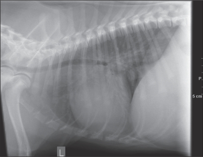
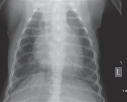
A 5-month-old, neutered male English springer spaniel was presented to the Veterinary Teaching Hospital, Ontario Veterinary College (OVC) on August 25th, 2009 for a sudden onset of lethargy. The owners reported a 2-day history of profound lethargy, exercise intolerance, coughing episodes, and decreased appetite. The dog had previously been presented to the referring clinic for routine puppy vaccinations on June 18th, July 2nd, and August 13th. On those 3 occasions, no abnormal findings were noted on physical examination and the heart rates were consistently around 150 beats/min (bpm). On physical examination, the dog was quiet and alert. Pale mucous membranes (CRT < 2 s) and bradycardia (HR 48 bpm) were noted. No heart murmurs were noted and the lung fields sounded clear. The heart rhythm was slow and regular. Femoral pulses were hyperdynamic. Complete blood (cell) count (CBC) and biochemistry profile revealed normal parameters for a puppy of this age. The Na+/K+ ratio was 25:1. Normal cell morphology was found on blood smear cytology. Thoracic radiographs revealed generalized cardiomegaly, pulmonary venous distension, and diffuse bronchointerstitial changes that were consistent with cardiogenic pulmonary edema (Figures 1, 2). An electrocardiogram (ECG) and echocardiogram were performed. A regular bradycardic ventricular rhythm with the absence of P waves was noted on ECG (Figure 3). Echocardiography revealed biatrial dilation, absence of left atrial contraction, weak residual right atrial contraction, mild biventricular dilation, and good contractility.
Figure 1.
Thoracic radiograph, lateral view. The heart is generally enlarged and there is distension of the pulmonary vein. There is a loss of cranial and caudal cardiac waist. Pulmonary edema is evident in the dorsocaudal lung field.
Figure 2.
Thoracic radiograph, dorsoventral view. Evidence of bronchointerstitial pattern. The heart is enlarged on both the right and left sides.
Figure 3.
Electrocardiogram showing a regular bradycardic ventricular rhythm with the absence of P waves. Heart rate is 38 bpm. Paper speed is 25.0 mm/s.
Discussion
The physical examination and diagnostic findings were consistent with persistent atrial standstill caused by atrioventricular muscular dystrophy. Atrioventricular muscular dystrophy is a putative familial disease (1) resulting in the replacement of cardiomyocytes with fibrous tissue, initially at the atria. This causes the atria to be unable to depolarize and mechanically contract. As a result, cardiac contractions are driven solely from ventricular escape beats that originate from the ventricular subsidiary pacemaker site. This is a progressive disease whereby the ventricles eventually become affected and the heart inevitably fails to function.
The disorder manifests clinically as bradycardia (< 60 bpm), with or without a heart murmur or syncopal events at a young age. In some cases the disease manifests as an initial 3rd degree heartblock that further progresses into persistent atrial standstill. A classical absence of P waves with a regular ventricular rhythm that is not alleviated by atropine administration or exercise is observed on ECG examination (2). There may also be muscular dystrophy of facial and shoulder muscles. Similar heritable disorders, such as facioscapulohumeral muscular dystrophy and Duchenne muscular dystrophy, have also been described in humans (2,3). Radiographic changes may include generalized cardiomegaly and cardiogenic pulmonary edema (4). Pulmonary edema is due to volume overload secondary to bradycardia. Affected animals are usually diagnosed at 1 to 3 years of age. This is the earliest diagnosis of atrioventricular muscular dystrophy in dogs that have been presented at the OVC.
Atrioventricular muscular dystrophy is a rare disorder and is commonly described in the literature in English springer spaniel and Old English sheepdog breeds, but has also been found to occur in other dog breeds, and rarely in cats (5). Over the past 19 y, 10 dogs have been presented to the OVC with persistent atrial standstill, including this case. Of the 10 dogs, 2 were English springer spaniels, 3 were Labrador retrievers, 3 were mixed breed dogs, 1 was an English cocker spaniel, and 1 was a Bernese mountain dog. These data suggest that although the condition may be heritable, there is no evidence of breed predilection for it.
The prognosis for atrioventricular muscular dystrophy is grave. Affected dogs usually die of congestive heart failure due to myocardial failure despite treatment. Treatment includes pacemaker implantation and management of congestive heart failure, which may prolong the lifespan from 6 mo to 2 y. Therapies to manage the congestive heart failure include furosemide and benazepril. Although theophylline is not a specific treatment for heart failure, it is often used as adjunct therapy in these cases in an effort to increase heart rate. Taking into consideration the sudden onset of clinical signs in this dog at a mere 5 mo of age, it is suggestive that the rapid progression of the disease carries an even graver prognosis.
The owners declined treatment with pacemaker implantation and the dog was discharged with medication to manage his congestive heart failure. The dog was euthanized the following day.
Acknowledgments
Many thanks to the cardiology department at the OVC Veterinary Teaching Hospital, especially to Dr. Lynne O’ Sullivan and Dr. Jeremy Orr for their patience and excellent coaching. A special acknowledgment to Dr. Wilson and Dr. Rex Crawford from Dufferin Animal Hospital for making this publication possible. CVJ
Footnotes
Ms. Lai’s current address is University of Guelph, Ontario Veterinary College, Mailbox 438, Guelph, Ontario N1G 2W1.
Ms. Lai will receive 50 free copies of her article courtesy of The Canadian Veterinary Journal.
Use of this article is limited to a single copy for personal study. Anyone interested in obtaining reprints should contact the CVMA office ( hbroughton@cvma-acmv.org) for additional copies or permission to use this material elsewhere.
References
- 1.Holland CT, Canfield PJ, Watson ADJ, Allan GS. Dyserythropoiesis, polymyopathy, and cardiac disease in three related English springer spaniels. J Vet Int Med. 1991;5:151–159. doi: 10.1111/j.1939-1676.1991.tb00942.x. [DOI] [PubMed] [Google Scholar]
- 2.Tilley LP. Essentials of Canine and Feline Electrocardiography. St. Louis, Missouri: Mosby; 1979. pp. 144–145. [Google Scholar]
- 3.Perloff JK, Moise NS, Stevenson WG, Gilmour RF. Cardiac electrophysiology in Duchenne muscular dystrophy: From basic science to clinical expression. J Cardiovasc Electr. 1992;3:394–409. [Google Scholar]
- 4.James Buchanan Cardiology Library [database on the Internet] Davis, California: Veterinary Information Network, Inc; c2000. [Last accessed October 14, 2009]. Available from http://www.vin.com/library/general/JB106atrialStandstill.htm. [Google Scholar]
- 5.Gavaghan BJ, Kittleson MD, McAloose D. Persistent atrial standstill in a cat. Aust Vet J. 1999;77:574–579. doi: 10.1111/j.1751-0813.1999.tb13192.x. [DOI] [PubMed] [Google Scholar]


