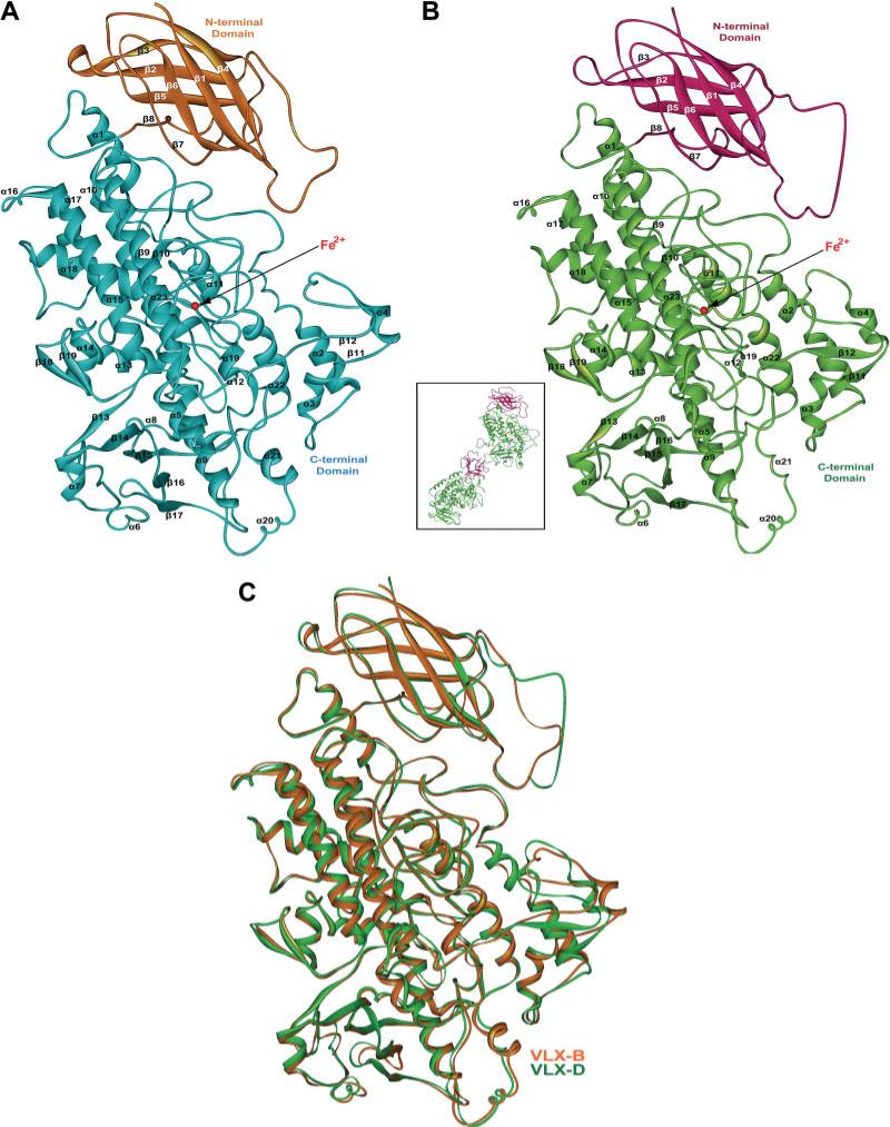
Fig. 3.
Crystal structure of VLX-B (A) and VLX-D (B). The asymmetric unit in the lattice of VLX-D is composed of two molecules (inset of (B)). The N-terminal and C-terminal domains are colored in orange and light blue for VLX-B and violet and light green for VLX-D, respectively. Secondary structural elements have been numbered sequentially as α1–α23 and β1–β19 for the α-helices and β-strands, respectively. The Fe2+ ion is indicated by the red sphere. (C) Superimposed views of VLX-B (orange) and VLX-D (green).

