

Abstract
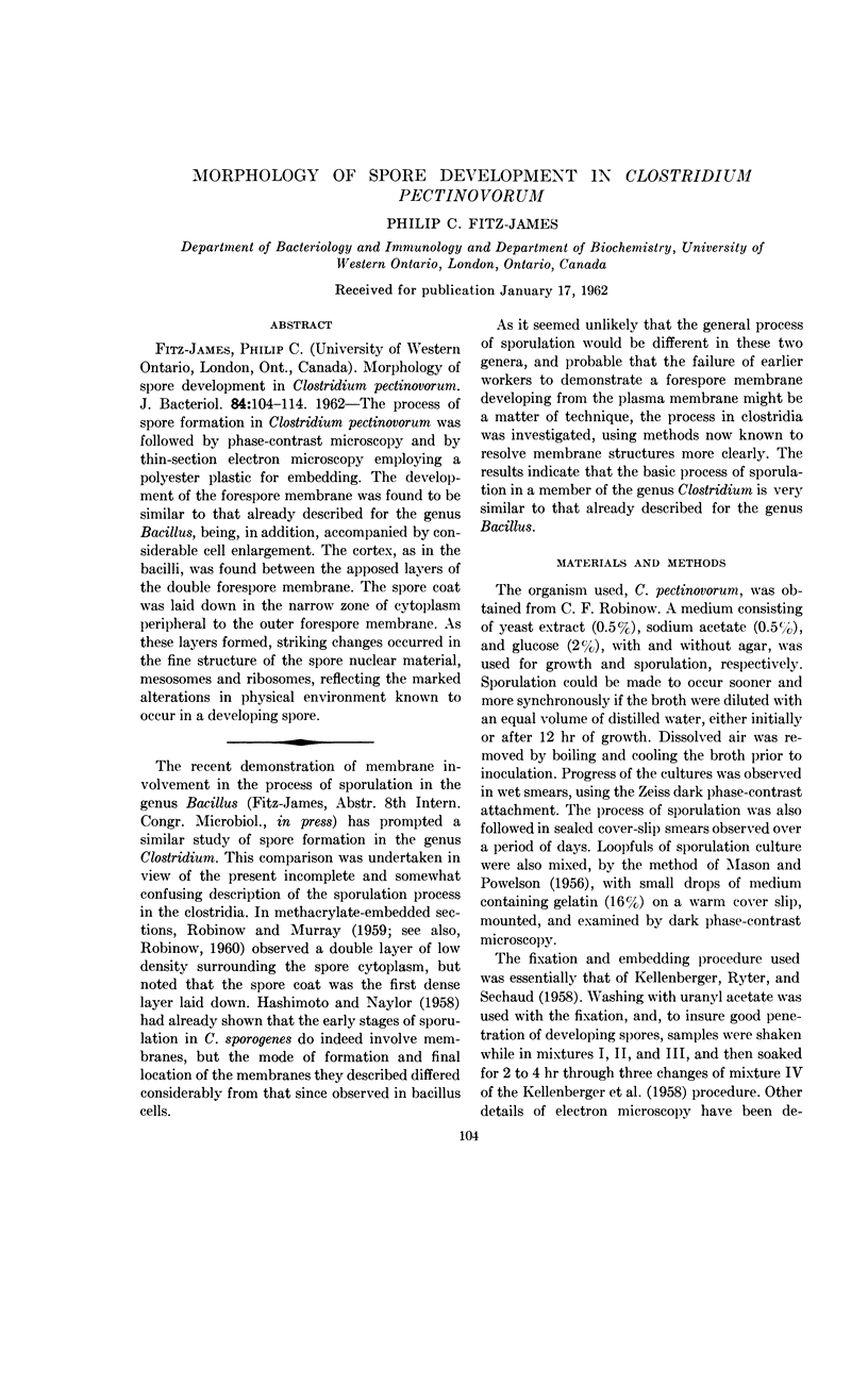
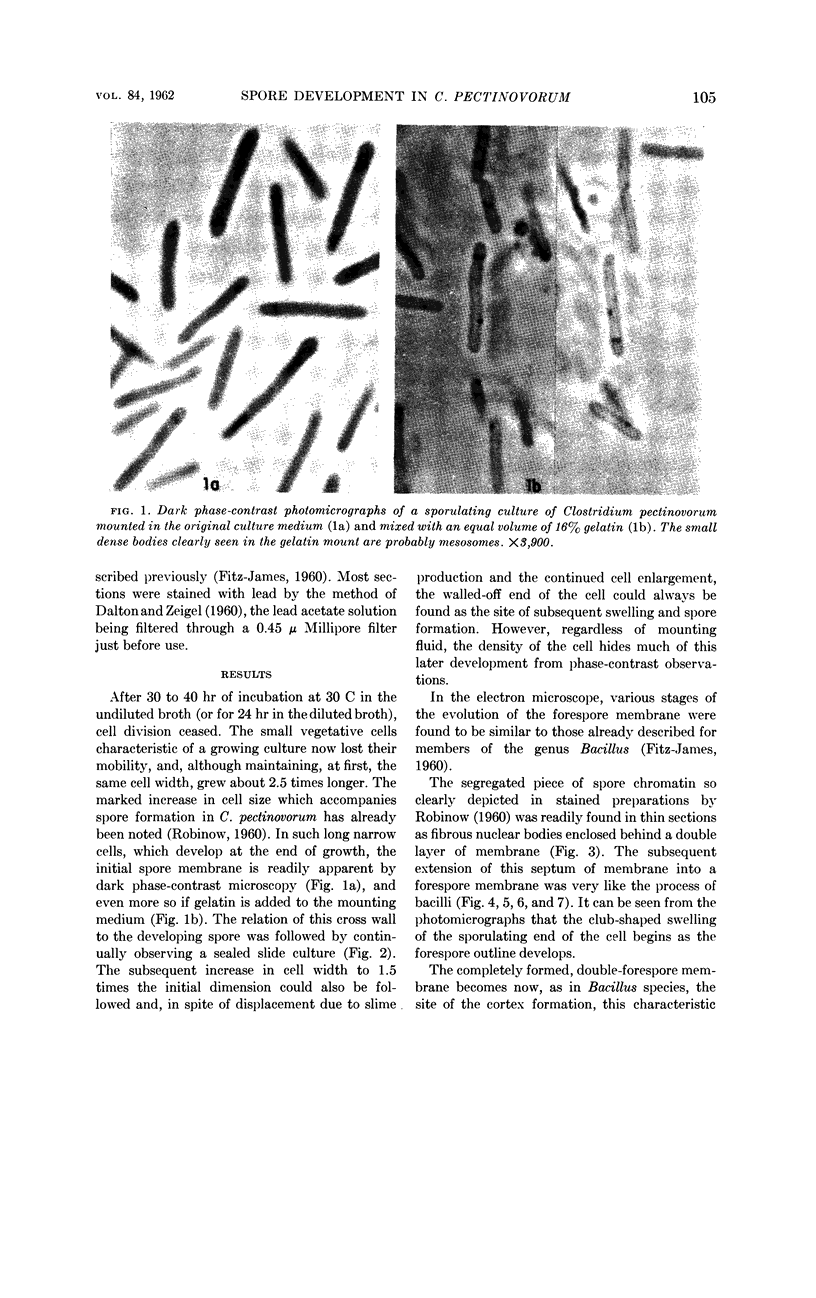
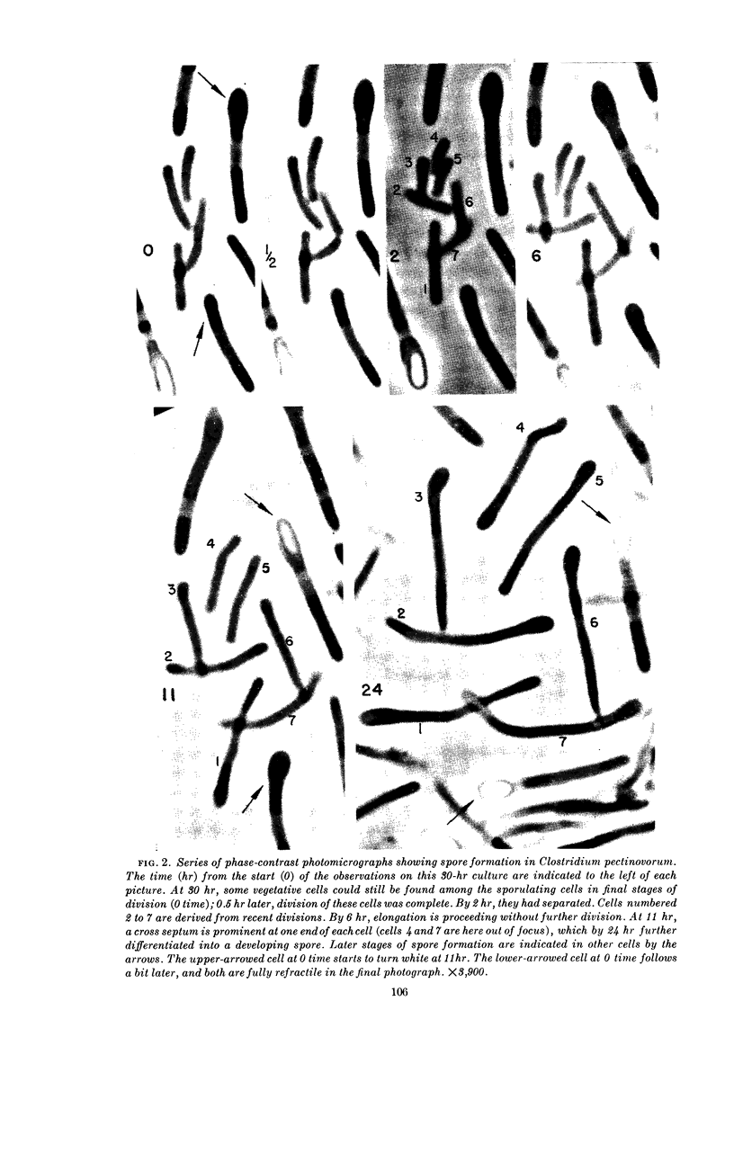
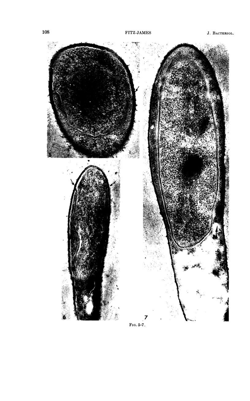
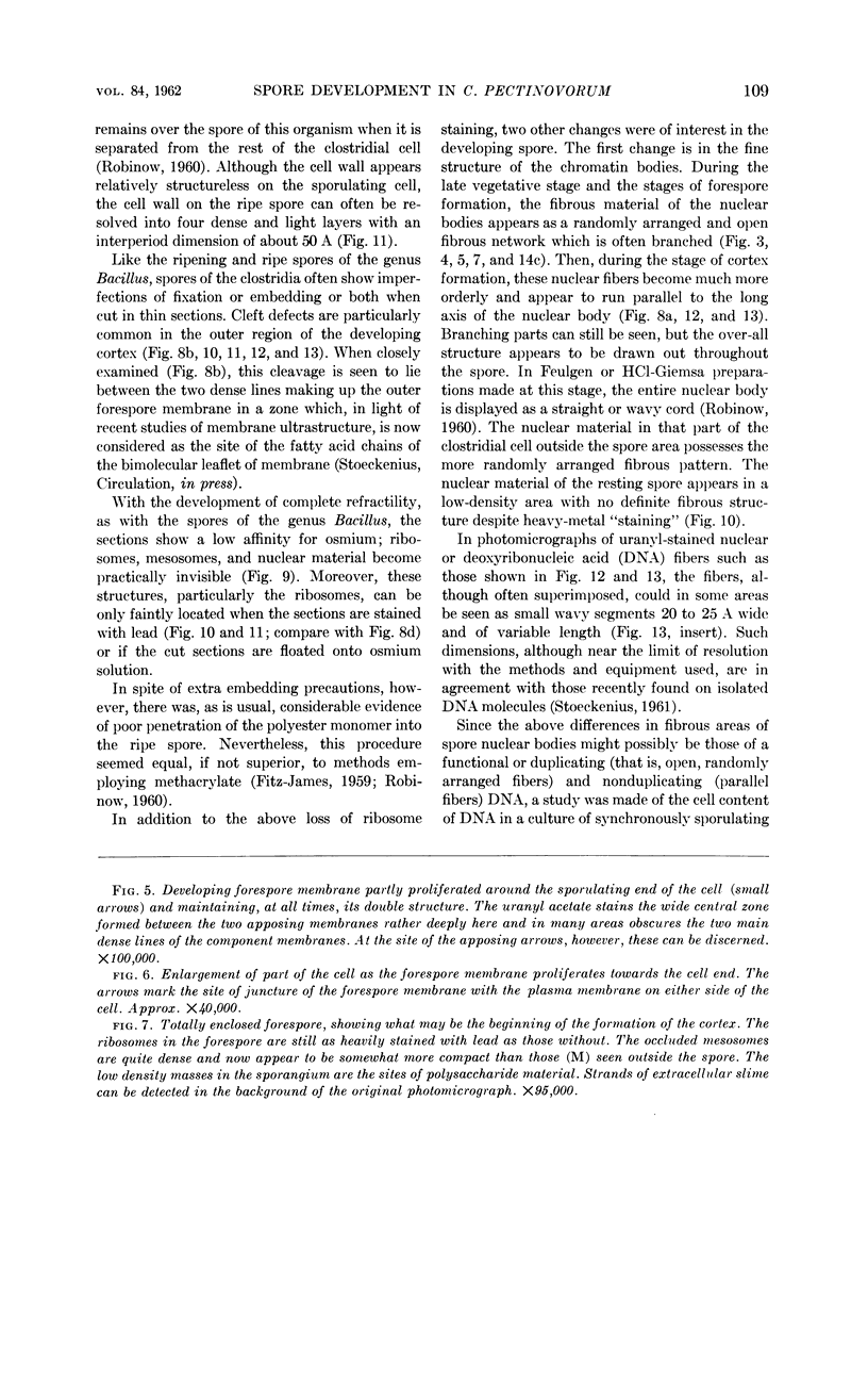
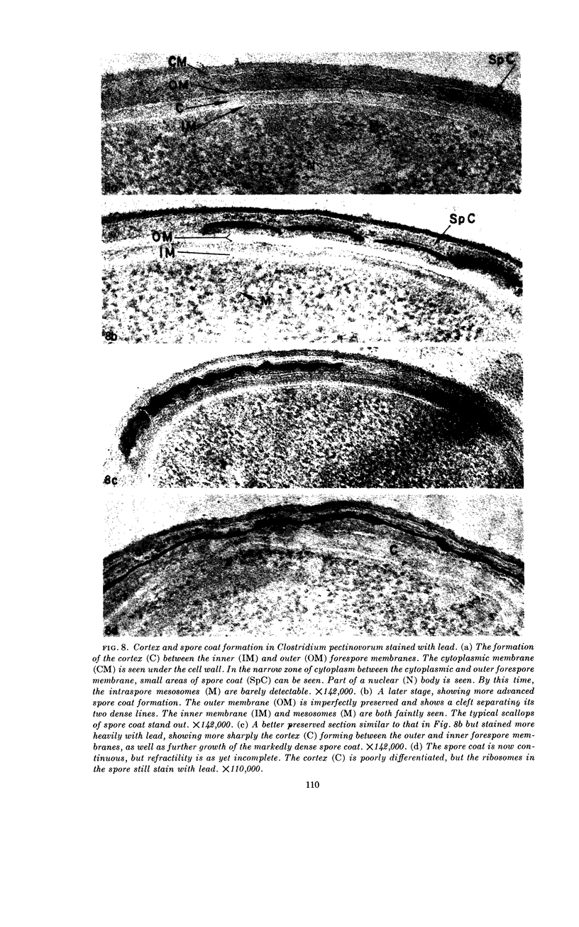
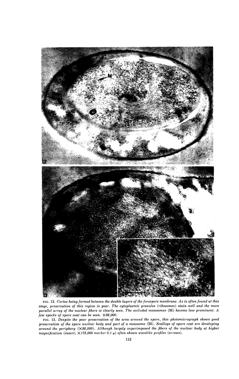
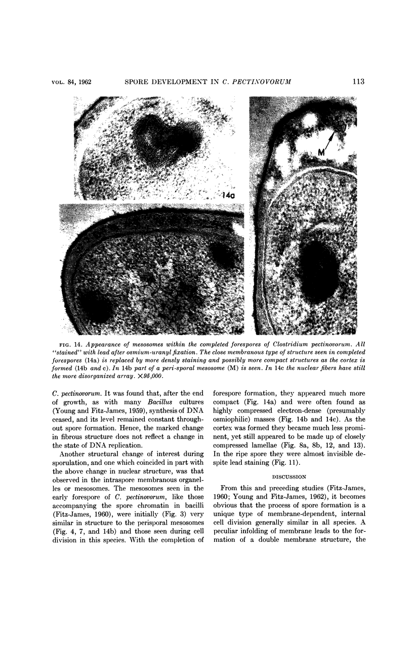
Fitz-James, Philip C. (University of Western Ontario, London, Ont., Canada). Morphology of spore development in Clostridium pectinovorum. J. Bacteriol. 84:104–114. 1962—The process of spore formation in Clostridium pectinovorum was followed by phase-contrast microscopy and by thin-section electron microscopy employing a polyester plastic for embedding. The development of the forespore membrane was found to be similar to that already described for the genus Bacillus, being, in addition, accompanied by considerable cell enlargement. The cortex, as in the bacilli, was found between the apposed layers of the double forespore membrane. The spore coat was laid down in the narrow zone of cytoplasm peripheral to the outer forespore membrane. As these layers formed, striking changes occurred in the fine structure of the spore nuclear material, mesosomes and ribosomes, reflecting the marked alterations in physical environment known to occur in a developing spore.
Full text
PDF
Images in this article
Selected References
These references are in PubMed. This may not be the complete list of references from this article.
- DALTON A. J., ZEIGEL R. F. A simplified method of staining thin sections of biolgical material with lead hydroxide for electron microscopy. J Biophys Biochem Cytol. 1960 Apr;7:409–410. doi: 10.1083/jcb.7.2.409. [DOI] [PMC free article] [PubMed] [Google Scholar]
- FITZ-JAMES P. C. Participation of the cytoplasmic membrane in the growth and spore fromation of bacilli. J Biophys Biochem Cytol. 1960 Oct;8:507–528. doi: 10.1083/jcb.8.2.507. [DOI] [PMC free article] [PubMed] [Google Scholar]
- Fitz-James P. C., Young I. E. CYTOLOGICAL COMPARISON OF SPORES OF DIFFERENT STRAINS OF BACILLUS MEGATERIUM. J Bacteriol. 1959 Dec;78(6):755–764. doi: 10.1128/jb.78.6.755-764.1959. [DOI] [PMC free article] [PubMed] [Google Scholar]
- HASHIMOTO T., NAYLOR H. B. Studies of the fine structure of microorganisms. II. Electron microscopic studies on sporulation of Clostridium sporogenes. J Bacteriol. 1958 Jun;75(6):647–653. doi: 10.1128/jb.75.6.647-653.1958. [DOI] [PMC free article] [PubMed] [Google Scholar]
- KELLENBERGER E., RYTER A., SECHAUD J. Electron microscope study of DNA-containing plasms. II. Vegetative and mature phage DNA as compared with normal bacterial nucleoids in different physiological states. J Biophys Biochem Cytol. 1958 Nov 25;4(6):671–678. doi: 10.1083/jcb.4.6.671. [DOI] [PMC free article] [PubMed] [Google Scholar]
- MASON D. J., POWELSON D. M. Nuclear division as observed in live bacteria by a new technique. J Bacteriol. 1956 Apr;71(4):474–479. doi: 10.1128/jb.71.4.474-479.1956. [DOI] [PMC free article] [PubMed] [Google Scholar]
- STOECKENIUS W. Electron microscopy of DNA molecules "stained" with heavy metal salts. J Biophys Biochem Cytol. 1961 Nov;11:297–310. doi: 10.1083/jcb.11.2.297. [DOI] [PMC free article] [PubMed] [Google Scholar]
- YOUNG I. E., JAMES P. C. Chemical and morphological studies of bacterial spore formation. IV. The development of spore refractility. J Cell Biol. 1962 Jan;12:115–133. doi: 10.1083/jcb.12.1.115. [DOI] [PMC free article] [PubMed] [Google Scholar]