

Abstract
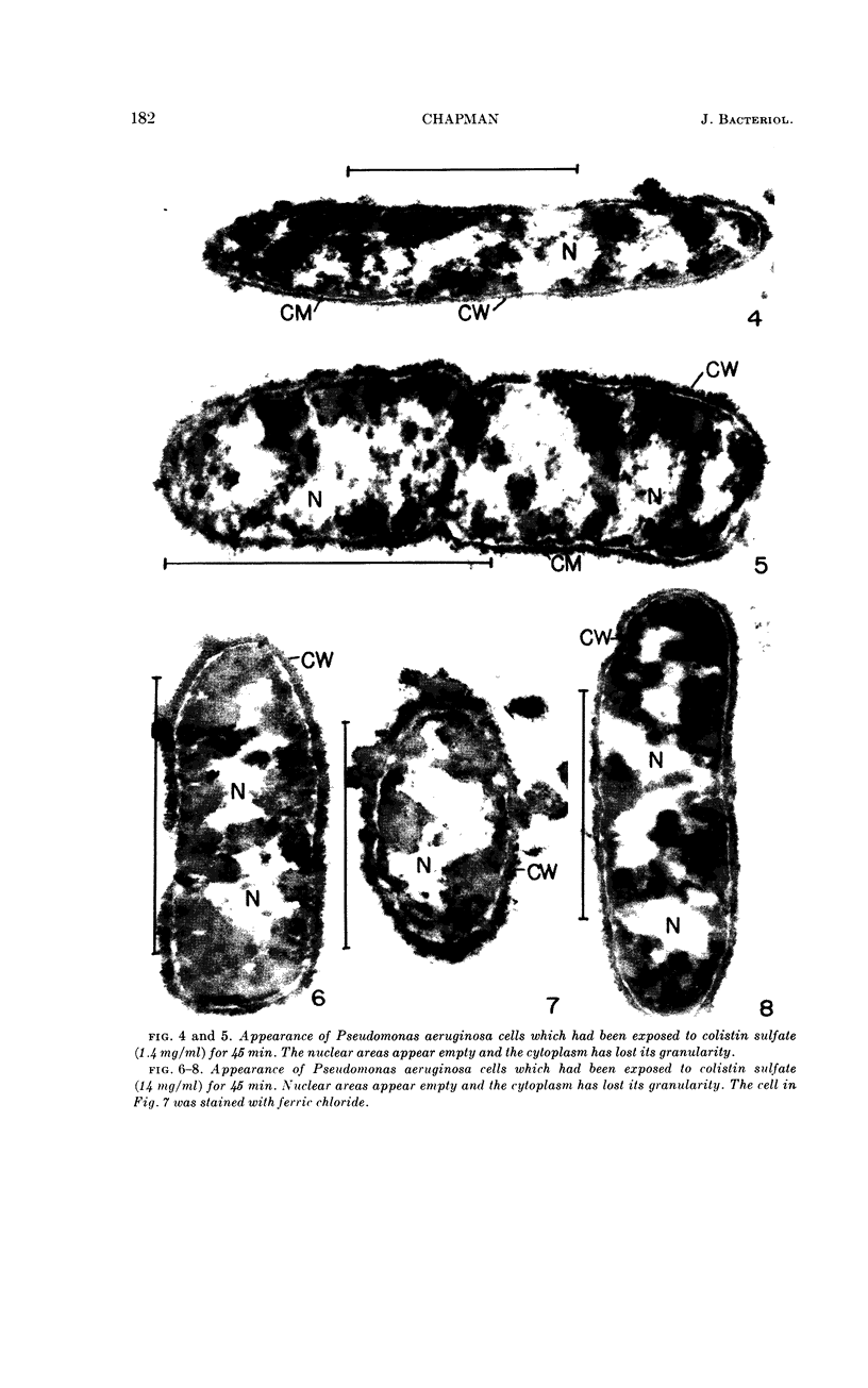
Chapman, George B. (Cornell University Medical College, New York, N.Y.). Cytological aspects of antimicrobial antibiosis. II. Cytological changes associated with the exposure of Pseudomonas aeruginosa and Bacillus megaterium to colistin sulfate. J. Bacteriol. 84:180–185. 1962—Broth cultures of Pseudomonas aeruginosa and Bacillus megaterium were exposed to the antibiotic colistin sulfate. Control (unexposed) and exposed cells were fixed, dehydrated, and embedded in methacrylate. Ultrathin sections were examined in an RCA EMU2-D electron microscope. Two conspicuous cytological changes were noted in P. aeruginosa. The nuclear material was no longer demonstrable in its normal sites, leaving an empty space, and the cytoplasm lost its granularity, becoming homogeneous. In B. megaterium, the latter change was also noted. The nuclear material, however, although no longer demonstrable, did not leave an empty space. Rather, it seemed that cytoplasmic material had engulfed and masked nuclear areas. Cells which showed these changes were nonviable.
Full text
PDF


Images in this article

Selected References
These references are in PubMed. This may not be the complete list of references from this article.
- BERNSTEIN M. H. Iron as a stain for nucleic acids in electron microscopy. J Biophys Biochem Cytol. 1956 Sep 25;2(5):633–634. doi: 10.1083/jcb.2.5.633. [DOI] [PMC free article] [PubMed] [Google Scholar]
- CHAPMAN G. B. Cytological aspects of antimicrobial antibiosis. I. Cytological changes associated with the exposure of Escherichia coli to colistin sulfate. J Bacteriol. 1962 Jul;84:169–179. doi: 10.1002/path.1700840118. [DOI] [PMC free article] [PubMed] [Google Scholar]
- CHAPMAN G. B. Electron microscopy of ultra-thin sections of bacteria. II. Sporulation of Bacillus megaterium and Bacillus cereus. J Bacteriol. 1956 Mar;71(3):348–355. doi: 10.1128/jb.71.3.348-355.1956. [DOI] [PMC free article] [PubMed] [Google Scholar]
- DALES S. A study of the fine structure of mammalian somatic chromosomes. Exp Cell Res. 1960 Apr;19:577–590. doi: 10.1016/0014-4827(60)90065-3. [DOI] [PubMed] [Google Scholar]
- GOLDBERG H. S., MORGAN B. S. Change in bacterial morphology as a result of low concentrations of streptomycin. J Bacteriol. 1954 Oct;68(4):507–508. doi: 10.1128/jb.68.4.507-508.1954. [DOI] [PMC free article] [PubMed] [Google Scholar]
- KELLENBERGER E., RYTER A., SECHAUD J. Electron microscope study of DNA-containing plasms. II. Vegetative and mature phage DNA as compared with normal bacterial nucleoids in different physiological states. J Biophys Biochem Cytol. 1958 Nov 25;4(6):671–678. doi: 10.1083/jcb.4.6.671. [DOI] [PMC free article] [PubMed] [Google Scholar]
- LIBENSON L., McILROY A. P. On the mechanism of the gram stain. J Infect Dis. 1955 Jul-Aug;97(1):22–26. doi: 10.1093/infdis/97.1.22. [DOI] [PubMed] [Google Scholar]
- SCHWARTZ B. S., WARREN M. R., BARKLEY F. A., LANDIS L. Microbiological and pharmacological studies of colistin sulfate and sodium colistinmethanesulfonate. Antibiot Annu. 1959;7:41–60. [PubMed] [Google Scholar]