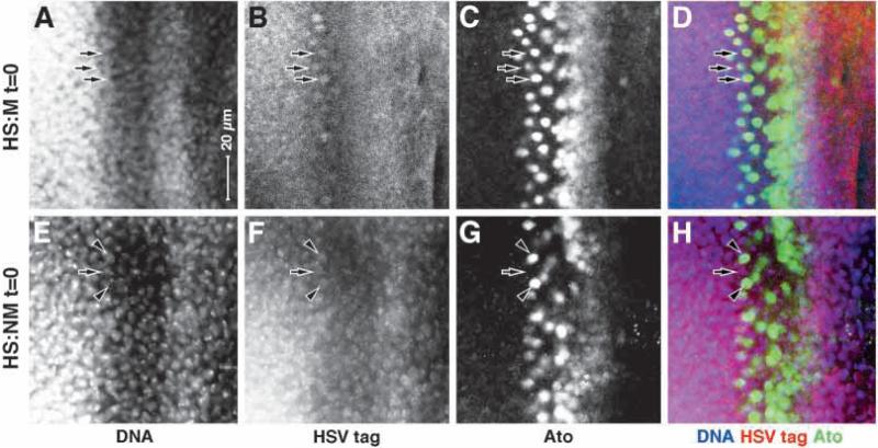
Fig. 9.
Nuclear localization of MAPK detected directly with an epitope tag. Eye discs are shown; anterior rightwards, dorsal upwards. All stained immediately after 1 hour of heat induction. (A-D) Homozygous HS:MG larvae; (E-H) HS:NM larvae. (A,E) Stained to show DNA; (B,F) stained for the HSV epitope tag; (C,G) stained for Atonal; (D,H) Merges. Note that in HS:M, the epitope is expressed at a general and low level and does not appear to be nuclear, except in the final two columns of Atonal-positive cells, where is does appear to be nuclear (three examples indicted by arrows in A-D). Also note that in HS:NM the epitope is expressed at a uniform, low level, and does appear to be nuclear in both Atonal-positive (arrowheads) and -negative (arrow) cells.

