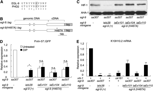
Figure 4.—
The egl-9(H487A) mutation does not abolish egl-9-mediated repression of HIF-1 activity. (A) Alignment of human PHD2 and C. elegans EGL-9 at the region around EGL-9 His487. (B) Diagrams of egl-9 minigenes. The minigenes are fusions of genomic and cDNA sequences. Boxes represent coding regions. GFP is fused in frame to egl-9. The H487A mutation disrupts the Fe(II) binding pocket in EGL-9 and impairs EGL-9 catalytic activity. (C) In egl-9(sa307) mutants, HIF-1 destabilization was rescued by the transgene coding for wild-type egl-9(iaIs38), but not by the egl-9(H487A) transgenes (iaEx101, iaEx104, or iaEx110). AHA-1 protein levels have been shown to be unaffected by severe loss-of-function mutations in egl-9 or vhl-1 (Shen et al. 2006), and AHA-1 serves as a loading control. (D and E) Transgenes expressing either wild-type egl-9 or egl-9(H487A) can suppress expression of HIF-1 targets in an egl-9(sa307) background. (D) In strains carrying the wild-type egl-9 transgene, repression of Pnhr-57∷GFP is more effective and is inhibited by DIP. In strains expressing egl-9(H487A), DIP does not have a significant effect. The bar graph shows averages of Pnhr-57∷GFP protein levels from three biological replicates, normalized to expression levels in an egl-9(sa307) mutant. The asterisk indicates DIP causes a statistically significant difference in the expression of the reporter (*P < 0.05) and NS is no significant difference. (E) The wild-type egl-9 transgene and the egl-9(H487) mutant transgene were able to repress K10H10.2 mRNA levels in an egl-9(sa307) mutant. K10H10.2 mRNA levels were also assayed in wild-type N2 and in vhl-1-deficient animals for comparison. The bar graph shows the relative K10H10.2 mRNA levels in each strain, compared to egl-9(sa307) and the error bars reflect standard error. NS, no significant difference. *P < 0.05; **P < 0.01.

