Abstract
The incidence of pulmonary disease caused by nontuberculous mycobacteria (NTM) appears to be increasing worldwide. In Korea, M. avium complex and M. abscessus account for most of the pathogens encountered, whilst M. kansasii is a relatively uncommon cause of NTM pulmonary diseases. NTM pulmonary disease is highly complex in terms of its clinical presentation and management. Because its clinical features are indistinguishable from those of pulmonary tuberculosis and NTMs are ubiquitous in the environment, the isolation and identification of causative organisms are mandatory for diagnosis, and some specific diagnostic criteria have been proposed. The treatment of NTM pulmonary disease depends on the infecting species, but decisions concerning the institution of treatment are never easy. Treatment requires the use of multiple drugs for 18 to 24 months. Thus, treatment is expensive, often has significant side effects, and is frequently not curative. Therefore, clinicians should be confident that there is sufficient pathology to warrant prolonged, multidrug treatment regimens. In all of the situations, outcomes can be best optimized only when clinicians, radiologists, and laboratories work cooperatively.
Keywords: Mycobacteria, Atypical; Mycobacterium avium Complex; Mycobacterium abscessus; Mycobacterium kansasii, Epidemiology; Diagnosis; Antibiotics, Antitubercular; Korea
INTRODUCTION
The genus Mycobacterium has more than 100 well-characterized species (1), and with the exception of Mycobacterium tuberculosis and M. leprae, the other members are normal inhabitants of natural waters, drinking waters, and soils. These mycobacteria are now generally called nontuberculous mycobacteria (NTM). Previous names for this group of organisms included environmental mycobacteria, opportunistic mycobacteria, atypical mycobacteria, or mycobacteria other than tuberculosis. Human disease due to NTMs is classified into four distinct clinical syndromes: chronic pulmonary disease, lymphadenitis, cutaneous disease, and disseminated disease. Of these, chronic pulmonary disease is most commonly encountered clinically (2, 3).
In Korea, NTM pulmonary disease shows an increasing prevalence due to the increasing numbers of immunocompetent patients with or without pre-existing lung disease (4, 5). In fact, many referral centers have reported increasing numbers of patients with NTM pulmonary disease over recent years. Moreover, in the future it appears that patients with NTM pulmonary disease will be as commonly encountered in daily clinical practice, as patients with pulmonary tuberculosis are today.
NTM infections are thus posing an increasing challenge to physicians in terms of diagnosis and treatment. The clinical and radiographic manifestations of NTM pulmonary disease are variable and often subtle, to the extent that they may be indistinguishable from those of tuberculosis. Moreover, the treatment of patients with NTM pulmonary disease requires more individualization than tuberculosis patients. This individualization is dependent on the species of mycobacteria recovered, the site and severity of infection, antimicrobial drug susceptibility test results, underlying diseases, and the patient's general condition. In addition, the requirement for protracted treatment, side effects, the cost of therapy, and frequent relapses all hinder successful treatment (2, 3).
In this article, we review the epidemiology, the clinical and radiographic manifestations, and the diagnosis and treatment of NTM pulmonary disease with an emphasis on human immunodeficiency virus (HIV)-negative patients.
EPIDEMIOLOGY
NTM organisms are ubiquitously distributed in the environment, and it had been well documented that reservoirs for most of these infections by M. avium, M. intracellulare, M. fortuitum, M. chelonae, and M. abscessus are probably soil, natural waters, and the aerosols of these (6, 7). In addition, tap water, ice prepared from tap water, processed tap water for dialysis, and swimming pools and their aerosols are all a source of NTM infection. Local tap water and hospital water tanks have been identified as sources of M. kansasii infection, which is not found in natural waters or soil (6, 7).
Unlike infection by M. tuberculosis, person-to-person transmission is thought not to occur, and thus the isolation of infected individuals is not required. It is generally assumed that most are infected by environmental NTM. Of the many possible sources of infection, airborne NTM may play an important role in respiratory disease, whereas ingestion is likely to be an important source of infection for children with NTM cervical lymphadenitis and for the majority of HIV-infected patients, in whom M. avium dissemination begins as gastrointestinal colonization. In addition, the direct inoculation of NTM organisms from water or some other material is likely to be a source of infection for those with a soft tissue infection (6, 7).
The most frequent pathogens are M. avium complex (MAC), M. kansasii, M. abscessus, M. xenopi, and M. malmoense in NTM pulmonary disease (2, 3, 8). Unlike tuberculosis, NTM diseases are not required to be reported to public health authorities in most countries, including Korea, and therefore, precise incidence and prevalence data are not available. However, on the basis of laboratory reports and surveillance studies, there appears to be marked geographic variability both in the prevalence of disease and in the mycobacterial species responsible for it (8). NTM pulmonary disease in the United States is most commonly due to MAC, followed by M. kansasii (2). In the United Kingdom, M. kansasii is the most common pathogen in England and Wales, whereas M. malmoense is most common in Scotland, and M. xenopi in southeast England (3). In Japan, MAC is the most common cause of NTM pulmonary disease, followed by M. kansasii (9).
It is well-known from studies in developed countries that the relative importance of NTM pulmonary disease tends to increase, as the incidence of tuberculosis decreases. And, the reported cases with NTM pulmonary disease has continued to rise in Korea since the early 1990s, whereas the incidence of tuberculosis has decreased remarkably over the past 30 yr (4). Before the early 1990s, more than 97-98% of clinical isolates from sputum specimens contained M. tuberculosis in Korea (10-12). However, after the late 1990s, NTM was isolated from 20-30% of all clinical specimens submitted to mycobacterial laboratory in some Korean referral hospitals (13, 14).
Traditionally, tuberculosis case detection is primarily based on the microscopic examination of sputum for acid-fast bacilli (AFB), although a definitive diagnosis of pulmonary tuberculosis normally requires the isolation of M. tuberculosis. In Korea, patients with AFB smear-positive sputum specimens were previously automatically diagnosed as having pulmonary tuberculosis, and antituberculosis treatment was promptly administered according to the guidelines set by the National Tuberculosis Program (15, 16). This is not the case nowadays, even in Korea, because AFB seen on a smear test may represent either M. tuberculosis or NTM. We recently analyzed data from 1,328 AFB smear-positive consecutive sputum specimens collected between 1998 and 2001 at the Samsung Medical Center. NTM were recovered from 9.1% (121/1,328) of these specimens, and from 8.1% (50/616) of patients with smear-positive sputum (17). Lee et al. also reported that NTM were recovered from 10.6% (76/719) AFB smear-positive respiratory specimens at the Asan Medical Center (a positive smear was defined as one with ≥1 AFB in 100 high-power fields) (14). In addition, NTM were isolated from 7.3% of 4,506 AFB smear-positive specimens that were submitted to the Korean Institute of Tuberculosis between 2001 and 2002 from various institutions, including public health centers and private hospitals (17). These results suggest that a substantial proportion of patients with AFB smear-positive sputum specimens in Korea actually have NTM pulmonary disease, rather than pulmonary tuberculosis.
The Korean Institute of Tuberculosis started testing for species identification of NTM from 1981, and the first case of MAC pulmonary disease in Korea was reported in 1981 (18) and other sporadic reports of lung disease caused by MAC (19), M. fortuitum (19), and M. abscessus (20) followed in the early 1980s. Subsequently, a national survey by the Korean Academy of Tuberculosis and Respiratory Diseases was performed in 1995 using the clinical and laboratory data of 158 patients from whom NTM had been isolated, and requested for species identification to the Korean Institute of Tuberculosis from 1981 to 1994 (21). This survey found that 66% of NTM isolates were MAC, 13% were M. fortuitum, 9% were M. chelonae complex, and 12% were other NTMs. Most isolates that were identified as M. chelonae complex are assumed to be M. abscessus, because the Korean Institute of Tuberculosis reported M. abscessus isolates as M. chelonae complex without further species identification until 2000. Unfortunately, this survey did not use specific diagnostic criteria to differentiate the disease from the colonization or contamination. In fact, only 61% of patients were confirmed to have ≥2 positive cultures in this survey (21). After the mid-1990s, the number of NTM isolates submitted to the Korean Institute of Tuberculosis for species identification dramatically increased and exceeded 1000 isolates per year in recent years (22). According to these results, MAC isolates were most frequent in clinical specimens, followed by M. abscessus, and M. fortuitum (22).
The most useful study type with respect to addressing the epidemiology of NTM pulmonary disease should combine information from a mycobacterial laboratory and a clinician's assessment regarding to the presence or absence of disease, because the isolation of an environmental NTM species from a respiratory sample provides insufficient evidence concerning the presence of NTM pulmonary disease (8). Several papers have detailed the relative frequencies of NTM isolates in patients with clinically significant NTM pulmonary disease, by using microbiological and clinical data (Table 1) (14, 23-26). We recently performed a 2-yr study of 794 patients with positive NTM cultures at the Samsung Medical Center over 2002 and 2003 (26). Approximately one quarter of patients with positive cultures were found to have clinical lung disease. The most commonly involved organisms were MAC (48%) and M. abscessus (33%) (26). This study confirms that MAC is the most common pathogen of NTM pulmonary disease in Korea. However, contrary to many other countries the second most common cause in Korea is M. abscessus, and M. kansasii has remained a relatively uncommon cause of NTM pulmonary disease. Actually, we reported the first three cases of M. kansasii pulmonary disease in immunocompetent patients in 2003 (27), and by using the Korean Institute of Tuberculosis database from 1997 to 2002, Yim et al. identified 15 patients with M. kansasii pulmonary disease (28).
Table 1.
Etiology of nontuberculous mycobacterial pulmonary disease in case series studies in Korea
*M. abscessus isolates were reported as M. chelonae complex without further species identification until 2000. Because M. chelonae is an extremely rare cause of pulmonary disease, most respiratory isolates identified as M. chelonae complex can be reasonably assumed to be M. abscessus isolates.
CLINICAL PRESENTATION AND DIAGNOSTIC CRITERIA
NTM organisms are commonly isolated from environmental sources, and thus their growths in culture raise the question of specimen contamination. Moreover, even a finding of mycobacteria in an uncontaminated clinical specimen raises the question as to whether the isolate is the cause of infection or disease. On the other hand, isolation from blood or other sterile sites, along with the clinical manifestations of the disease, is reasonable grounds for considering that an NTM isolate is the causative agent. Decisions on diagnoses are more complex in cases of pulmonary disease, because respiratory secretions from patients with underlying lung disease may be colonized by these organisms without overt clinical manifestations.
Thus, the presence of compatible symptoms and signs is a necessary clinical criterion, with the reasonable exclusion of other etiologies of pulmonary disease (2). However, the signs and symptoms of NTM pulmonary disease are often variable and nonspecific. Patients often present with a chronic cough, productive sputum, and fatigue. NTM infection of the lungs often occurs in the context of a preexisting lung disease, especially chronic obstructive pulmonary disease, bronchiectasis, pneumoconiosis, and previous tuberculosis. As a result, the clinical manifestations of NTM pulmonary disease are often similar to those of underlying disease (2). Sometimes, it is very difficult to separate symptoms that may be due to NTM infection from those that may be due to an underlying disease that has increased the individual's susceptibility to NTM.
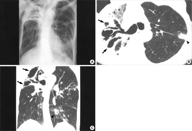
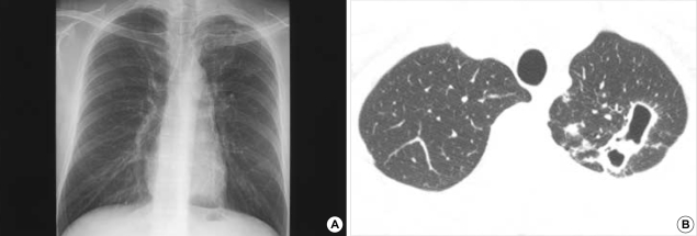
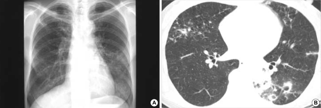
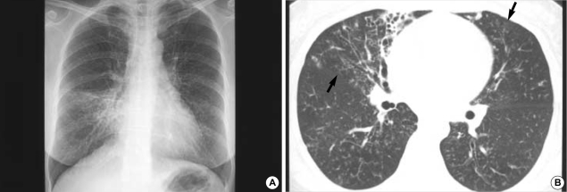
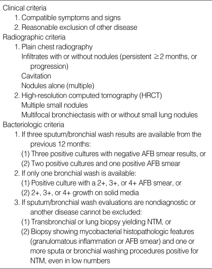
The radiographic criteria required are the presence of infiltrates, cavitation, or multiple nodules on plain chest radiographs, and/or multiple small nodules of less than 10 mm in diameter or multifocal bronchiectasis on high-resolution computed tomography (HRCT) of the lungs (Fig. 1-4) (2). The well-known radiographic features of NTM pulmonary disease caused by MAC or M. kansasii are similar to those of postprimary tuberculosis (29-33). In these classic upper lung zone diseases, the most common findings are linear and nodular areas of increased opacity in the apical and posterior segments of the upper lobes. These opacities can progress or remain stable over many years. Cavitation occurs in many cases and there is frequent pleural thickening (Fig. 1, 4). Recent studies on HRCT of the chest have shown that many patients with non-cavitary disease of the middle and lower lung zones caused by NTM infection, show both multifocal bronchiectasis and clusters of small nodules and branching linear structures are present (Fig. 2, 3) (34-39).
Fig. 1.
M. intracellulare pulmonary disease of the upper lobe cavitary form in a 73-yr-old man. Chest radiograph shows parenchymal opacity which is containing multiple cavities in the right upper lobe. Also note nodular lesions in left middle and lower lung zones (A). Transaxial lung window CT image (2.5-mm section thickness, 70 mA) obtained at the level of the right upper lobar bronchus shows dilated bronchi in an opacified right upper lobe. Also note associated multiple cavitary lesions (arrows). A lobular consolidative lesion (arrowhead) is seen in the left upper lobe (B). Coronal reformation (2.0-mm section thickness) image demonstrates dilated bronchi and multiple thin walled cavities (arrows) in the right upper lobe. Also note multiple variable-sized nodules and consolidation (arrowhead) in the left lung (C).
Fig. 4.
M. kansasii pulmonary disease in a 30-yr-old man. Chest radiograph shows cavitary and small nodular lesions in the left upper lobe (A). Transaxial lung window CT thin-section images (1.0-mm section thickness, 170 mA) obtained at the thoracic inlet level show multiple thin-walled cavities in the left upper lobe. Also note small nodular lesions and tree-in-bud opacities in the left upper lobe (B).
Fig. 2.
M. intracellulare pulmonary disease of the nodular bronchiectatic form in a 63-yr-old woman. Chest radiograph shows a multifocal patchy distribution of small nodular clusters in both lungs (A). Transaxial lung window CT image (2.5-mm section thickness, 70 mA) obtained at the level of basal trunk (B) show small centrilobular nodules and bronchiectasis in the right middle lobe and in the lingular division of the left upper lobe. Also note small cavitating nodules and lobular consolidation in the left lower lobe.
Fig. 3.
M. abscessus pulmonary disease in a 49-yr-old woman. Chest radiograph shows multifocal patchy areas of small nodular clusters in both lungs. Also note parenchymal opacity in the right middle lobe (A). Transaxial lung window CT image (2.5-mm section thickness, 70 mA) obtained at the basal trunk (B) show bronchiectasis and small centrilobular nodules or tree-in-bud opacities (arrows) in the right middle lobe and in the lingular division of the left upper lobe. Also note bronchiolitis of small centrilobular nodules and tree-in-bud opacities in both lower lobes.
In the absence of the diagnostic specificities of clinical manifestations or chest radiographic findings, a diagnosis of NTM pulmonary disease requires microbiologic confirmation. However, sputum cultures positive for NTM must be interpreted cautiously. The discovery of NTM in a single sputum sample is not proof of NTM disease, especially when the AFB smear is negative and NTMs are cultured in small numbers. The distinctions between colonization, contamination, and true infection are not always clear-cut for isolates from a given individual.
In 1997, the American Thoracic Society revised its diagnostic criteria for NTM pulmonary disease (Table 2) (2). The American Thoracic Society diagnostic criteria put much greater emphasis on multiple cultures and identification using at least three sputum samples. HRCT and the invasive bronchoscopic approach including bronchial washing and transbronchial lung biopsy were recommended by the American Thoracic Society, especially in patients without cavitary infiltrates (2).
Table 2.
Criteria for the diagnosis of nontuberculous mycobacterial lung disease in non-immunocompromised patients*
AFB, acid-fast bacilli; NTM, nontuberculous mycobacteria.
*reprinted with permission from American Thoracic Society (2).
Because NTM pulmonary disease can be indolent, the importance of appropriate follow-up to determine the significance of potentially pathogenic NTMs isolated from sputum cannot be overemphasized. Delays in diagnosis are frequent, and radiographs may remain unchanged for years. When NTM cultures are positive, stable chest radiographic findings, especially at relatively short intervals, are not sufficient grounds to exclude true infection. In the absence of a lung biopsy, months to years of clinical, radiographic, and microbiological follow-up may be required to reliably determine the significance of NTM respiratory isolates (2).
LABORATORY METHODS
In general, the AFB staining and culture techniques adopted for M. tuberculosis also work satisfactory for NTM. The microscopic appearance of NTM is generally indistinguishable from that of M. tuberculosis. Thus, AFB seen on a smear test may represent either M. tuberculosis or NTM. Therefore, in regions with higher rates of NTM infection, such as the United States, nucleic acid amplification (NAA) tests are routinely recommended to differentiate M. tuberculosis and NTM in AFB smear-positive sputum samples (40).
NAA tests are also reported to be useful for the differentiation of pulmonary tuberculosis and NTM pulmonary disease in patients with AFB smear-positive sputum in Korea (41, 42). However, these tests cannot be routinely performed in many laboratories in Korea, because they are expensive and necessitate advanced facilities. Further studies are needed to evaluate the clinical utility and cost-effectiveness of applying NAA tests for the rapid differentiation of NTM and M. tuberculosis in AFB smear-positive sputum samples in Korea, especially in patients with specific risk factors of NTM pulmonary disease, such as an advanced age or a previous history of pulmonary tuberculosis (17).
With regard to mycobacterial culture methods, it is now recommended that, when possible, samples should be inoculated onto one or more solid medias (Löwenstein-Jensen or Middlebrook 7H10 and 7H11) and into a liquid medium (BACTEC, MGIT, ESP). The latter allows more rapid culture and the isolation of a greater range of species than does solid media alone (2, 7, 40). The use of solid media as the primary or sole means of culture is no longer recommended in developed countries, such as the United States (2, 7, 40). However, at present most microbiology laboratories in Korea use only Löwenstein-Jensen solid media (43, 44), which is an excellent medium for M. tuberculosis recovery, but is generally inferior to Middlebrook agar as an all-purpose medium for both M. tuberculosis and NTM (2, 7, 40).
NTM organisms are identified by their patterns of pigmentation, growth characteristics and microscopic appearances, and by biochemical tests such as niacin production, nitrate reduction, Tween-80 hydrolysis etc. However, this strategy is time consuming and inconclusive for many isolates with variable characters. More rapid discriminatory systems are being developed; these include nucleic acid probe techniques, polymerase chain reaction restriction enzyme analysis, high-performance liquid chromatography, and nucleic acid sequencing (2, 7). Nowadays, polymerase chain reaction and restriction fragment length polymorphism analysis (PRA) using the rpoB gene or the hsp65 gene has been developed for use in clinical practice in Korea (45, 46).
Antimicrobial susceptibility testing of NTMs is both more difficult and controversial than that of M. tuberculosis (2, 3). In general, the results of standard susceptibility tests are of little or no value in terms of predicting clinical efficacy in cases of NTM infection, and the provision of in vitro susceptibility results to clinicians is more likely to confuse rather than help (2, 3). Currently, M. kansasii is routinely tested only against rifampin, and MAC only against a newer macrolide (usually clarithromycin), whereas rapidly growing mycobacteria are generally tested against multiple drugs (2, 3).
In clinical practice, unnecessary or inappropriate antituberculosis treatment has been instituted in many patients with NTM pulmonary disease in Korea (17). Some patients have even been treated for presumed multidrug-resistant tuberculosis (47, 48). We consider that this is probably related to insufficient efforts being made to detect such patients, and that this is mainly due to bacteriological testing practices. In some laboratories, the specimen examination is limited to direct sputum smear microscopy, whilst in other laboratories, even when culture isolation is a part of the routine laboratory protocol, physicians may be unaware of the differentiation of NTM and M. tuberculosis (43, 44). Another major problem that may limit NTM recovery is that culture isolation is performed only on an egg-based medium in the majority of Korean laboratories, which is not an optimal choice for NTM isolation (43, 44). In addition, in an area with a high incidence of tuberculosis such as Korea, patients with AFB smear-positive sputum and an abnormal chest radiograph are likely to be administered empiric antituberculosis therapy, which is also partially active against MAC and highly active against M. kansasii (2, 3). This makes attempts to obtain further confirmatory isolates from patients with MAC or M. kansasii disease less likely to be successful. A solution to these problems would be of particular value to clinicians in Korea.
Mycobacterium avium COMPLEX LUNG DISEASE
The two species of MAC, i.e., M. avium and M. intracellulare, are the most frequently encountered cause of NTM pulmonary disease. Although they are distinct species and can be identified and distinguished by using nucleic acid probes, they are very similar phenotypically. Moreover, demographics, symptoms, radiographic features, and responses to therapy are indistinguishable for patients infected with these two species (2, 3).
The symptoms and signs of MAC lung disease are variable and nonspecific. In addition, the natural history of MAC lung disease in HIV-negative patients is unpredictable. Some patients maintain a stable clinical and radiographic picture for years, whereas others experience rapid disease progression. This feature appears to be related in part to the existence of two types of clinical disease and presentation.
MAC lung disease has been recently differentiated into two distinct subtypes, the upper lobe cavitary form and the nodular bronchiectatic form (2). The best known of these is upper lobe cavitary lung disease which radiographically and clinically mimics reactivation tuberculosis (Fig. 1). These patients are predominantly middle-aged or elderly male alcoholics and/or smokers with coexistent chronic obstructive pulmonary disease. Chest radiography frequently demonstrates apical cavitary change, similar to that seen in reactivated tuberculosis (29-33). Endobronchial disease spread is also common and manifests as unilateral or bilateral scattered nodular areas of increased opacity. Adenopathy and pleural effusion are uncommon, though progressive fibrosis with volume loss and traction bronchiectasis in the upper lobes occurs in one-third of patients (29-33). This form of disease is generally progressive, and if left untreated can lead to extensive lung destruction and death (2).
A second clinical presentation, the so-called nodular bronchiectatic form, has recently been recognized (Fig. 2) (2). These patients are predominantly nonsmoking middle-aged or elderly women without previous or underlying lung disease (49, 50). The disease is often focused in the lingular and right middle lobes, a feature referred to as Lady Windermere's syndrome (50). The lungs typically show nodular disease and cylindrical bronchiectasis, often in the same lung segment. In the nodular bronchiectatic form, the isolation of MAC from sputum specimens is less consistent than from the upper lobe cavitary form. Sputum may be intermittently positive or positive with low numbers of organisms (51). Because of the high false-negative rates of sputum cultures in such a population, bronchoscopy or lung biopsy is required in many patients to diagnose active MAC infection (52). This low sensitivity of sputum cultures may be the result of the non-cavitary nature of the disease. Therefore, this type of disease has often been referred to as airway "colonization" in the past, and the real disease has been considered to be bronchiectasis (53).
However, this concept has been continuously challenged. Recent studies involving the use of HRCT scanning have indicated that these patients show specific radiographic features of parenchymal disease in addition to multifocal areas of bronchiectasis. Typical HRCT findings are multiple small nodules (<5 mm) and branching linear structures combined with bronchiectasis in the same lobe of the lung, which are usually confined to or most severe in the right middle lobe and lingular segment of the left upper lobe (Fig. 2) (34-39). In addition, some studies of biopsy specimens show granulomatous inflammation, suggesting lung tissue invasion by the organisms involved (38, 54, 55). Both HRCT and pathologic findings are considered to be due to the presence of mycobacterial "disease", and the term "colonization" may, therefore, be inappropriate (2). Patients with this new clinical presentation of MAC lung disease account for approximately 50% of cases in Korea and in the United States and Japan (47, 51, 56).
Then, the appropriate distinction is not between colonization and invasive disease but between those with nodular bronchiectasis who require immediate therapy directed at MAC, and those in whom such a decision can be delayed (2, 57, 58). Treatment requires careful assessment on an individual basis, and the risks and benefits of therapy should be weighed before starting. Specifically, one should recognize that MAC lung disease may be present, but be only minimally progressive over months or years. In such instances, the strategy of prolonged treatment with multiple, poorly tolerated antibiotics may be replaced by an observational strategy. If a decision is made to observe such a patient, it is incumbent upon the treating physician to continue collecting respiratory specimens for AFB analysis and to follow-up with chest radiographs and/or HRCTs over a relatively long period of time, as MAC disease will probably progress at some time. On the other hand, if progression is noted based on clinical, radiographic, and microbiologic criteria, prompt treatment will probably help stabilize symptoms, pulmonary function, and radiographic abnormalities (2, 57, 58).
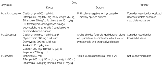
Historically, therapy for MAC lung disease, until the introduction of macrolides, was only partially effective (2, 3). The newer macrolides, clarithromycin and azithromycin, have had a great impact on the treatment of this disease, and recent studies show good in vitro and clinical results (59-61). For the treatment of adults without HIV infection, the American Thoracic Society recommended a daily regimen of clarithromycin or azithromycin, with rifampin or rifabutin in combination with ethambutol, and intermittent streptomycin for the first 2 to 3 months of therapy was recommended for extensive disease (2). We have typically prescribed initial daily dosing with clarithromycin (500 mg twice a day), rifampin (600 mg for body weight >50 kg, 450 mg for body weight ≤50 kg), and ethambutol (25 mg/kg per day for 2 months followed by 15 mg/kg per day). Streptomycin has been used in patients with radiographically extensive or cavitary disease, especially when this is accompanied by strongly positive sputum smears (Table 3) (62). Clarithromycin is likely to be more effective than azithromycin, and rifampin is better tolerated than rifabutin (63). If intolerance evolves (typically taste perversion or a gastrointestinal upset), the dose of clarithromycin should be reduced or azithromycin substituted. Toxicity screening should be performed to detect liver function abnormalities, visual changes, and hearing alterations. Dose adjustments or the discontinuation of one or more antibiotics are frequently required on an individual basis. Generally, therapy should be continued until sputum cultures are consecutively negative for at least one year (typically 18 to 24 months) (2). However, treatment success rates are unsatisfactory. If success is defined as eradication of the organism without relapse over a period of several years after treatment has been discontinued, the reported treatment success rate for macrolide containing regimens is less than 60% (58). Surgery may be useful in cases of localized disease, especially upper lobe cavitary disease, if drug response is poor, if a patient's isolate has become macrolide resistant, or if a patient is drug intolerant (2). We reported the first Korean case of refractory MAC pulmonary disease treated successfully by surgery in 2003 (64).
Table 3.
Treatment protocol for nontuberculous mycobacterial lung diseases (Samsung Medical Center)*
b.i.d., twice daily; t.i.d., three times daily. *Adapted from the reference (2).
Routine susceptibility testing is unnecessary, as all untreated MAC isolates are macrolide susceptible (2, 3). For those who do not respond or who relapse on clarithromycin or azithromycin, susceptibility testing to these agents becomes critical. In one study, sputum converted in only 25% of patients with strains that had resistance or intermediate sensitivity to clarithromycin strains compared with 84% of patients with susceptible strains before treatment (61). No effective drug regimen for clarithromycin-resistant isolates has been established. Alternative second-line agents include fluoroquinolone, cycloserine, ethionamide, clofazimine and prolonged use of streptomycin or amikacin (2, 57).
With regard to the macrolide resistance of MAC infections, we are concerned about the current clinical practice used for bronchiectasis treatment. In practice, macrolides have been frequently used in Korea to treat patients with diffuse bronchiectasis (65), as prolonged treatment with low-lose erythromycin has been reported to be effective in diffuse panbronchiolitis and in some cases of bronchiectasis (66). However, we strongly recommend that the long-term use of low-dose macrolide therapy in patients with diffuse bronchiectasis should only be commenced after carefully considering and evaluating the possibility of NTM pulmonary disease. This rationale is recommended to avoid macrolide monotherapy and the development of macrolide-resistant strains in patients with MAC lung disease. HRCT findings in patients with diffuse panbronchiolitis are similar to those reported for patients with NTM pulmonary disease (34-39). We recently demonstrated that NTM infection is the most common and important cause of, or condition associated with, bilateral bronchiectasis and bronchiolitis in Korean patients (39).
Mycobacterium abscessus LUNG DISEASE
The nomenclature used to describe rapidly growing mycobacteria has changed much and is a source of confusion for clinicians. For instance, over the past 20 yr, M. abscessus, the most common respiratory pathogen among the rapidly growing mycobacteria, has been labeled as M. cheloneii subspecies abscessus, M. chelonae subspecies abscessus, and finally, in 1992, as M. abscessus (67). Species identification is important because therapy differs significantly depending on the rapidly growing mycobacteria species obtained.
M. abscessus accounts for approximately 80% of rapidly growing mycobacterial pulmonary disease isolates, and M. fortuitum accounts for 15% (68). Rarely, M. chelonae and other rapidly growing mycobacteria are identified as respiratory pathogens. Overall, M. abscessus appears to be a more virulent respiratory pathogen than M. fortuitum (2, 67) and a single respiratory isolate of M. abscessus is more likely to indicate significant disease than a single isolate of M. fortuitum. In addition, patients with multiple M. abscessus respiratory isolates almost invariably have significant and progressive M. abscessus pulmonary disease. However, even after obtaining multiple M. fortuitum respiratory isolates, there may still be doubt about the significance of these isolates in the absence of a clear predisposing condition, such as achalasia or recurrent aspiration (67).
M. abscessus pulmonary disease patients tend to occur in middle-aged or older, female nonsmokers (67, 68). Approximately 40% of patients have an underlying medical condition, such as prior mycobacterial infection, lipoid pneumonia, or gastroesophageal disorders with chronic vomiting (67, 68). Patients with the above risk factors often develop disease at a younger age compared with patients without underlying medical conditions (67).
The most frequent patterns seen at chest radiography are multilobar, patchy, reticulonodular or mixed interstitial-alveolar infiltrates. Cavitation occurs in less than 20% of patients. The most common HRCT findings in patients with M. abscessus pulmonary disease are diffuse bronchiectasis, multiple small lung nodules, focal areas of consolidation, and bronchial wall thickening (Fig. 3) (69, 70), which are very similar to those reported for MAC lung disease (38, 39). In most patients with M. abscessus without an underlying disorder, the disease progresses very slowly, and some patients show little radiographic change over a period of years (67, 68).
M. abscessus pulmonary disease clinically and radiographically resembles MAC pulmonary disease, especially the nodular bronchiectatic form of MAC pulmonary disease (67, 70). In addition, comparable percentages of patients with the nodular bronchiectatic form of MAC infection or M. abscessus infection also had M. abscessus or MAC, respectively, in sputum (51, 68). Not surprisingly, it can be difficult to determine whether MAC or M. abscessus is the most important pathogen in these patients. These findings suggest that the risk factor for the two diseases may be similar. Studies by Japanese investigators have demonstrated that the association between specific human leukocyte-associated antigen phenotypes and MAC lung disease could raise the possibility of genetic defects in patients with NTM pulmonary disease (71, 72). Recently, we found a significant association between polymorphisms of NRAMP1 gene and the nodular bronchiectatic form of NTM pulmonary disease caused by MAC or M. abscessus (73). These findings suggested that a genetic susceptibility might play a role in the development of NTM pulmonary disease.
The antimicrobial susceptibility testing results of rapidly growing mycobacteria differ from those of other NTMs. M. abscessus, M. chelonae, and M. fortuitum are resistant to all antituberculosis drugs, including isoniazid, rifampin, ethambutol, and pyrazinamide. Thus, routine susceptibility testing to antituberculous drugs is not recommended (2, 67). However, they are susceptible to a number of traditional antibacterial agents. M. abscessus is usually susceptible to clarithromycin, amikacin, and cefoxitin and is moderately susceptible to imipenem. Thus, all clinically significant isolates should be tested against selected antibacterial agents, such as, amikacin, cefoxitin, imipenem, clarithromycin, ciprofloxacin, doxycycline, and sulfamethoxazole (2, 67).
The optimal therapeutic regimens and treatment duration for M. abscessus pulmonary disease have not been established. Clarithromycin was administered in a small study as monotherapy for M. abscessus pulmonary disease, but was found to be insufficient to produce microbiologic cure (74). The American Thoracic Society recommends treating patients with clarithromycin in combination with high-dose cefoxitin and low-dose amikacin (2). This approach usually requires the placement of long-term indwelling intravenous access, and introduces the risk of attendant morbidity. Short courses (2 to 6 weeks) of amikacin, cefoxitin, and clarithromycin in combination can provide symptomatic, bacteriologic, and radiographic improvements but may not be curative (2, 67). Typically, we have treated patients with progressive M. abscessus pulmonary disease with long-term clarithromycin (1,000 mg/day), ciprofloxacin (1,000 mg/day), and doxycycline (200 mg/day) together with parenteral amikacin (15 mg/kg/day) and cefoxitin (200 mg/kg/day) initially for the first four weeks (Table 3) (75). Symptomatic and radiographic improvements were observed in almost all patients, and negative sputum conversion was achieved and maintained in about 60% of patients for more than 6 months (75). Unfortunately, adverse therapy-related reactions, such as leukopenia, thrombocytopenia, and hepatotoxicity were frequently observed which were attributed to cefoxitin, and these necessitated changing to imipenem in about 60% of patients during initial antibiotic therapy (75). Unfortunately, the long-term consequences of the above study are unclear.
The only reliably curative therapy for M. abscessus pulmonary disease is surgical resection of the involved lung (67, 68). Patients with focal lung disease who can withstand lung resection should be treated surgically after an initial period on antimicrobials to lessen the microbial burden. We also reported on the successful surgical treatment of M. abscessus pulmonary disease (76). Unfortunately, suppressive therapy, including periodic parenteral antibiotic or oral macrolide therapy, may be all that can be realistically administered to control the symptoms and progression of this disease (2, 67). Moreover, because M. abscessus pulmonary disease may show little progression in many immunocompetent patients, a decision to withhold treatment may be made in some patients. For example, in an elderly patient with minimal symptoms and noncavitary disease, the best approach may be to only follow closely for evidence of more rapid progression instead of immediately starting therapy (67).
Mycobacterium kansasii LUNG DISEASE
Unlike other NTMs, M. kansasii is only rarely isolated from natural water supplies (2, 6). Tap water is likely to be the major source for M. kansasii causing human infections and the organism has been isolated from tap water in communities where patients with M. kansasii disease have been identified. Previous studies have demonstrated that M. kansasii cases are significantly more likely to come from urban than rural areas, which supports a possible association between clinical disease and the presence of the organism in potable water supplies (2, 6).
The clinical and radiological features of pulmonary disease caused by M. kansasii usually resemble those of pulmonary tuberculosis (2, 3, 77) and is more likely to occur in older men. Risk factors for M. kansasii infection include chronic obstructive pulmonary disease, pneumoconiosis, previous mycobacterial disease, malignancy, and alcoholism. However, approximately 40% of immunocompetent patients with M. kansasii pulmonary disease were found to have no identifiable predisposing condition (77). Its chest radiographic changes closely resemble those of reactivation pulmonary tuberculosis. Many patients have fibronodular or fibrocavitary upper lobe disease (Fig. 4), whereas the others may have only noncavitary (nodular/bronchiectasis) infiltrates similar to those in patients with MAC or M. abscessus pulmonary disease (77).
Untreated strains of M. kansasii are inhibited by rifampin, isoniazid, ethambutol, ethionamide, streptomycin, clarithromycin, and probably ciprofloxacin at concentrations readily achievable in serum using usual therapeutic doses (2, 77). The concentrations of antituberculous drugs used in susceptibility testing were chosen for their usefulness with M. tuberculosis. Because M. kansasii is less susceptible to these drugs in vitro (but still susceptible to achievable blood levels), some isolates may be reported resistant to isoniazid at 0.2 or 1.0 µg/mL and to streptomycin at 2.0 µg/mL. These isolates are susceptible to slightly higher drug concentrations, so that laboratory reports of resistance to low concentrations of these two drugs have no clinical or therapeutic significance as long as a rifampin-containing regimen is being used. Thus, when clinically indicated, isoniazid and/or streptomycin should be used against M. kansasii regardless of the in vitro susceptibility results (2, 77).
The use of single-drug concentrations of rifampin chosen for susceptibility testing of M. tuberculosis has predictive clinical value for M. kansasii. Then, all initial clinically significant M. kansasii isolates should be tested against rifampin. Moreover, as acquired rifampin resistance may develop during therapy and because prior therapy history may not be determinable, all initial isolates of M. kansasii and those from patients with known prior therapy should be tested against rifampin. Also, testing should be performed when a patient's sputum fails to convert from smear and/or culture positive or when a relapse occurs during therapy (2, 77).
The recommended treatment regimen for patients whose M. kansasii isolates are susceptible to rifampin, is a three-drug combination of isoniazid, rifampin, and ethambutol (2, 77). Medication is usually administered daily for 18 months with a minimum of 12 months culture negativity. Sputum conversion rates for rifampin-containing regimens are almost 100%, and treatment failure and long-term relapse rates are low. Surgery is now considered to have no role in the management of routine pulmonary disease cases (2, 77).
CONCLUSION
It is likely that the incidence of pulmonary disease caused by NTM will continue to rise in Korea, although it should be noted that changes in clinician awareness have increases the numbers of investigations, and the availabilities of laboratory methods, which allow the isolation and identification of previously unnoticed organisms, have all certainly contributed to this trend. However, the incidences of NTM pulmonary disease are also expected to rise because increasing proportions of the population are aging or are subject to some type of immunosuppression. It appears that most pulmonary physicians will be increasingly challenged by this mysterious epidemic in the future.
Pulmonary diseases caused by NTM are highly complex in terms of clinical presentation and management. The important components for establishing a diagnosis of NTM pulmonary disease are: 1) the patient must have a compatible clinical presentation including the time course; 2) the radiographic picture must be consistent with a diagnosis of NTM pulmonary disease; and 3) there must be a clear demonstration that the NTM recovered is there either in sufficient numbers or in pulmonary tissue. It should be emphasized that all three elements must be fulfilled to establish a diagnosis of NTM pulmonary disease.
Treatment varies depending on the infecting species. However, the decisions about the institution of treatment for NTM pulmonary disease are never easy. Treatments require the use of multiple drugs for 18 to 24 months, and thus are expensive. Moreover, they frequently have significant side effects, and are often not curative. Therefore, clinicians should be confident that there is sufficient pathology to warrant prolonged, multidrug treatment regimens. In all of the situations, outcomes can be best optimized only when clinicians, radiologists, and laboratories work cooperatively
Footnotes
This work was supported by the SRC/ERC program of Ministry of Science and Technology/Korea Science and Engineering Foundation (R11-2002-103), Republic of Korea Government.
References
- 1.Hale YM, Pfyffer GE, Salfinger M. Laboratory diagnosis of mycobacterial infections: new tools and lessons learned. Clin Infect Dis. 2001;33:834–846. doi: 10.1086/322607. [DOI] [PubMed] [Google Scholar]
- 2.American Thoracic Society. Diagnosis and treatment of disease caused by nontuberculous mycobacteria. Am J Respir Crit Care Med. 1997;156:S1–S25. doi: 10.1164/ajrccm.156.2.atsstatement. [DOI] [PubMed] [Google Scholar]
- 3.British Thoracic Society. Management of opportunist mycobacterial infections: Joint Tuberculosis Committee Guidelines 1999. Thorax. 2000;55:210–218. doi: 10.1136/thorax.55.3.210. [DOI] [PMC free article] [PubMed] [Google Scholar]
- 4.Koh WJ, Kwon OJ, Lee KS. Nontuberculous mycobacterial pulmonary diseases in immunocompetent patients. Korean J Radiol. 2002;3:145–157. doi: 10.3348/kjr.2002.3.3.145. [DOI] [PMC free article] [PubMed] [Google Scholar]
- 5.Koh WJ, Kwon OJ. Treatment of nontuberculous mycobacterial pulmonary diseases. Tuberc Respir Dis. 2004;56:5–17. [Google Scholar]
- 6.Falkinham JO., 3rd Nontuberculous mycobacteria in the environment. Clin Chest Med. 2002;23:529–551. doi: 10.1016/s0272-5231(02)00014-x. [DOI] [PubMed] [Google Scholar]
- 7.Heifets L. Mycobacterial infections caused by nontuberculous mycobacteria. Semin Respir Crit Care Med. 2004;25:283–295. doi: 10.1055/s-2004-829501. [DOI] [PubMed] [Google Scholar]
- 8.Marras TK, Daley CL. Epidemiology of human pulmonary infection with nontuberculous mycobacteria. Clin Chest Med. 2002;23:553–567. doi: 10.1016/s0272-5231(02)00019-9. [DOI] [PubMed] [Google Scholar]
- 9.Sakatani M. The non-tuberculous mycobacteriosis. Kekkaku. 2005;80:25–30. [PubMed] [Google Scholar]
- 10.Kim SC, Kim SC. A study on unclassified mycobacteria isolated from human sputa. Tuberc Respir Dis. 1970;17:33–42. [Google Scholar]
- 11.Choi CS, Shin SS, Chung SI, Yang YT, Kim SJ, Bai GH. Species identification of mycobacteria of group II isolated from sputa of patients with pulmonary tuberculosis and tuberculosis-like diseases. J Korean Soc Microbiol. 1985;20:25–34. [Google Scholar]
- 12.Chin DI, Woo KJ, Chong Y, Lee SY. Species of Mycobacterium other than Mycobacterium tuberculosis isolated from clinical materials. J Lab Med Qual Assur. 1990;12:267–272. [Google Scholar]
- 13.Koh WJ, Kwon OJ, Yu CM, Jeon KM, Suh GY, Chung MP, Kim HJ, Han SW, Park SY, Lee NY. Recovery rate of nontuberculous mycobacteria from acid-fast-bacilli smear-positive sputum specimens. Tuberc Respir Dis. 2003;54:22–32. [Google Scholar]
- 14.Lee JY, Choi HJ, Lee H, Joung EY, Huh JW, Oh YM, Lee SD, Kim WS, Kim DS, Kim WD, Shim TS. Recovery rate and characteristics of nontuberculous mycobacterial isolates in a university hospital in Korea. Tuberc Respir Dis. 2005;58:385–391. [Google Scholar]
- 15.Korean Academy of Tuberculosis and Respiratory Diseases. Diagnostic standards of pulmonary tuberculosis, 1997. Tuberc Respir Dis. 1997;44:1447–1453. [Google Scholar]
- 16.Korean Center for Disease Control and Prevention. Guidelines for the control of tuberculosis 2005. Seoul: 2005. [Google Scholar]
- 17.Jeon K, Koh WJ, Kwon OJ, Suh GY, Chung MP, Kim H, Lee NY, Park YK, Bai GH. Recovery rate of NTM from AFB smear-positive sputum specimens at a medical centre in South Korea. Int J Tuberc Lung Dis. 2005;9:1046–1051. [PubMed] [Google Scholar]
- 18.Kim SJ, Hong YP, Kim SC, Bai GH, Jin BW, Park CD. A case of pulmonary disease due to Mycobacterium avium-intracelluare complex. Tuberc Respir Dis. 1981;28:121–124. [Google Scholar]
- 19.Kim SJ, Hong YP, Bai GH, Kim SC, Jin BW. Nontuberculous pulmonary infection in two patients with Mycobacterium avium-intracellulare complex and a patient with M. fortuitum. J Korean Soc Microbiol. 1982;17:87–93. [Google Scholar]
- 20.Kim HJ, Oh SH, Lee WY, Kim SJ. Report of a case of pulmonary mycobacteriosis caused by Mycobacterium chelonei subsp. abscessus. Tuberc Respir Dis. 1985;32:54–57. [Google Scholar]
- 21.Korean Academy of Tuberculosis and Respiratory Diseases. National survey of mycobacterial diseases other than tuberculosis in Korea. Tuberc Respir Dis. 1995;42:277–294. [Google Scholar]
- 22.Lew WJ. Clinical manifestations and epidemiology of nontuberculous mycobacterial infection in Korea. Tuberc Respir Dis. 2002;53(Suppl 2):83–87. [Google Scholar]
- 23.Bai GH, Park KS, Kim SJ. Clinically isolated Mycobacteria other than Mycobacterium tuberculosis from 1980 to 1990 in Korea. J Korean Soc Microbiol. 1993;28:1–5. [Google Scholar]
- 24.Lew WJ, Ahn DI, Yoon YJ, Cho JS, Kwon DW, Kim SJ, Hong YP. Clinical experience on mycobacterial diseases other than tuberculosis. Tuberc Respir Dis. 1992;39:425–432. [Google Scholar]
- 25.Koh WJ, Kwon OJ, Ham HS, Suh GY, Chung MP, Kim HJ, Han D, Kim TS, Lee KS, Lee NY, Park EM, Park YK, Bai GH. Clinical significance of nontuberculous mycobacteria isolated from respiratory specimens. Korean J Intern Med. 2003;65:10–21. [Google Scholar]
- 26.Koh WJ, Kwon OJ, Jeon K, Kim TS, Lee KS, Park YK, Bai GH. Clinical significance of nontuberculous mycobacteria isolated from respiratory specimens in Korea. Chest. doi: 10.1378/chest.129.2.341. (in press) [DOI] [PubMed] [Google Scholar]
- 27.Koh WJ, Kwon OJ, Suh GY, Chung MP, Kim H, Lee NY, Kim TS, Lee KS, Park EM, Park YK, Bai GH. A case report of three patients with nontuberculous mycobacterial pulmonary disease caused by Mycobacterium kansasii. Tuberc Respir Dis. 2003;54:459–466. [Google Scholar]
- 28.Yim JJ, Park YK, Lew WJ, Bai GH, Han SK, Shim YS. Mycobacterium kansasii pulmonary diseases in Korea. J Korean Med Sci. 2005;20:957–960. doi: 10.3346/jkms.2005.20.6.957. [DOI] [PMC free article] [PubMed] [Google Scholar]
- 29.Christensen EE, Dietz GW, Ahn CH, Chapman JS, Murry RC, Anderson J, Hurst GA. Pulmonary manifestations of Mycobacterium intracellularis. AJR Am J Roentgenol. 1979;133:59–66. doi: 10.2214/ajr.133.1.59. [DOI] [PubMed] [Google Scholar]
- 30.Christensen EE, Dietz GW, Ahn CH, Chapman JS, Murry RC, Anderson J, Hurst GA. Initial roentgenographic manifestations of pulmonary Mycobacterium tuberculosis, M. kansasii, and M. intracellularis infections. Chest. 1981;80:132–136. doi: 10.1378/chest.80.2.132. [DOI] [PubMed] [Google Scholar]
- 31.Ahn CH, McLarty JW, Ahn SS, Ahn SI, Hurst GA. Diagnostic criteria for pulmonary disease caused by Mycobacterium kansasii and Mycobacterium intracellulare. Am Rev Respir Dis. 1982;125:388–391. doi: 10.1164/arrd.1982.125.4.388. [DOI] [PubMed] [Google Scholar]
- 32.Miller WT., Jr Spectrum of pulmonary nontuberculous mycobacterial infection. Radiology. 1994;191:343–350. doi: 10.1148/radiology.191.2.8153304. [DOI] [PubMed] [Google Scholar]
- 33.Erasmus JJ, McAdams HP, Farrell MA, Patz EF., Jr Pulmonary nontuberculous mycobacterial infection: radiologic manifestations. Radiographics. 1999;19:1487–1505. doi: 10.1148/radiographics.19.6.g99no101487. [DOI] [PubMed] [Google Scholar]
- 34.Hartman TE, Swensen SJ, Williams DE. Mycobacterium avium-intracellulare complex: evaluation with CT. Radiology. 1993;187:23–26. doi: 10.1148/radiology.187.1.8451419. [DOI] [PubMed] [Google Scholar]
- 35.Swensen SJ, Hartman TE, Williams DE. Computed tomographic diagnosis of Mycobacterium avium-intracellulare complex in patients with bronchiectasis. Chest. 1994;105:49–52. doi: 10.1378/chest.105.1.49. [DOI] [PubMed] [Google Scholar]
- 36.Lynch DA, Simone PM, Fox MA, Bucher BL, Heinig MJ. CT features of pulmonary Mycobacterium avium complex infection. J Comput Assist Tomogr. 1995;19:353–360. doi: 10.1097/00004728-199505000-00003. [DOI] [PubMed] [Google Scholar]
- 37.Primack SL, Logan PM, Hartman TE, Lee KS, Muller NL. Pulmonary tuberculosis and Mycobacterium avium-intracellulare: a comparison of CT findings. Radiology. 1995;194:413–417. doi: 10.1148/radiology.194.2.7824720. [DOI] [PubMed] [Google Scholar]
- 38.Jeong YJ, Lee KS, Koh WJ, Han J, Kim TS, Kwon OJ. Nontuberculous mycobacterial pulmonary infection in immunocompetent patients: comparison of thin-section CT and histopathologic findings. Radiology. 2004;231:880–886. doi: 10.1148/radiol.2313030833. [DOI] [PubMed] [Google Scholar]
- 39.Koh WJ, Lee KS, Kwon OJ, Jeong YJ, Kwak SH, Kim TS. Bilateral bronchiectasis and bronchiolitis at thin-section CT: diagnostic implications in nontuberculous mycobacterial pulmonary infection. Radiology. 2005;235:282–288. doi: 10.1148/radiol.2351040371. [DOI] [PubMed] [Google Scholar]
- 40.American Thoracic Society. Diagnostic standards and classification of tuberculosis in adults and children. Am J Respir Crit Care Med. 2000;161:1376–1395. doi: 10.1164/ajrccm.161.4.16141. [DOI] [PubMed] [Google Scholar]
- 41.Yu CM, Koh WJ, Ryu YJ, Jeon K, Choi JC, Kang EH, Suh GY, Chung MP, Kim H, Kwon OJ, Lee JH, Ki CS, Lee NY. Usefulness of PCR test for M. tuberculosis for the differentiation of pulmonary tuberculosis and nontuberculous mycobacterial lung diseases in patients with smear-positive sputum. Tuberc Respir Dis. 2004;57:528–534. [Google Scholar]
- 42.Lee JS, Ji HS, Hong SB, Oh YM, Lim CM, Lee SD, Koh Y, Kim WS, Kim DS, Kim WD, Shim TS. Clinical utility of polymerase chain reaction for the differentiation of nontuberculous mycobacteria in patients with acid-fast bacilli smear-positive specimens. Tuberc Respir Dis. 2005;58:452–458. [Google Scholar]
- 43.Kim MN, Lee SH, Yang SE, Pai CH. Mycobacterial testing in hospital laboratories in Korea: results of a survey of 40 university or tertiary-care hospitals. Korean J Clin Pathol. 1999;19:86–91. [Google Scholar]
- 44.Chang C, Park TS, Kim MN, Lee NY, Lee HJ, Suh JT. Survey on changes in mycobacterial testing practices in Korean laboratories. Korean J Clin Microbiol. 2001;4:108–114. [Google Scholar]
- 45.Lee H, Park HJ, Cho SN, Bai GH, Kim SJ. Species identification of mycobacteria by PCR-restriction fragment length polymorphism of the rpoB gene. J Clin Microbiol. 2000;38:2966–2971. doi: 10.1128/jcm.38.8.2966-2971.2000. [DOI] [PMC free article] [PubMed] [Google Scholar]
- 46.Kim H, Kim SH, Shim TS, Kim MN, Bai GH, Park YG, Lee SH, Cha CY, Kook YH, Kim BJ. PCR restriction fragment length polymorphism analysis (PRA)-algorithm targeting 644 bp Heat Shock Protein 65 (hsp65) gene for differentiation of Mycobacterium spp. J Microbiol Methods. 2005;62:199–209. doi: 10.1016/j.mimet.2005.02.010. [DOI] [PubMed] [Google Scholar]
- 47.Koh WJ, Kwon OJ, Kang EH, Jeon IS, Pyun YJ, Ham HS, Suh GY, Chung MP, Kim HJ, Han DH, Kim TS, Lee KS. Clinical characteristics of the patients with Mycobacterium avium complex pulmonary disease. Tuberc Respir Dis. 2003;54:33–44. [Google Scholar]
- 48.Koh WJ, Kwon OJ, Kang EH, Jeon IS, Pyun YJ, Ham HS, Suh GY, Chung MP, Kim HJ, Han DH, Kim TS, Lee KS. Clinical and radiographic characteristics of 12 patients with Mycobacterium abscessus pulmonary disease. Tuberc Respir Dis. 2003;54:45–56. [Google Scholar]
- 49.Prince DS, Peterson DD, Steiner RM, Gottlieb JE, Scott R, Israel HL, Figueroa WG, Fish JE. Infection with Mycobacterium avium complex in patients without predisposing conditions. N Engl J Med. 1989;321:863–868. doi: 10.1056/NEJM198909283211304. [DOI] [PubMed] [Google Scholar]
- 50.Reich JM, Johnson RE. Mycobacterium avium complex pulmonary disease presenting as an isolated lingular or middle lobe pattern. The Lady Windermere syndrome. Chest. 1992;101:1605–1609. doi: 10.1378/chest.101.6.1605. [DOI] [PubMed] [Google Scholar]
- 51.Wallace RJ, Jr, Zhang Y, Brown BA, Dawson D, Murphy DT, Wilson R, Griffith DE. Polyclonal Mycobacterium avium complex infections in patients with nodular bronchiectasis. Am J Respir Crit Care Med. 1998;158:1235–1244. doi: 10.1164/ajrccm.158.4.9712098. [DOI] [PubMed] [Google Scholar]
- 52.Huang JH, Kao PN, Adi V, Ruoss SJ. Mycobacterium avium-intracellulare pulmonary infection in HIV-negative patients without preexisting lung disease: diagnostic and management limitations. Chest. 1999;115:1033–1040. doi: 10.1378/chest.115.4.1033. [DOI] [PubMed] [Google Scholar]
- 53.Wallace RJ., Jr Mycobacterium avium complex lung disease and women. Now an equal opportunity disease. Chest. 1994;105:6–7. doi: 10.1378/chest.105.1.6. [DOI] [PubMed] [Google Scholar]
- 54.Tanaka E, Amitani R, Niimi A, Suzuki K, Murayama T, Kuze F. Yield of computed tomography and bronchoscopy for the diagnosis of Mycobacterium avium complex pulmonary disease. Am J Respir Crit Care Med. 1997;155:2041–2046. doi: 10.1164/ajrccm.155.6.9196113. [DOI] [PubMed] [Google Scholar]
- 55.Jeon K, Koh WJ, Kwon OJ, Kang EH, Suh GY, Chung MP, Kim H, Kim TS, Lee KS, Lee NY, Han J. Usefulness of bronchoscopy for the diagnosis of nontuberculous mycobacterial pulmonary disease. Tuberc Respir Dis. 2004;57:242–249. [Google Scholar]
- 56.Sakatani M. Nontuberculous mycobacteriosis; the present status of epidemiology and clinical studies. Kekkaku. 1999;74:377–384. [PubMed] [Google Scholar]
- 57.Iseman MD. Medical management of pulmonary disease caused by Mycobacterium avium complex. Clin Chest Med. 2002;23:633–641. doi: 10.1016/s0272-5231(02)00023-0. [DOI] [PubMed] [Google Scholar]
- 58.Field SK, Fisher D, Cowie RL. Mycobacterium avium complex pulmonary disease in patients without HIV infection. Chest. 2004;126:566–581. doi: 10.1378/chest.126.2.566. [DOI] [PubMed] [Google Scholar]
- 59.Wallace RJ, Jr, Brown BA, Griffith DE, Girard WM, Murphy DT, Onyi GO, Steingrube VA, Mazurek GH. Initial clarithromycin monotherapy for Mycobacterium avium-intracellulare complex lung disease. Am J Respir Crit Care Med. 1994;149:1335–1341. doi: 10.1164/ajrccm.149.5.8173775. [DOI] [PubMed] [Google Scholar]
- 60.Wallace RJ, Jr, Brown BA, Griffith DE, Girard WM, Murphy DT. Clarithromycin regimens for pulmonary Mycobacterium avium complex. The first 50 patients. Am J Respir Crit Care Med. 1996;153:1766–1772. doi: 10.1164/ajrccm.153.6.8665032. [DOI] [PubMed] [Google Scholar]
- 61.Tanaka E, Kimoto T, Tsuyuguchi K, Watanabe I, Matsumoto H, Niimi A, Suzuki K, Murayama T, Amitani R, Kuze F. Effect of clarithromycin regimen for Mycobacterium avium complex pulmonary disease. Am J Respir Crit Care Med. 1999;160:866–872. doi: 10.1164/ajrccm.160.3.9811086. [DOI] [PubMed] [Google Scholar]
- 62.Koh WJ, Kwon OJ, Kang EH, Suh GY, Chung MP, Kim H, Chung MJ, Kim TS, Lee KS, Lee NY, Park YK, Bai GH. Treatment of Mycobacterium avium complex (MAC) pulmonary disease. Tuberc Respir Dis. 2004;57:234–241. [Google Scholar]
- 63.Griffith DE, Brown BA, Cegielski P, Murphy DT, Wallace RJ., Jr Early results (at 6 months) with intermittent clarithromycin-including regimens for lung disease due to Mycobacterium avium complex. Clin Infect Dis. 2000;30:288–292. doi: 10.1086/313644. [DOI] [PubMed] [Google Scholar]
- 64.Koh WJ, Kwon OJ, Kang EH, Suh GY, Chung MP, Kim HK, Kim K, Lee NY, Han J, Kim TS, Lee KS. Successful pulmonary resection combined with chemotherapy for the treatment of Mycobacterium avium pulmonary disease: a case report. Tuberc Respir Dis. 2003;54:621–627. [Google Scholar]
- 65.Kim YW, Oh YM, Chung MP, Yoo CG, Han SK, Shim YS, Kim KY, Han YC. The effect of low-dose long-term erythromycin on bronchiectasis. Tuberc Respir Dis. 1993;40:390–394. [Google Scholar]
- 66.Tsang KW, Tipoe GL. Bronchiectasis: not an orphan disease in the East. Int J Tuberc Lung Dis. 2004;8:691–702. [PubMed] [Google Scholar]
- 67.Daley CL, Griffith DE. Pulmonary disease caused by rapidly growing mycobacteria. Clin Chest Med. 2002;23:623–632. doi: 10.1016/s0272-5231(02)00021-7. [DOI] [PubMed] [Google Scholar]
- 68.Griffith DE, Girard WM, Wallace RJ., Jr Clinical features of pulmonary disease caused by rapidly growing mycobacteria. An analysis of 154 patients. Am Rev Respir Dis. 1993;147:1271–1278. doi: 10.1164/ajrccm/147.5.1271. [DOI] [PubMed] [Google Scholar]
- 69.Han D, Lee KS, Koh WJ, Yi CA, Kim TS, Kwon OJ. Radiographic and CT findings of nontuberculous mycobacterial pulmonary infection caused by Mycobacterium abscessus. AJR Am J Roentgenol. 2003;181:513–517. doi: 10.2214/ajr.181.2.1810513. [DOI] [PubMed] [Google Scholar]
- 70.Chung MJ, Lee KS, Koh WJ, Lee JH, Kim TS, Kwon OJ, Kim S. Thin-section CT findings of nontuberculous mycobacterial pulmonary diseases: comparison between Mycobacterium avium-intracellulare complex and Mycobacterium abscessus infection. J Korean Med Sci. 2005;20:777–783. doi: 10.3346/jkms.2005.20.5.777. [DOI] [PMC free article] [PubMed] [Google Scholar]
- 71.Kubo K, Yamazaki Y, Hanaoka M, Nomura H, Fujimoto K, Honda T, Ota M, Kamijou Y. Analysis of HLA antigens in Mycobacterium avium-intracellulare pulmonary infection. Am J Respir Crit Care Med. 2000;161:1368–1371. doi: 10.1164/ajrccm.161.4.9906094. [DOI] [PubMed] [Google Scholar]
- 72.Takahashi M, Ishizaka A, Nakamura H, Kobayashi K, Nakamura M, Namiki M, Sekita T, Okajima S. Specific HLA in pulmonary MAC infection in a Japanese population. Am J Respir Crit Care Med. 2000;162:316–318. doi: 10.1164/ajrccm.162.1.9908071. [DOI] [PubMed] [Google Scholar]
- 73.Koh WJ, Kwon OJ, Kim EJ, Lee KS, Ki CS, Kim JW. NRAMP1 gene polymorphism and susceptibility to nontuberculous mycobacterial lung diseases. Chest. 2005;128:94–101. doi: 10.1378/chest.128.1.94. [DOI] [PubMed] [Google Scholar]
- 74.Griffith BE, Brown BA, Blinkhorn RJ. Treatment of Mycobacterium abscessus lung disease with clarithromycin. Am J Respir Crit Care Med. 1993;147:A917. (abstract) [Google Scholar]
- 75.Koh WJ, Kwon OJ, Suh GY, Chung MP, Kim H, Lee NY, Kim TS, Lee KS, Park YK, Bai GH. Treatment outcome of Mycobacterium abscessus pulmonary disease. Tuberc Respir Dis. 2003;55(Suppl 2):107. (abstract) [Google Scholar]
- 76.Kwon YS, Koh WJ, Kwon OJ, Lee NY, Han J, Lee KS, Shim YM. Successful treatment of Mycobacterium abscessus lung disease with pneumonectomy combined with antibiotic therapy: a case report. Korean J Intern Med. 2005;60:424–427. [Google Scholar]
- 77.Griffith DE. Management of disease due to Mycobacterium kansasii. Clin Chest Med. 2002;23:613–621. doi: 10.1016/s0272-5231(02)00016-3. [DOI] [PubMed] [Google Scholar]


