

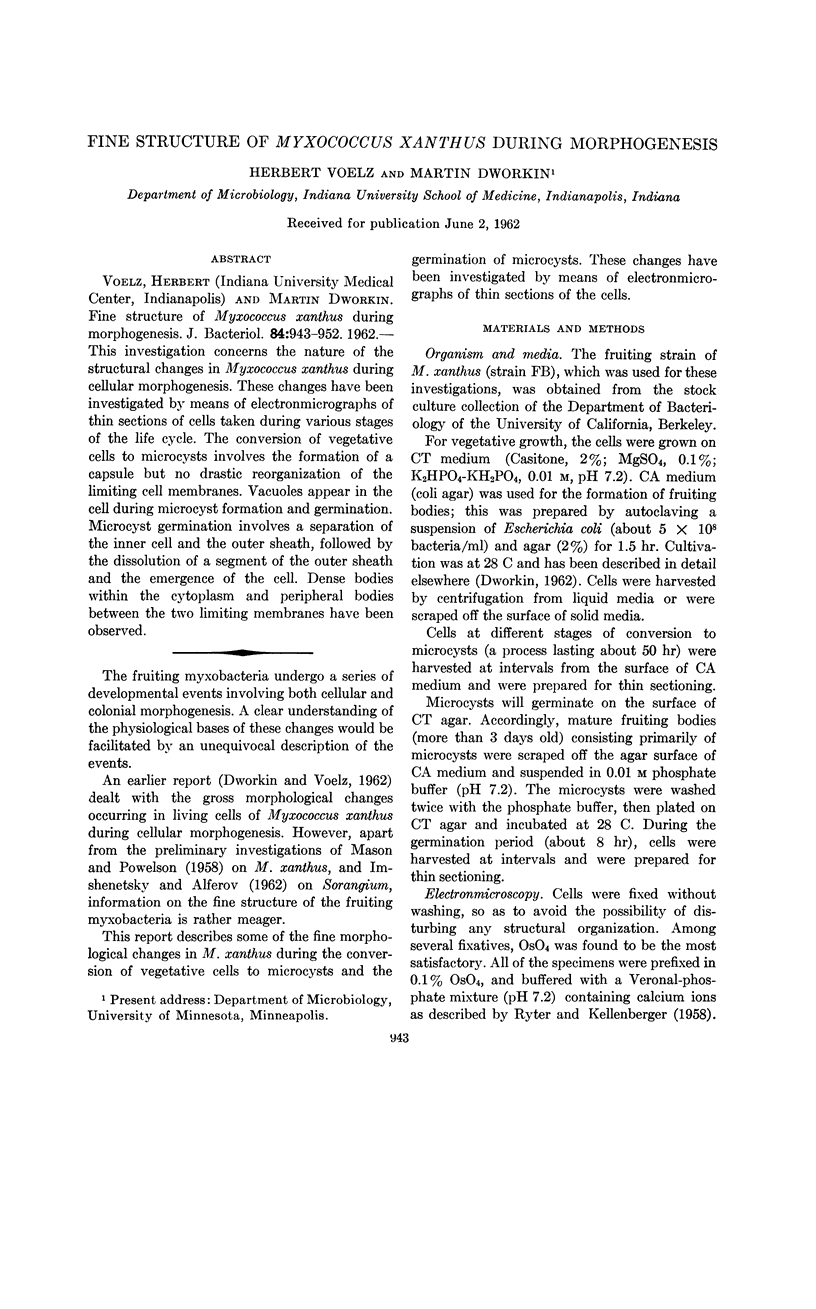
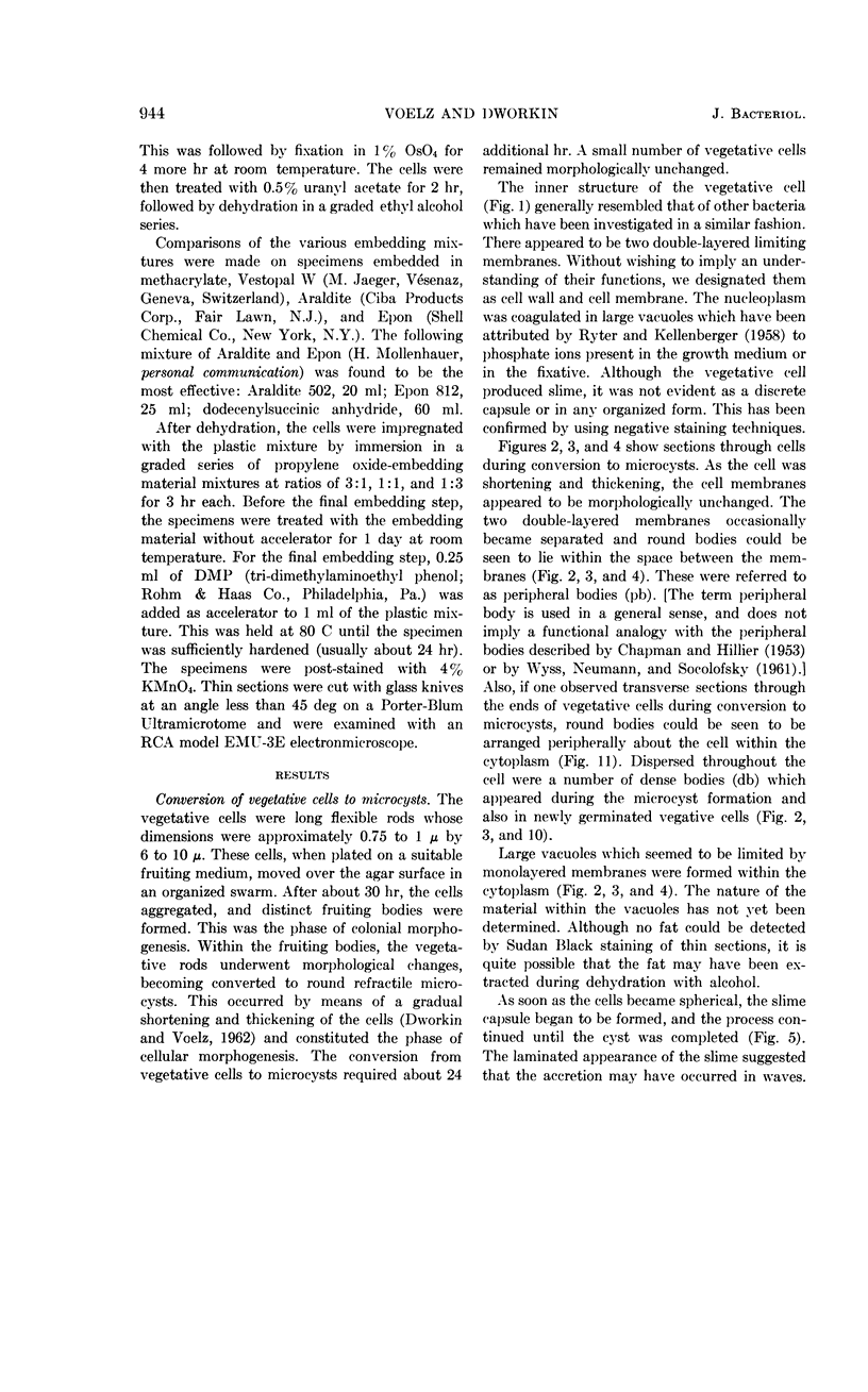
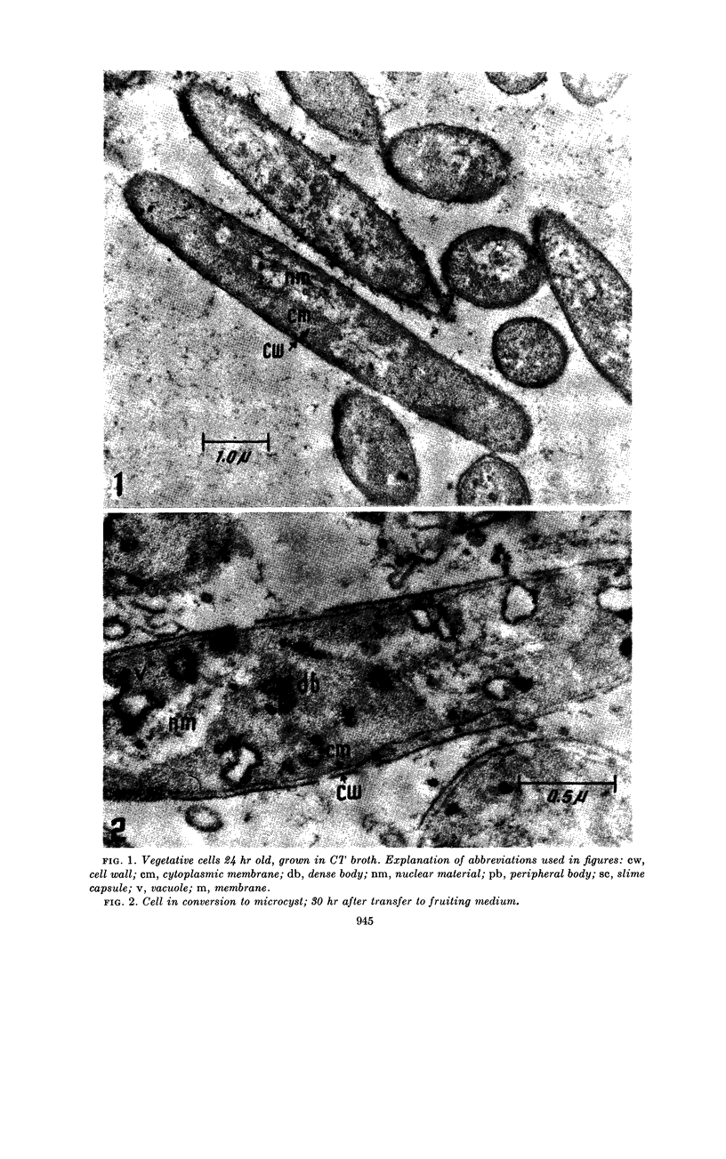
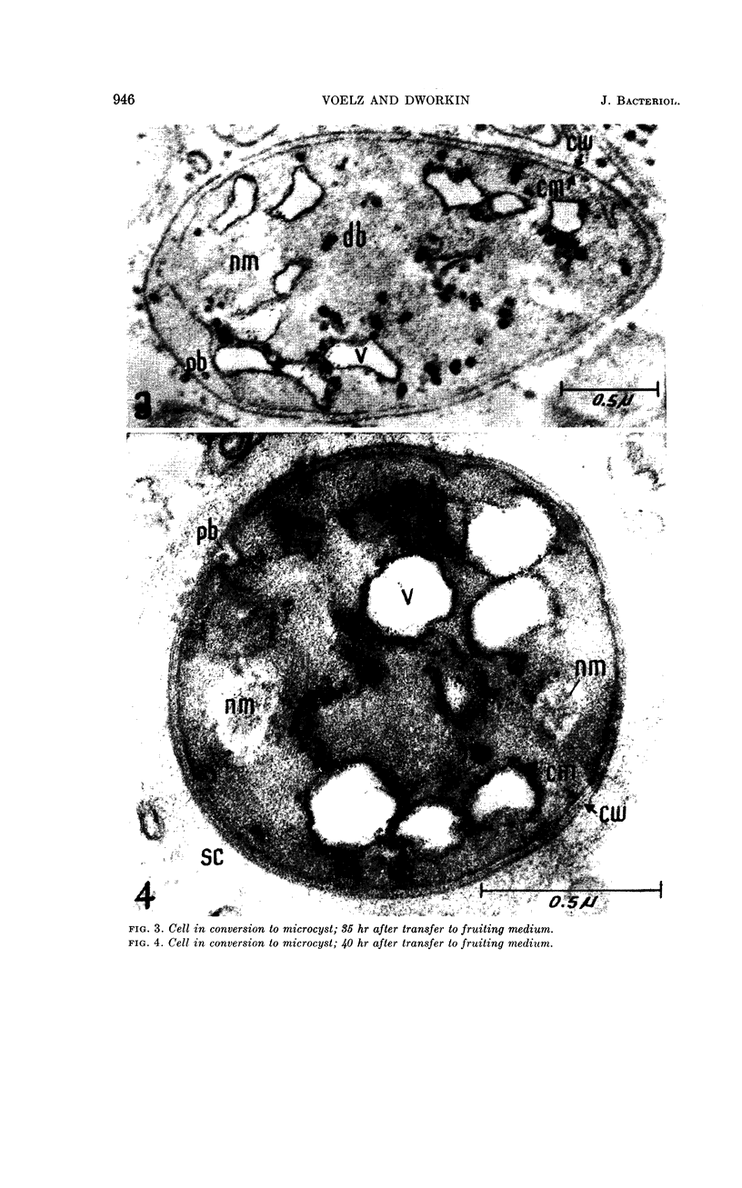
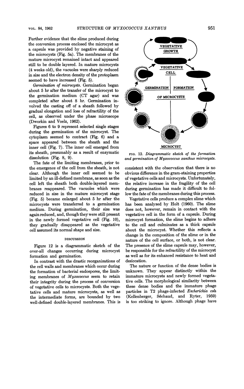
Abstract
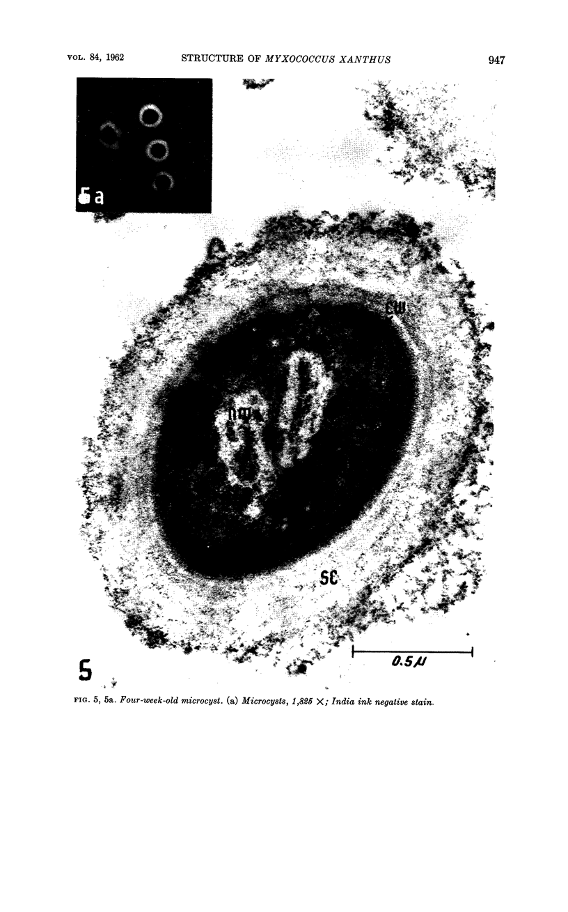
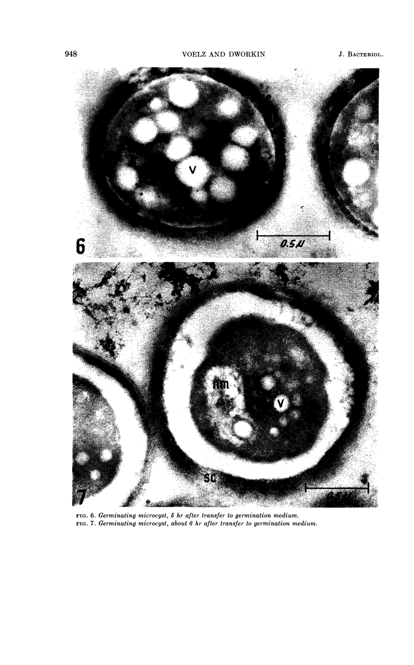
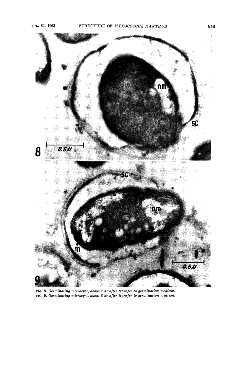
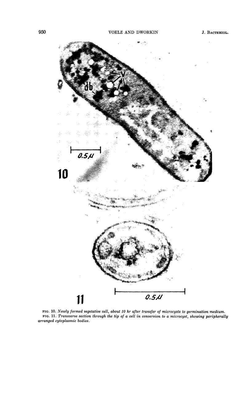
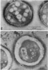
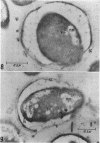
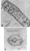
Voelz, Herbert (Indiana University Medical Center, Indianapolis) and Martin Dworkin. Fine structure of Myxococcus xanthus during morphogenesis. J. Bacteriol. 84:943–952. 1962.—This investigation concerns the nature of the structural changes in Myxococcus xanthus during cellular morphogenesis. These changes have been investigated by means of electromicrographs of thin sections of cells taken during various stages of the life cycle. The conversion of vegetative cells to microcysts involves the formation of a capsule but no drastic reorganization of the limiting cell membranes. Vacuoles appear in the cell during microcyst formation and germination. Microcyst germination involves a separation of the inner cell and the outer sheath, followed by the dissolution of a segment of the outer sheath and the emergence of the cell. Dense bodies within the cytoplasm and peripheral bodies between the two limiting membranes have been observed.
Full text
PDF
Images in this article

Selected References
These references are in PubMed. This may not be the complete list of references from this article.
- ANACKER R. L., ORDAL E. J. Study of a bacteriophage infecting the myxobacterium Chondrococcus columnaris. J Bacteriol. 1955 Dec;70(6):738–741. doi: 10.1128/jb.70.6.738-741.1955. [DOI] [PMC free article] [PubMed] [Google Scholar]
- CHAPMAN G. B., HILLIER J. Electron microscopy of ultra-thin sections of bacteria I. Cellular division in Bacillus cereus. J Bacteriol. 1953 Sep;66(3):362–373. doi: 10.1128/jb.66.3.362-373.1953. [DOI] [PMC free article] [PubMed] [Google Scholar]
- DWORKIN M. Nutritional requirements for vegetative growth of Myxococcus xanthus. J Bacteriol. 1962 Aug;84:250–257. doi: 10.1128/jb.84.2.250-257.1962. [DOI] [PMC free article] [PubMed] [Google Scholar]
- DWORKIN M., VOELZ H. The formation and germination of microcysts in Myxococcus xanthus. J Gen Microbiol. 1962 Apr;28:81–85. doi: 10.1099/00221287-28-1-81. [DOI] [PubMed] [Google Scholar]
- KELLENBERGER E., SECHAUD J., RYTER A. Electron microscopical studies of phage multiplication. IV. The establishment of the DNA pool of vegetative phage and the maturation of phage particles. Virology. 1959 Aug;8:478–498. doi: 10.1016/0042-6822(59)90050-9. [DOI] [PubMed] [Google Scholar]
- MASON D. J., POWELSON D. The cell wall of Myxococcus xanthus. Biochim Biophys Acta. 1958 Jul;29(1):1–7. doi: 10.1016/0006-3002(58)90138-0. [DOI] [PubMed] [Google Scholar]
- RYTER A., KELLENBERGER E., BIRCHANDERSEN A., MAALOE O. Etude au microscope électronique de plasmas contenant de l'acide désoxyribonucliéique. I. Les nucléoides des bactéries en croissance active. Z Naturforsch B. 1958 Sep;13B(9):597–605. [PubMed] [Google Scholar]
- Stanier R. Y., Van Niel C. B. The Main Outlines of Bacterial Classification. J Bacteriol. 1941 Oct;42(4):437–466. doi: 10.1128/jb.42.4.437-466.1941. [DOI] [PMC free article] [PubMed] [Google Scholar]
- WYSS O., NEUMNN M. G., SOCOLOFSKY M. D. Development and germination of the Azotobacter cyst. J Biophys Biochem Cytol. 1961 Aug;10:555–565. doi: 10.1083/jcb.10.4.555. [DOI] [PMC free article] [PubMed] [Google Scholar]