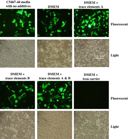
Fig. 2.
An iron supplement in Medium A inhibits PEI-mediated transfection. CHO DXB11 cells were plated onto a six well plate in DMEM with 10% FBS. The next day, the medium in one of the wells was replaced with medium A with no supplements, and the remaining wells contained DMEM and 10% FBS with the indicated supplement. The cells were transfected with the vector pEGFP-N1 with PEI as detailed in “Materials and methods”. After 48 h, the cells were examined by fluorescent microscopy to determine expression of GFP. The experiment was performed three times with similar results

