Abstract
Age-related hearing loss (AHL), known as presbycusis, is a universal feature of mammalian aging and is the most common sensory disorder in the elderly population. The molecular mechanisms underlying AHL are unknown, and currently there is no treatment for the disorder. Here we report that C57BL/6J mice with a deletion of the mitochondrial pro-apoptotic gene Bak exhibit reduced age-related apoptotic cell death of spiral ganglion neurons and hair cells in the cochlea, and prevention of AHL. Oxidative stress induces Bak expression in primary cochlear cells, and Bak deficiency prevents apoptotic cell death. Furthermore, a mitochondrially targeted catalase transgene suppresses Bak expression in the cochlea, reduces cochlear cell death, and prevents AHL. Oral supplementation with the mitochondrial antioxidants α-lipoic acid and coenzyme Q10 also suppresses Bak expression in the cochlea, reduces cochlear cell death, and prevents AHL. Thus, induction of a Bak-dependent mitochondrial apoptosis program in response to oxidative stress is a key mechanism of AHL in C57BL/6J mice.
Keywords: aging, antioxidant, cochlea, oxidative stress, presbycusis
Age-related hearing loss (AHL), also known as presbycusis, is characterized by an age-dependent decline of auditory function associated with loss of sensory hair cells, spiral ganglion (SG) neurons, and stria vascularis cells in the cochlea of the inner ear (1, 2). Hair cells and SG neurons do not regenerate in mammals, and loss of these long-lived cochlear cells leads to permanent hearing impairment. AHL affects more than 40% of people greater than 65 years of age in the United States (1, 2) and is projected to afflict more than 28 million Americans by 2030 (1, 3). Because of its high prevalence, AHL is a major social and health problem.
Most inbred mouse strains display at least some degree of AHL, and the age of onset of AHL varies from 3 months in DBA/2J mice to older than 20 months in CBA/CaJ mice (4). The C57BL/6J mouse strain, which is widely used for aging research, displays the classic pattern of AHL by 12 to 15 months of age (5–7). The onset of AHL in this strain begins in the high-frequency region and spreads toward the low frequencies with age; these functional deficits are accompanied by the loss of hair cells and neurons that begins in the base and spread toward the apex of the cochlea with age (5, 7). Strains susceptible to early-onset AHL, including C57BL/6J, are known to carry a specific mutation in the cadherin 23 gene (Cdh23), which encodes a component of the hair-cell tip link (5, 8, 9).
The mitochondrial theory of aging postulates that reactive oxygen species (ROS) generated inside mitochondria damage key mitochondrial components, including mtDNA and respiratory chain complex proteins (10). Such damage accumulates with time and ultimately leads to permanent age-related mitochondrial dysfunction, which in turn contributes to the aging phenotypes (10, 11). In support of the relevance of this hypothesis to AHL, hearing loss is a common symptom in individuals with inherited mtDNA mutations (12–14), and patients with AHL have a significant load of acquired mtDNA mutations in their auditory tissues (15). Moreover, accumulation of mtDNA mutations plays a causal role in AHL in mice (16). Therefore, mitochondrial damage and dysfunction are thought to play a central role in AHL.
We have previously reported that caloric restriction (CR), which retards several aspects of the aging process in multiple species (17), slows the progression of AHL in C57BL/6J mice, reduces the levels of apoptosis in the cochlea, and reduces the levels of the mitochondrial proapoptotic Bcl-2 family member Bak (6). Apoptosis may play a key role in the age-related decline of physiological function in multiple organs (18), including aging in the cochlea (6, 19). CR attenuates the age-related induction of apoptotic factors in skeletal muscle (20), and may prevent stress-induced apoptotic cell death through the induction of the deacetylase SIRT1 (21). Apoptosis can occur through 2 major pathways: the intrinsic pathway, also known as the mitochondrial pathway, which is initiated when the outer mitochondrial membrane loses its integrity; or the extrinsic pathway, which is initiated through ligand binding to cell surface receptors (18, 22). In mammals, mitochondria play a major role in apoptosis that is regulated by Bcl-2 family members (18). Of the Bcl-2 family members, the proapoptotic proteins Bak and Bax play a central role in promoting mitochondrial-mediated apoptosis (18, 22). These Bcl-2 proteins promote permeabilization of the outer mitochondrial membrane, leading to caspase activation and cell death (18). We therefore tested whether the cell-intrinsic, mitochondrial pathway of apoptosis contributes to AHL in C57BL/6J mice.
Results
Bak Deficiency Reduces Apoptotic Cochlear Cell Death and Delays the Onset of AHL.
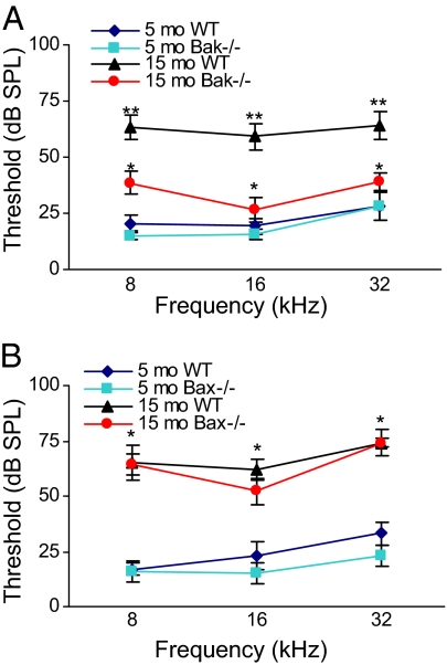
To investigate whether the proapoptotic Bak and/or Bax proteins contribute to the onset of AHL, we conducted hearing tests in 5- and 15-month old C57BL/6J Bak−/− and WT mice. The auditory brainstem response (ABR), an objective electrophysiological test of hearing function, was used to monitor the progression of AHL (4). ABR hearing thresholds from middle-aged Bak−/− mice were significantly lower than those of age-matched WT mice at all frequencies tested, but were not significantly different from those of young WT mice at the middle and high frequencies (Fig. 1A), indicating that Bak is required for the development of AHL. There were no differences in body weight between middle-aged WT and Bak−/− groups [supporting information (SI) Fig. S1A]. Because Bax and Bak are thought to have redundant functions in the control of mitochondrial apoptosis (22), we also tested the role of Bax in AHL. Surprisingly, no significant differences were observed in ABR thresholds between middle-aged WT and Bax−/− mice (Fig. 1B).
Fig. 1.
Bak deficiency delays the onset of AHL, whereas Bax deficiency does not. (A) ABR hearing thresholds were measured from Bak−/− and WT mice at 5 and 15 months of age (n = 10). (B) ABR hearing thresholds were measured from Bax−/− and WT mice at 5 and 15 months of age (n = 10). *Significantly different from 15-month-old WT mice (P < 0.05), **Significantly different from 5-month-old WT mice (P < 0.05). Error bars represent SEM.
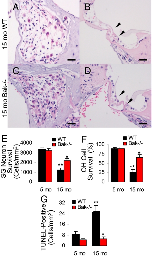
Decreased SG neuron density and hair cell numbers are hallmarks of AHL (5). The hair cells that transduce sound relay their electrical response postsynaptically to SG neurons, which in turn transmit the neural signal through the auditory neuraxis to the auditory cortex, where the information is interpreted (23). In agreement with the ABR results, middle-aged Bak−/− mice displayed only minor loss of SG neurons (Fig. 2C) and hair cells (Fig. 2D). Cell counting also demonstrated that Bak deficiency increased SG neuron survival (Fig. 2E and Fig. S2A) and outer hair (OH) cell survival (Fig. 2F and Fig. S3A), whereas Bax deficiency did not increase SG neuron survival (Fig. S2B). There were no differences in SG neuron and hair cell survival between and young WT and Bak−/− mice (Fig. 2E and Fig. S2A; Fig. 2F and Fig. S3 A and B). To confirm that age-related cochlear cell death was apoptotic, we conducted TUNEL staining to measure nuclear DNA fragmentation, a key feature of apoptosis (16, 24), in 5- and 15-month-old Bak−/− and WT mice. Consistent with the SG neuron and OH cell counts, aging resulted in increased levels of TUNEL-positive cells in the WT cochlea, whereas levels of TUNEL-positive cells in the Bak−/− cochlea did not increase with age (Fig. 2G). Moreover, no significant difference was observed in levels of TUNEL-positive cells between young WT and middle-aged Bak−/− cochlea (Fig. 2G). Collectively, our results indicate that Bak is required for the development of AHL through the induction of apoptosis in mice.
Fig. 2.
Bak deficiency reduces cochlear pathology. SG neurons (A and C) and hair cells (B and D) in the basal cochlear regions from 15-month-old WT and Bak−/− mice. Arrows indicate the hair cell regions. (Scale bar, 200 μm.) (E) SG neuron survival (i.e., SG neuron density) of basal cochlear regions was measured from WT and Bak−/− mice at 5 and 15 months of age (n = 5). (F) OH cell survival (%) of basal cochlear regions was measured from WT and Bak−/− mice at 5 and 15 months of age (n = 5). (G) TUNEL-positive cells were counted in the cochlea of WT and Bak−/− mice at 5 and 15 months of age (n = 5). **Significantly different from 15-month-old WT mice (P < 0.05), **Significantly different from 5-month-old WT mice (P < 0.05). Error bars represent SEM.
Oxidative Stress Triggers Bak Expression in Primary Cochlear Cells and Bak Deficiency Reduces Apoptotic Cell Death.
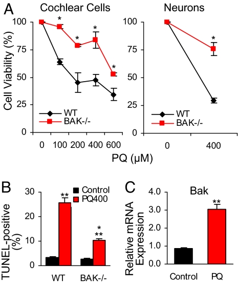
To uncover the pathogenic mechanisms that trigger Bak-mediated apoptosis in the cochlea, we conducted in vitro studies to investigate whether primary cochlear cells lacking Bak are resistant to oxidative stress-induced cell death using paraquat (PQ), which damages neurons and cochlear cells by generating ROS, such as superoxide anions (25, 26). We performed oxidative stress tests, followed by cell viability tests using primary cochlear cells derived from 4-d-old WT and Bak−/− mice. We found that PQ reduced cochlear cell viability in a dose-dependent manner in WT cells (Fig. 3A); however, cochlear cells lacking Bak were more resistant to PQ-induced cell death at all PQ concentrations measured (Fig. 3A). Bak-deficient cochlear primary neurons were also more resistant to PQ-induced cell death than WT neurons (Fig. 3A). These results indicate that Bak promotes cochlear cell death in response to oxidative stress. Next, to confirm that PQ-induced cochlear cell death was apoptotic in nature, we measured TUNEL staining following PQ treatment. Consistent with the in vivo TUNEL test results, PQ-induced cell death was apoptotic and cochlear cells lacking Bak were resistant to PQ-induced apoptosis (Fig. 3B). Finally, to examine whether PQ-induced oxidative stress can trigger the expression of Bak mRNA in cochlear cells, we conducted oxidative stress tests, followed by quantitative RT-PCR (QRT-PCR) to measure relative Bak mRNA level in WT cells. Mean relative Bak mRNA levels in PQ-treated cells were significantly higher than in controls (Fig. 3C). Thus, oxidative stress induces Bak expression in cochlear cells, and cochlear cells lacking Bak are resistant to oxidative stress-induced apoptotic cell death.
Fig. 3.
Cochlear cells lacking Bak are resistant to PQ-induced apoptotic cell death. (A) Cochlear cell viability (%) and neuron viability (%) were measured after PQ treatment of cells derived from 4-d-old WT and Bak−/− mice (n = 3). (B) TUNEL-positive cells were counted after PQ treatment (400 μM) of cells derived from 4-d-old WT and Bak−/− mice (n = 3). (C) Relative cochlear mRNA expression of Bak was measured after PQ treatment (400 μM) of cells derived from 4-d-old WT and Bak−/− mice (n = 3). *Significantly different from WT (P < 0.05). **Significantly different from control (P < 0.05). Error bars represent SEM.
Mitochondrially-Targeted Catalase Suppresses Bak Expression in the Cochlea, Reduces ROS-Induced Cochlear DNA Damage, and Delays the Onset of AHL.
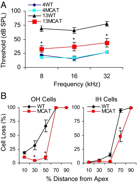
To test the hypothesis that mitochondria-derived ROS play a causal role in AHL, we conducted hearing tests in C57BL/6J transgenic mice that overexpress catalase localized to mitochondria (MCAT) (27). At 13 months of age the mean ABR hearing thresholds of MCAT transgenic mice were significantly lower than those of age-matched WT mice at all the frequencies tested (Fig. 4A). In agreement with the ABR test results, middle-aged MCAT mice displayed only minor age-related loss of SG neurons (Fig. S4E) and hair cells (Fig. S4F). Cell counting also demonstrated that catalase overexpression reduced OH cell and inner hair (IH) cell loss (Fig. 4B). These results indicate that mitochondria-derived ROS play a causal role in AHL
Fig. 4.
Overexpression of catalase targeted to mitochondria delays the onset of AHL and reduces cochlear pathology. (A) ABR hearing thresholds were measured from WT and MCAT mice at 4 and 13 months of age (n = 6–9). (B) OH cell and IH cell loss (%) of cochleae were measured from WT and MCAT mice at 13 months of age (n = 5–6). *Significantly different from WT mice (P < 0.05). Error bars represent SEM.
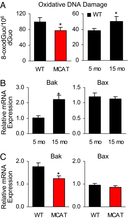
To investigate whether oxidative damage to nucleic acids is blocked by catalase overexpression, we measured oxidative damage to DNA and RNA in the cochleae of WT and MCAT mice at 13 months of age. We found that oxidative DNA damage, but not RNA damage, was reduced in the cochleae of MCAT mice (Fig. 5A and Fig. S4G). We also found that cochlear oxidative DNA damage, but not RNA damage, increased during aging in WT mice (Fig. 5A and Fig. S5A). Cochlear oxidative DNA damage also increased during aging in Bak−/− mice, suggesting that oxidative stress acts upstream of Bak (Fig. S5B). Next, we measured relative mRNA levels of the proapoptotic Bcl-2 family members Bak, Bax, Bid, and Bim, and anti-apoptotic Bcl-2 (18) in the cochleae of WT mice at 5 and 15 months of age; the mean relative mRNA expression of Bak increased with age (Fig. 5B). No significant age-related changes were observed in relative mRNA levels of Bax, Bid, and Bim with aging (Fig. 5B and Fig. S5C). Interestingly, the mean relative mRNA expression of Bcl-2 decreased with age (Fig. S5C). Moreover, we found that the mean relative mRNA expression of Bak (but not Bax) in the cochleae of MCAT transgenic mice was significantly lower than that of WT mice (Fig. 5C). Together, these results provide compelling evidence that enhancing mitochondrial antioxidant defenses reduces oxidative DNA damage, Bak expression, and cochlear cell death, and also delays the onset of AHL in mice.
Fig. 5.
Enhancing antioxidant defenses reduce oxidative DNA damage and reduce Bak expression in the cochlea. (A) Oxidative damage to DNA (8-oxodGuo) was measured in the cochleae from WT and MCAT mice at 13 months of age and from WT mice at 5 and 15 months of age (n = 5–10). (B) Relative cochlear mRNA expression of Bak and Bax was measured in the cochleae from WT mice at 5 and 15 months of age (n = 5). (C) Relative cochlear mRNA expression of Bak and Bax was measured in the cochleae from WT and MCAT mice at 13 months of age (n = 5). *Significantly different from control diet or WT mice (P < 0.05). Error bars represent SEM.
Supplementation with Mitochondrial Antioxidants Suppresses Bak Expression in the Cochlea and Delays the Onset of AHL.
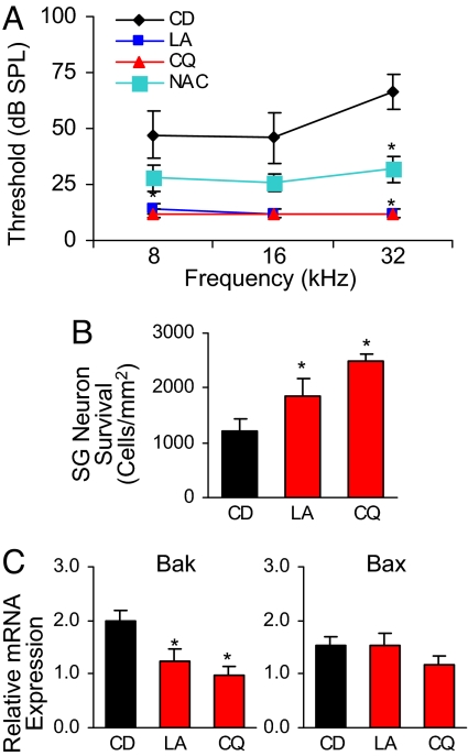
We tested the effects of 17 antioxidant compounds on AHL in C57BL/6J mice (Table S1). Animals were fed the compounds orally under conditions of controlled caloric intake, and the dietary regimen was maintained from 4 months of age until 15 months of age. We found that at 15 months of age the mean ABR hearing thresholds from mice fed α-lipoic acid (LA), coenzyme Q10 (CQ), or N-acetyl-L-cysteine (NAC) were significantly lower at the high frequency than those of control diet-fed mice (Fig. 6A and Fig. S6). In addition, the mean ABR hearing threshold at the low frequency was significantly lower in CQ-treated mice than in controls (Fig. 6A and Fig. S6). There were no differences in body weight among control, LA, CQ, and NAC diet-fed groups (Fig. S7). LA and NAC are thiol compounds that have been shown to reduce mitochondrial ROS production and associated dysfunction (28–30), whereas CQ is an essential component of the mitochondrial electron transfer chain and acts as a mitochondrial antioxidant (31). Interestingly, antioxidants that do not selectively target mitochondria did not delay the onset of AHL at all the frequencies tested (Fig. S6). Consistent with the hearing test results, LA and CQ diet-fed mice displayed only minor loss of SG neurons (Fig. S8 E and F) and hair cells (Fig. S8 H and J). SG neuron counting also revealed that supplementation with LA and CQ increased SG neuron survival (Fig. 6B). Furthermore, we found that the mean relative mRNA expression of Bak, but not Bax, in the cochleae of LA and CQ diet-fed mice was significantly lower than in control diet-fed mice (Fig. 6C). Together, these results provide evidence that enhancing mitochondrial antioxidant defenses through mitochondrial antioxidant supplementation reduces pro-apoptotic Bak expression, reduces cochlear cell death, and delays the onset of AHL in mice.
Fig. 6.
Supplementation of antioxidants that targets mitochondria delays the onset of AHL and reduces cochlear pathology. (A) ABR hearing thresholds were measured at 8, 16, and 32 kHz in 15-month-old mice fed control diet (CD) or diets supplemented with LA, CQ, or NAC compounds (n = 5–7). (B) SG neuron survival (i.e., SG neuron density) of basal cochlear regions was measured from control, LA, and CQ diet-fed mice at 15 months of age (n = 5). (C) Relative cochlear mRNA expression of Bak and Bax was measured in the cochleae from 15-month-old control, LA, and CQ diet-fed mice (n = 5). *Significantly different from control diet or WT mice (P < 0.05). Error bars represent SEM.
Discussion
Previous studies have shown that the apoptotic protein Bak is up-regulated in the human aging brain (32), as well as in the hippocampus of patients with Alzheimer disease (33). Therefore, our finding that mitochondrial oxidative stress triggers Bak-mediated apoptosis in the cochlea may have general implications for aging and cell death in the brain and other sensory organs in mammals. It is well established that the tumor suppressor and nuclear transcription factor p53 is activated by cell stress and DNA damage and that activation of p53 can trigger apoptosis in a wide range of cell types including neurons (34). In response to cell stress, p53 rapidly translocates to mitochondria (35) and directly binds to Bak and induces its oligomerization, leading to cytochrome c release (36, 37). Therefore, we speculate that, in response to increased oxidative DNA damage in the aged cochlea, p53 translocates to mitochondria and activates Bak, leading to Bak-mediated apoptosis and eventually to cochlear cell death. We also note that based on our cochlear cell culture experiments, the age-related increase in oxidative stress is likely to account for the increased Bak gene expression in the aged cochlea, promoting apoptosis.
The C57BL/6J mouse strain is a long-lived strain (mean lifespan of approximately 30 months) and is the most widely used mouse model for the study of aging and age-associated diseases (17). It is also well known that C57BL/6J mice respond to CR with a robust extension of lifespan (17), in agreement with our observation that CR prevents AHL in this strain (6). C57BL/6J and many mouse strains that display AHL carry a specific mutation (Cdh23753A) in the Cdh23 gene, which encodes a component of the hair-cell tip link (5, 8, 9). The mutation is a hypomorphic allele that leads to a disorganized hair bundle (9). Because the Cdh23753A mutation is known to promote early onset of AHL in C57BL/6J mice (4–6, 8, 9), it is possible that hair cells in mice carrying the Cdh23753A mutation are more susceptible to oxidative stress and apoptosis, thereby limiting the general implication of our findings. However, the C57BL/6J strain displays the classic pattern of AHL (4, 5, 7), similar to that reported in humans (1, 38) and in the CBA/J mouse strain that does not possess the Cdh23753A mutation and displays late onset of AHL by 18 months of age (4, 7, 39). In both strains, the onset of AHL begins in the high-frequency region and spreads toward the low frequencies with age, and the loss of hair cells and neurons begins in the base and spreads toward the apex of the cochlea with age (4, 5, 7, 39). Thus, the Cdh23753A allele affects age of onset of AHL, but the basic mechanisms of cochlear aging are likely to be similar in C57BL/6J and CBA/J strains.
Our study also establishes that oxidative DNA damage increases with age in the cochlea of C57BL/6J mice, and that such oxidative damage plays a causal role in AHL through the induction of apoptosis. Consistent with the generality of these findings, oxidative protein damage also increases with age in the cochlea of CBA/J mice (40), and aging is associated with increased expression of apoptosis-associated genes in the cochlea of the CBA/CaJ mouse strain that also does not carry the Cdh23753A mutation (41). Importantly, CR slows the progression of AHL in both the C57BL/6J and CBA/J strains (6, 42), suggesting common age-related mechanisms of AHL.
CR is thought to slow the aging process by reducing levels of ROS in a wide variety of cell types (17, 43). Indeed, CR reduces oxidative damage to DNA, protein, and lipids (17, 43). In mice, most of the attenuation of the oxidative damage by CR occurs in post-mitotic tissues such as brain (17, 43). In agreement with these reports, CR reduces levels of mtDNA deletions in the cochlea (44) and delays the onset of AHL in rodents (6, 42, 44). Thus, we propose that CR slows the cochlear aging process by reducing oxidative DNA damage, thereby reducing levels of Bak-mediated apoptosis in the cochlea (6).
It is thought that the proapoptotic Bcl-2 family members Bak and Bax function in concert as a gateway to the mitochondrial-mediated cell death pathway, and that they are redundant in function in programmed apoptosis that is required for proper development (18, 22). However, recent studies suggest that Bak acts upstream of Bax in promoting mitochondrial apoptosis (25, 45), and that Bak plays the central role in apoptosis of differentiated neurons (24). Moreover, Bak may play an exclusive role in oxidative stress-mediated apoptosis in the brain (25), and also in mitochondrial fragmentation (45). In the current study, Bak deficiency prevented AHL whereas Bax deficiency did not. Levels of Bak mRNA were reduced in the cochleae of MCAT transgenic mice and LA and CQ treated mice, whereas levels of Bax mRNA were not altered. We also note that aging resulted in increased expression of Bak, but not Bax, in the WT cochlea. Thus, our data provide evidence that the mitochondrial apoptotic function of Bak is not redundant with that of Bax in the adult cochlea. In summary, our findings strongly support the hypothesis that mitochondrial ROS plays a role in limiting the mammalian health span and provide evidence that the induction of a Bak-mediated apoptosis program may be a central mechanism linking ROS, aging, and AHL.
Materials and Methods
An extended description of the materials and methods used in the present study is provided as SI Materials and Methods.
Animals.
Male and female Bak−/−, Bax−/−, and C57BL/6J mice were purchased from Jackson Laboratory. Bak−/− mice have been backcrossed for 5 generations onto the C57BL/6J background whereas Bax−/− mice have been backcrossed for 8 generations onto the C57BL/6J background. C57BL/6J mice were used as controls for the Bak−/− and Bax−/− mouse studies. The mice that overexpress catalase targeted to mitochondria (MCAT) on the C57BL/6J background were a gift from Peter S. Rabinovitch (Seattle, WA). Antioxidants studies were conducted using C57BL/6J mice at the American Association for the Accreditation of Laboratory Animal Care International-approved Animal Research Facility at William S. Middleton Memorial Veterans Affairs Medical Center. All other animal studies were conducted at the American Association for the Accreditation of Laboratory Animal Care International-approved Animal Facility in the Genetics and Biotechnology Center of the University of Wisconsin-Madison. Experiments were performed in accordance with protocols approved by the University of Wisconsin-Madison Institutional Animal Care and Use Committee and the Institutional Animal Care and Use Committee of the William. S. Middleton Memorial Veterans Affairs Hospital (Madison, WI).
ABR Hearing Screening, Histopathology, SG Neuron Counting, Hair Cell Counting, QRT-PCR, and TUNEL Staining.
ABR hearing tests, histopathological analysis, SG neuron counting, hair cell counting, QRT-PCR analysis, and TUNEL staining have been described previously (6, 19, 46–48). After ABR hearing measurements were performed, the same mice were used to conduct histopathological, SG neuron counting, hair cell counting, TUNEL staining, oxidative DNA and RNA damage, or QRT-PCR analyses. For detailed methods, see SI Materials and Methods.
Measurement of RNA and DNA Oxidation Levels Using HPLC and Electrochemical Detection.
Total RNA and DNA oxidation levels of cochleae were analyzed simultaneously using a HPLC/electrochemical detection (ECD) method (49, 50). For detailed methods, see SI Materials and Methods.
Antioxidant Dietary Study.
Starting at 4 months of age, C57BL/6J mice were randomly divided into 18 groups: control diet and 17 antioxidant diet groups. The antioxidant diet groups were fed the same caloric intake as controls, but were supplemented with one of 17 antioxidant compounds (Table S1) from 4 months of age until 15 months of age. For detailed methods, see SI Materials and Methods.
Primary Cochlear Cell Culture, Cell Viability Test, and Neuron Viability Test.
The procedures for preparing primary cochlear cell cultures were similar to those described previously (26, 51). For detailed methods of TUNEL staining, QRT-PCR analysis, cell viability test, and neuron viability test using primary cochlear cells, see SI Materials and Methods.
Statistical Analysis.
All statistical analyses were carried out by one-way ANOVA with post-Tukey multiple comparison tests using the Prism 4.0 statistical analysis program (GraphPad). All tests were 2-sided with statistical significance set at P < 0.05.
Supplementary Material
Acknowledgments.
We thank J. M. Vann and K. Papez for technical assistance, and S. Kinoshita for histological processing. This research was supported by National Institutes of Health grants AG021905 (to T.A.P.), AG189222 (to R.W.), AG17994 (to C.L.), and AG21042 (to C.L.); the National Projects on Protein Structural and Functional Analyses from the Ministry of Education, Culture, Sports, Science, and Technologies of Japan; and Marine Bio Foundation.
Footnotes
Conflict of interest statement: S.S. and T.A.P. have filed a patent covering the general approach of Bak inhibition as a therapeutic approach for age-related hearing loss.
This article is a PNAS Direct Submission.
This article contains supporting information online at www.pnas.org/cgi/content/full/0908786106/DCSupplemental.
References
- 1.Gates GA, Mills JH. Presbycusis. Lancet. 2005;366:1111–1120. doi: 10.1016/S0140-6736(05)67423-5. [DOI] [PubMed] [Google Scholar]
- 2.Yamasoba T, et al. Role of mitochondrial dysfunction and mitochondrial DNA mutations in age-related hearing loss. Hear Res. 2007;226:185–193. doi: 10.1016/j.heares.2006.06.004. [DOI] [PubMed] [Google Scholar]
- 3.Administration of Aging. Aging statistics. 2009. [Accessed July 1, 2009]. Available at http://www.aoa.gov/AoARoot/Aging_Statistics/index.aspx.
- 4.Zheng QY, Johnson KR, Erway LC. Assessment of hearing in 80 inbred strains of mice by ABR threshold analyses. Hear Res. 1999;130:94–107. doi: 10.1016/s0378-5955(99)00003-9. [DOI] [PMC free article] [PubMed] [Google Scholar]
- 5.Keithley EM, Canto C, Zheng QY, Fischel-Ghodsian N, Johnson KR. Age-related hearing loss and the ahl locus in mice. Hear Res. 2004;188:21–28. doi: 10.1016/S0378-5955(03)00365-4. [DOI] [PMC free article] [PubMed] [Google Scholar]
- 6.Someya S, Yamasoba T, Weindruch R, Prolla TA, Tanokura M. Caloric restriction suppresses apoptotic cell death in the mammalian cochlea and leads to prevention of presbycusis. Neurobiol Aging. 2007;28:1613–1622. doi: 10.1016/j.neurobiolaging.2006.06.024. [DOI] [PubMed] [Google Scholar]
- 7.Hunter KP, Willott JF. Aging and the auditory brainstem response in mice with severe or minimal presbycusis. Hear Res. 1987;30:207–218. doi: 10.1016/0378-5955(87)90137-7. [DOI] [PubMed] [Google Scholar]
- 8.Ohlemiller KK. Contributions of mouse models to understanding of age- and noise-related hearing loss. Brain Res. 2006;1091:89–102. doi: 10.1016/j.brainres.2006.03.017. [DOI] [PubMed] [Google Scholar]
- 9.Noben-Trauth K, Zheng QY, Johnson KR. Association of cadherin 23 with polygenic inheritance and genetic modification of sensorineural hearing loss. Nat Genet. 2003;35:21–23. doi: 10.1038/ng1226. [DOI] [PMC free article] [PubMed] [Google Scholar]
- 10.Loeb LA, Wallace DC, Martin. GM. The mitochondrial theory of aging and its relationship to reactive oxygen species damage and somatic mtDNA mutations. Proc Natl Acad Sci USA. 2005;102:18769–18770. doi: 10.1073/pnas.0509776102. [DOI] [PMC free article] [PubMed] [Google Scholar]
- 11.Wallace DC. A mitochondrial paradigm of metabolic and degenerative diseases, aging, and cancer: A dawn for evolutionary medicine. Annu Rev Genet. 2005;39:359–407. doi: 10.1146/annurev.genet.39.110304.095751. [DOI] [PMC free article] [PubMed] [Google Scholar]
- 12.Fischel-Ghodsian N. Mitochondrial deafness. Ear Hear. 2003;24:303–313. doi: 10.1097/01.AUD.0000079802.82344.B5. [DOI] [PubMed] [Google Scholar]
- 13.Chen J, et al. Mutations at position 7445 in the precursor of mitochondrial tRNA (Ser(UCN)) gene in three maternal Chinese pedigrees with sensorineural hearing loss. Mitochondrion. 2008;8:285–292. doi: 10.1016/j.mito.2008.05.002. [DOI] [PubMed] [Google Scholar]
- 14.Van Camp G, Smith RJ. Maternally inherited hearing impairment. Clin Genet. 2000;57:409–414. doi: 10.1034/j.1399-0004.2000.570601.x. [DOI] [PubMed] [Google Scholar]
- 15.Fischel-Ghodsian N, et al. Temporal bone analysis of patients with presbycusis reveals high frequency of mitochondrial mutations. Hear Res. 1997;110:147–154. doi: 10.1016/s0378-5955(97)00077-4. [DOI] [PubMed] [Google Scholar]
- 16.Kujoth GC, et al. Mitochondrial DNA mutations, oxidative stress, and apoptosis in mammalian aging. Science. 2005;309:481–484. doi: 10.1126/science.1112125. [DOI] [PubMed] [Google Scholar]
- 17.Weindruch R, Walford RL. Springfield: Charles C Thomas; 1988. The retardation of aging and disease by dietary restriction. [Google Scholar]
- 18.Youle RJ, Strasser A. The BCL-2 protein family: Opposing activities that mediate cell death. Nat Rev Mol Cell Biol. 2008;9:47–59. doi: 10.1038/nrm2308. [DOI] [PubMed] [Google Scholar]
- 19.Someya S, et al. The role of mtDNA mutations in the pathogenesis of age-related hearing loss in mice carrying a mutator DNA polymerase gamma. Neurobiol Aging. 2008;29:1080–1092. doi: 10.1016/j.neurobiolaging.2007.01.014. [DOI] [PMC free article] [PubMed] [Google Scholar]
- 20.Dirks AJ, Leeuwenburgh C. Aging and lifelong calorie restriction result in adaptations of skeletal muscle apoptosis repressor, apoptosis-inducing factor, X-linked inhibitor of apoptosis, caspase-3, and caspase-12. Free Radic Biol Med. 2004;36:27–39. doi: 10.1016/j.freeradbiomed.2003.10.003. [DOI] [PubMed] [Google Scholar]
- 21.Cohen HY, et al. Calorie restriction promotes mammalian cell survival by inducing the SIRT1 deacetylase. Science. 2004;305:390–392. doi: 10.1126/science.1099196. [DOI] [PubMed] [Google Scholar]
- 22.Lindsten T, et al. The combined functions of proapoptotic Bcl-2 family members bak and bax are essential for normal development of multiple tissues. Mol Cell. 2000;6:1389–1399. doi: 10.1016/s1097-2765(00)00136-2. [DOI] [PMC free article] [PubMed] [Google Scholar]
- 23.Hudspeth AJ. How hearing happens. Neuron. 1997;19:947–950. doi: 10.1016/s0896-6273(00)80385-2. [DOI] [PubMed] [Google Scholar]
- 24.Martin LJ, Liu Z, Pipino J, Chestnut B, Landek MA. Molecular Regulation of DNA Damage-Induced Apoptosis in Neurons of Cerebral Cortex. Cereb Cortex. 2009;19:1273–1293. doi: 10.1093/cercor/bhn167. [DOI] [PMC free article] [PubMed] [Google Scholar]
- 25.Fei Q, McCormack AL, Di Monte DA, Ethell DW. Paraquat neurotoxicity is mediated by a Bak-dependent mechanism. J Biol Chem. 2008;283:3357–3364. doi: 10.1074/jbc.M708451200. [DOI] [PubMed] [Google Scholar]
- 26.Nicotera TM, Ding D, McFadden SL, Salvemini D, Salvi R. Paraquat-induced hair cell damage and protection with the superoxide dismutase mimetic m40403. Audiol Neurootol. 2004;9:353–362. doi: 10.1159/000081284. [DOI] [PubMed] [Google Scholar]
- 27.Schriner SE, et al. Extension of murine life span by overexpression of catalase targeted to mitochondria. Science. 2005;308:1909–1911. doi: 10.1126/science.1106653. [DOI] [PubMed] [Google Scholar]
- 28.Palaniappan AR, Dai A. Mitochondrial ageing and the beneficial role of alpha-lipoic acid. Neurochem Res. 2007;32:1552–1558. doi: 10.1007/s11064-007-9355-4. [DOI] [PubMed] [Google Scholar]
- 29.Banaclocha MM. Therapeutic potential of N-acetylcysteine in age-related mitochondrial neurodegenerative diseases. Med Hypotheses. 2001;56:472–477. doi: 10.1054/mehy.2000.1194. [DOI] [PubMed] [Google Scholar]
- 30.Hart AM, Terenghi G, Kellerth JO, Wiberg M. Sensory neuroprotection, mitochondrial preservation, and therapeutic potential of N-acetyl-cysteine after nerve injury. Neuroscience. 2004;125:91–101. doi: 10.1016/j.neuroscience.2003.12.040. [DOI] [PubMed] [Google Scholar]
- 31.Sohal RS, Forster MJ. Coenzyme Q, oxidative stress and aging. Mitochondrion. 2007;(7 Suppl):S103–111. doi: 10.1016/j.mito.2007.03.006. [DOI] [PMC free article] [PubMed] [Google Scholar]
- 32.Kitamura Y, et al. Alteration of proteins regulating apoptosis, Bcl-2, Bcl-x, Bax, Bak, Bad, ICH-1 and CPP32, in Alzheimer's disease. Brain Res. 1998;780:260–269. doi: 10.1016/s0006-8993(97)01202-x. [DOI] [PubMed] [Google Scholar]
- 33.Obonai T, Mizuguchi M, Takashima S. Developmental and aging changes of Bak expression in the human brain. Brain Res. 1998;783:167–170. doi: 10.1016/s0006-8993(97)01361-9. [DOI] [PubMed] [Google Scholar]
- 34.Culmsee C, Mattson MP. p53 in neuronal apoptosis. Biochem Biophys Res Commun. 2005;331:761–777. doi: 10.1016/j.bbrc.2005.03.149. [DOI] [PubMed] [Google Scholar]
- 35.Erster S, Mihara M, Kim RH, Petrenko O, Moll UM. In vivo mitochondrial p53 translocation triggers a rapid first wave of cell death in response to DNA damage that can precede p53 target gene activation. Mol Cell Biol. 2004;24:6728–6741. doi: 10.1128/MCB.24.15.6728-6741.2004. [DOI] [PMC free article] [PubMed] [Google Scholar]
- 36.Mihara M, et al. p53 has a direct apoptogenic role at the mitochondria. Mol Cell. 2003;11:577–590. doi: 10.1016/s1097-2765(03)00050-9. [DOI] [PubMed] [Google Scholar]
- 37.Leu JI, Dumont P, Hafey M, Murphy ME, George DL. Mitochondrial p53 activates Bak and causes disruption of a Bak-Mcl1 complex. Nat Cell Biol. 2004;6:443–450. doi: 10.1038/ncb1123. [DOI] [PubMed] [Google Scholar]
- 38.Johnson KR, Zheng QY, Erway LC. A major gene affecting age-related hearing loss is common to at least ten inbred strains of mice. Genomics. 2000;70:171–180. doi: 10.1006/geno.2000.6377. [DOI] [PubMed] [Google Scholar]
- 39.Sha SH, et al. Age-related auditory pathology in the CBA/J mouse. Hear Res. 2008;243:87–94. doi: 10.1016/j.heares.2008.06.001. [DOI] [PMC free article] [PubMed] [Google Scholar]
- 40.Jiang H, Talaska AE, Schacht J, Sha SH. Oxidative imbalance in the aging inner ear. Neurobiol Aging. 2007;28:1605–1612. doi: 10.1016/j.neurobiolaging.2006.06.025. [DOI] [PMC free article] [PubMed] [Google Scholar]
- 41.Tadros SF, D'Souza M, Zhu. X, Frisina. RD. Apoptosis-related genes change their expression with age and hearing loss in the mouse cochlea. Apoptosis. 2008;13:1303–1321. doi: 10.1007/s10495-008-0266-x. [DOI] [PMC free article] [PubMed] [Google Scholar]
- 42.Sweet RJ, Price JM, Henry KR. Dietary restriction and presbyacusis: Periods of restriction and auditory threshold losses in the CBA/J mouse. Audiology. 1988;27:305–312. doi: 10.3109/00206098809081601. [DOI] [PubMed] [Google Scholar]
- 43.Weindruch R, Sohal RS. Seminars in medicine of the Beth Israel Deaconess Medical Center. Caloric intake and aging. N Engl J Med. 1997;337:986–994. doi: 10.1056/NEJM199710023371407. [DOI] [PMC free article] [PubMed] [Google Scholar]
- 44.Seidman MD. Effects of dietary restriction and antioxidants on presbyacusis. Laryngoscope. 2000;110:727–738. doi: 10.1097/00005537-200005000-00003. [DOI] [PubMed] [Google Scholar]
- 45.Brooks C, et al. Bak regulates mitochondrial morphology and pathology during apoptosis by interacting with mitofusins. Proc Natl Acad Sci USA. 2007;104:11649–11654. doi: 10.1073/pnas.0703976104. [DOI] [PMC free article] [PubMed] [Google Scholar]
- 46.Yamasoba T. Cochlear damage due to germanium-induced mitochondrial dysfunction in guinea pigs. Neurosci Lett. 2006;395:18–22. doi: 10.1016/j.neulet.2005.10.045. [DOI] [PubMed] [Google Scholar]
- 47.Someya S, Yamasoba T, Prolla TA, Tanokura M. Genes encoding mitochondrial respiratory chain components are profoundly down-regulated with aging in the cochlea of DBA/2J mice. Brain Res. 2007;1182:26–33. doi: 10.1016/j.brainres.2007.08.090. [DOI] [PubMed] [Google Scholar]
- 48.Ding D, Jiang H, Wang P, Salvi R. Cell death after co-administration of cisplatin and ethacrynic acid. Hear Res. 2007;226:129–139. doi: 10.1016/j.heares.2006.07.015. [DOI] [PubMed] [Google Scholar]
- 49.Hofer T, Seo AY, Prudencio M, Leeuwenburgh C. A method to determine RNA and DNA oxidation simultaneously by HPLC-ECD: Greater RNA than DNA oxidation in rat liver after doxorubicin administration. Biol Chem. 2006;387:103–111. doi: 10.1515/BC.2006.014. [DOI] [PubMed] [Google Scholar]
- 50.Xu J, Knutson MD, Carter CS, Leeuwenburgh C. Iron accumulation with age, oxidative stress and functional decline. PLoS One. 2008;3:e2865. doi: 10.1371/journal.pone.0002865. [DOI] [PMC free article] [PubMed] [Google Scholar]
- 51.Cheng AG, et al. Calpain inhibitors protect auditory sensory cells from hypoxia and neurotrophin-withdrawal induced apoptosis. Brain Res. 1999;850:234–243. doi: 10.1016/s0006-8993(99)01983-6. [DOI] [PubMed] [Google Scholar]
Associated Data
This section collects any data citations, data availability statements, or supplementary materials included in this article.


