Abstract
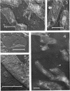
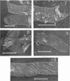
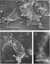
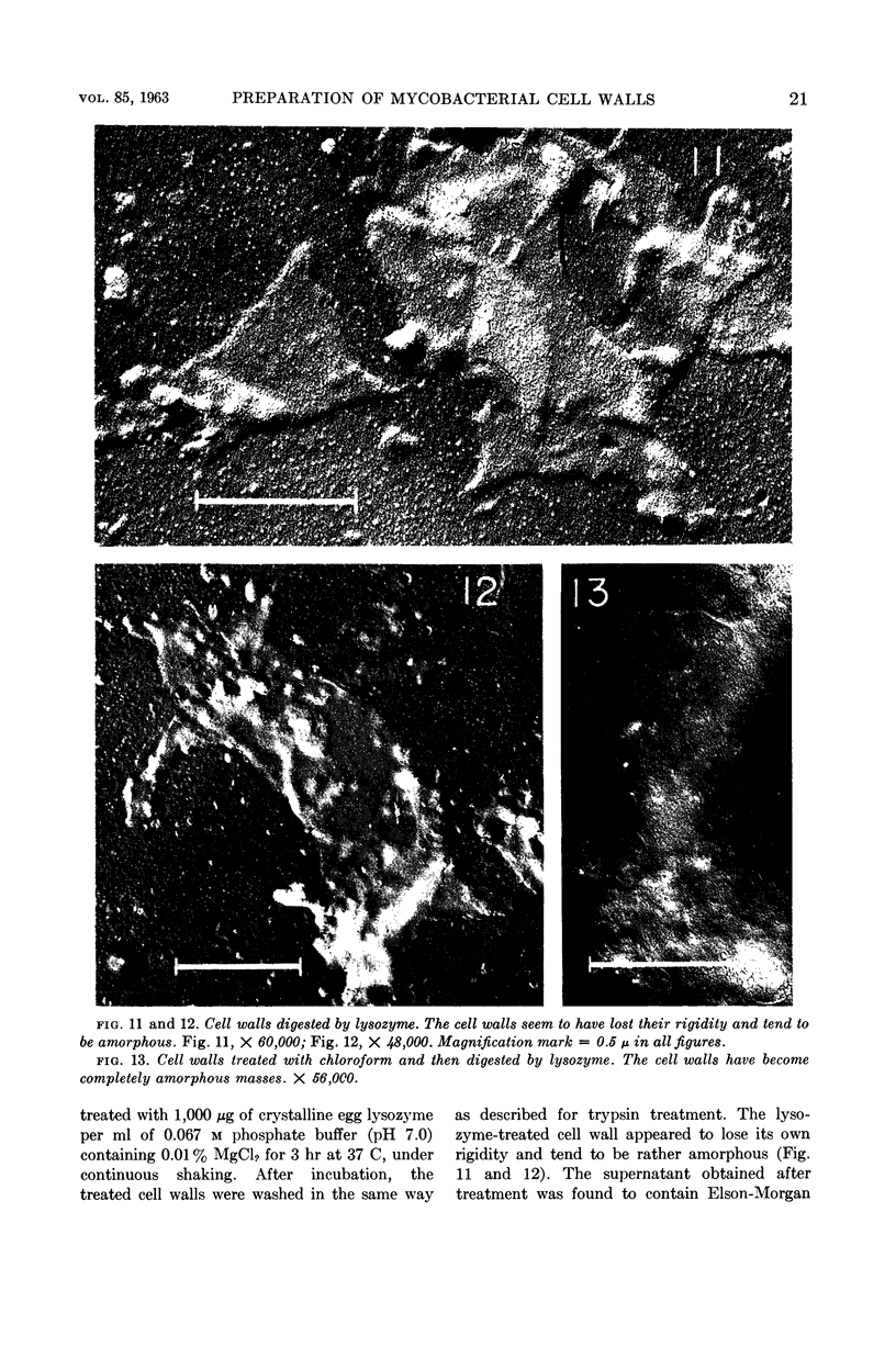
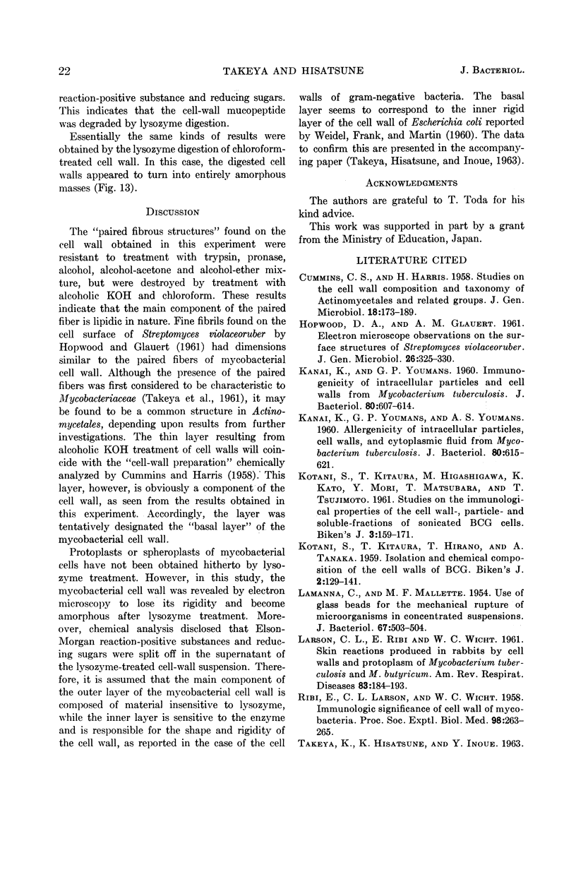
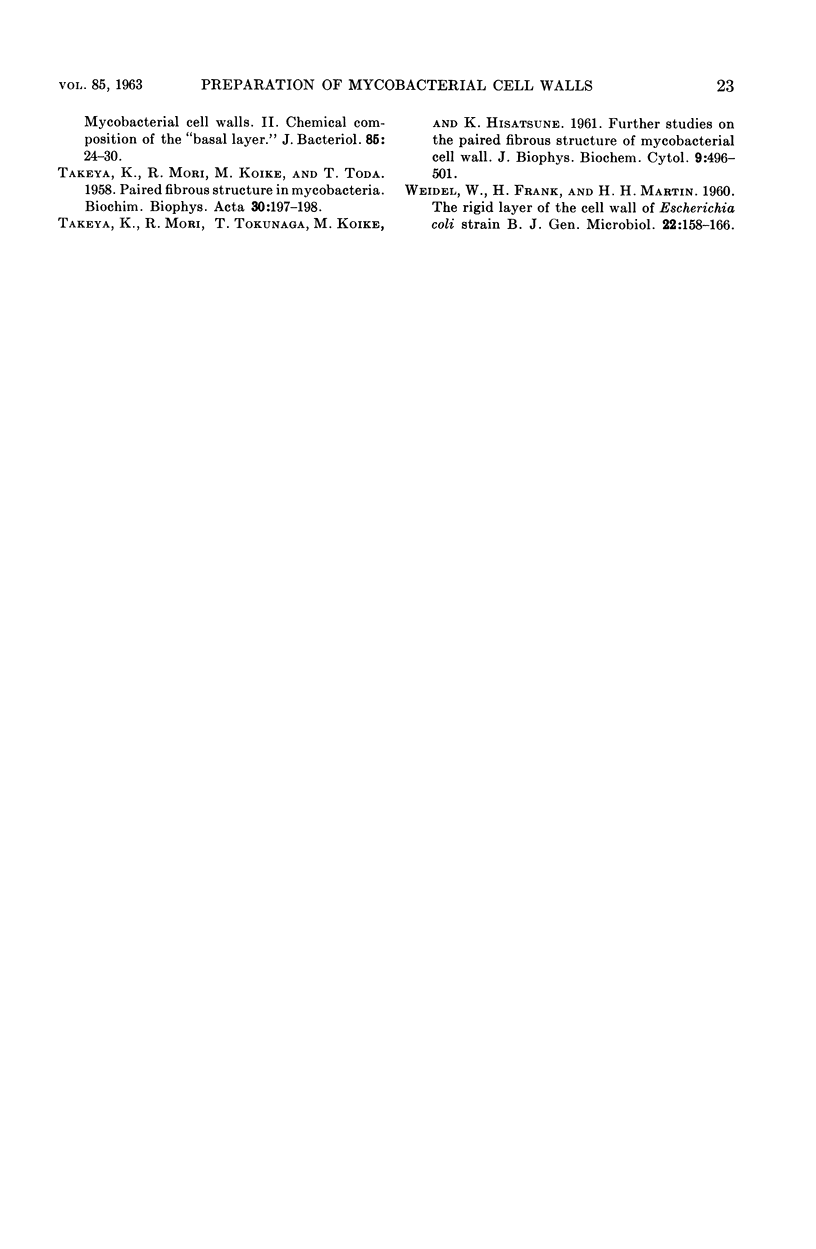
Takeya, Kenji (Kyushu University, Fukuoka, Japan) and Kazuhito Hisatsune. Mycobacterial cell walls. I. Methods of preparation and treatment with various chemicals. J. Bacteriol. 85:16–23. 1963.—Several methods of preparation of mycobacterial cell walls were examined, and the grinding method with glass powder, using Dry Ice, was found to give fairly good cell-wall preparations. “Paired fibrous structures” were clearly seen on the purified cell wall. The appearance of the cell wall as revealed by the electron microscope was not altered by digestion with trypsin, pronase, or pronase in 5% alcoholic solution, nor by treatment with 95% alcohol, acetone-alcohol mixture, or ether-alcohol mixture. By treatment with alcoholic KOH solution, the fibrous structure was removed. The remaining thin layer of the cell wall was tentatively designated the “basal layer” of the mycobacterial cell wall. The fibers appeared also to be removed by chloroform treatment. Nagarse digestion seemed to solubilize some constituents of the cell wall. The cell wall lost its shape and rigidity after lysozyme digestion.
Full text
PDF
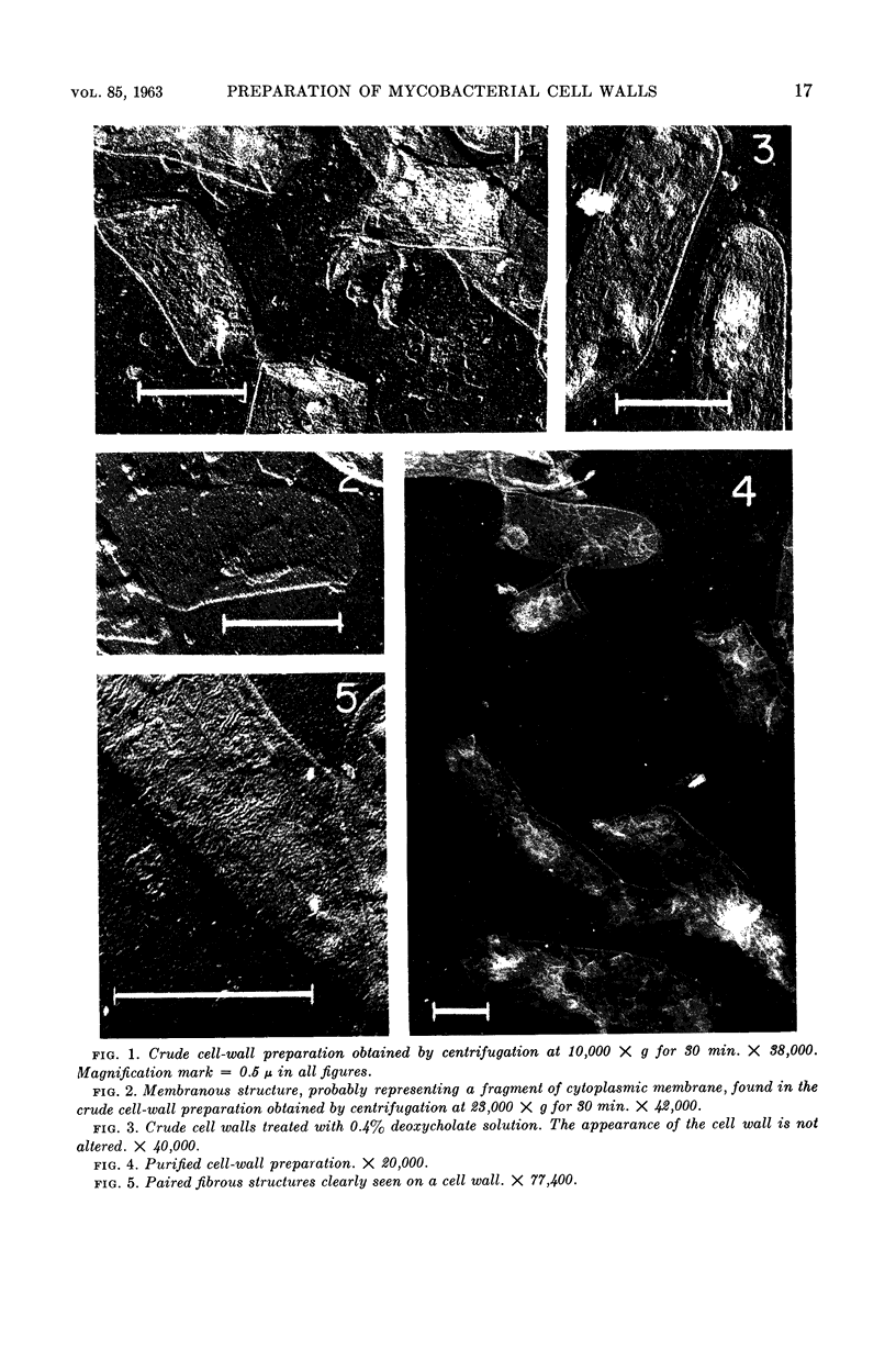
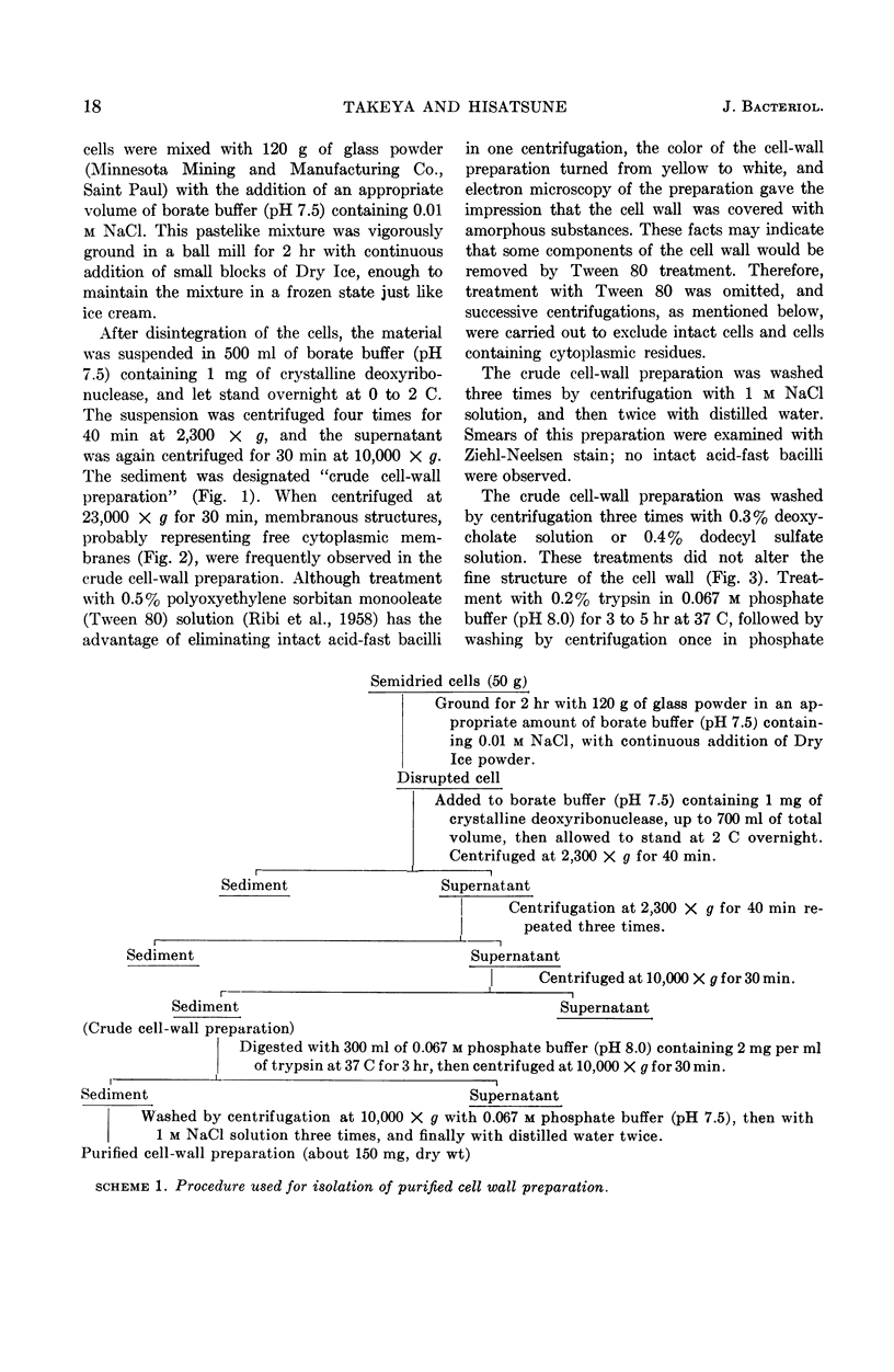
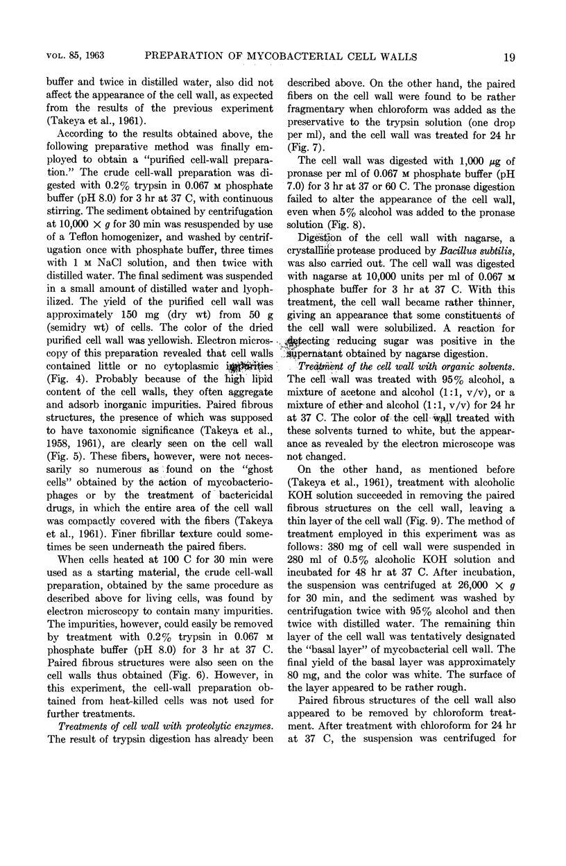
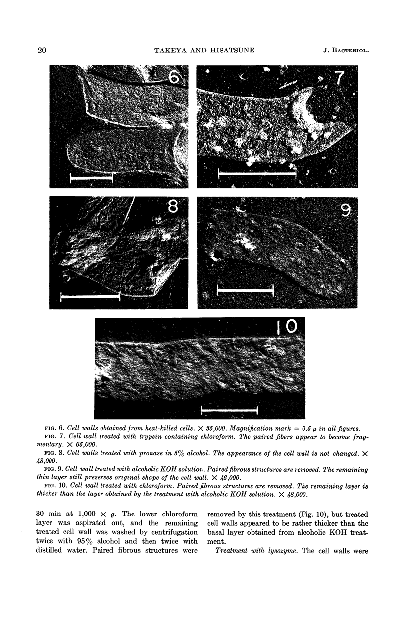
Images in this article
Selected References
These references are in PubMed. This may not be the complete list of references from this article.
- CUMMINS C. S., HARRIS H. Studies on the cell-wall composition and taxonomy of Actinomycetales and related groups. J Gen Microbiol. 1958 Feb;18(1):173–189. doi: 10.1099/00221287-18-1-173. [DOI] [PubMed] [Google Scholar]
- HOPWOOD D. A., GLAUERT A. M. Electron microscope observations on the surface structures of Streptomyces violaceoruber. J Gen Microbiol. 1961 Oct;26:325–330. doi: 10.1099/00221287-26-2-325. [DOI] [PubMed] [Google Scholar]
- KANAI K., YOUMANS G. P. Immunogenicity of intracellular particles and cell walls from Mycobacterium tuberculosis. J Bacteriol. 1960 Nov;80:607–614. doi: 10.1128/jb.80.5.607-614.1960. [DOI] [PMC free article] [PubMed] [Google Scholar]
- KANAI K., YOUMANS G. P., YOUMANS A. S. Allergenicity of intracellular particles, cell walls, and cytoplasmic fluid from Mycobacterium tuberculosis. J Bacteriol. 1960 Nov;80:615–621. doi: 10.1128/jb.80.5.615-621.1960. [DOI] [PMC free article] [PubMed] [Google Scholar]
- LAMANNA C., MALLETTE M. F. Use of glass beads for the mechanical rupture of microorganisms in concentrated suspensions. J Bacteriol. 1954 Apr;67(4):503–504. doi: 10.1128/jb.67.4.503-504.1954. [DOI] [PMC free article] [PubMed] [Google Scholar]
- LARSON C. L., RIBI E., WICHT W. C., LIST R. Skin reactions produced in rabbits by cell walls and protoplasm of Mycobacterium tuberculosis and M. butyricum. Am Rev Respir Dis. 1961 Feb;83:184–193. doi: 10.1164/arrd.1961.83.2P1.184. [DOI] [PubMed] [Google Scholar]
- RIBI E., LARSON C. L., LIST R., WICHT W. Immunologic significance of the cell wall of mycobacteria. Proc Soc Exp Biol Med. 1958 Jun;98(2):263–265. doi: 10.3181/00379727-98-24011. [DOI] [PubMed] [Google Scholar]
- TAKEYA K., HISATSUNE K., INOUE Y. Mycobacterial cell walls. II. Chemical composition of the "basal layer". J Bacteriol. 1963 Jan;85:24–30. doi: 10.1128/jb.85.1.24-30.1963. [DOI] [PMC free article] [PubMed] [Google Scholar]
- TAKEYA K., MORI R., KOIKE M., TODA T. Paired fibrous structure in mycobacteria. Biochim Biophys Acta. 1958 Oct;30(1):197–198. doi: 10.1016/0006-3002(58)90265-8. [DOI] [PubMed] [Google Scholar]
- TAKEYA K., MORI R., TOKUNAGA T., KOIKE M., HISATSUNE K. Further studies on the paired fibrous structure of mycobacterial cell wall. J Biophys Biochem Cytol. 1961 Feb;9:496–501. doi: 10.1083/jcb.9.2.496. [DOI] [PMC free article] [PubMed] [Google Scholar]
- WEIDEL W., FRANK H., MARTIN H. H. The rigid layer of the cell wall of Escherichia coli strain B. J Gen Microbiol. 1960 Feb;22:158–166. doi: 10.1099/00221287-22-1-158. [DOI] [PubMed] [Google Scholar]


