

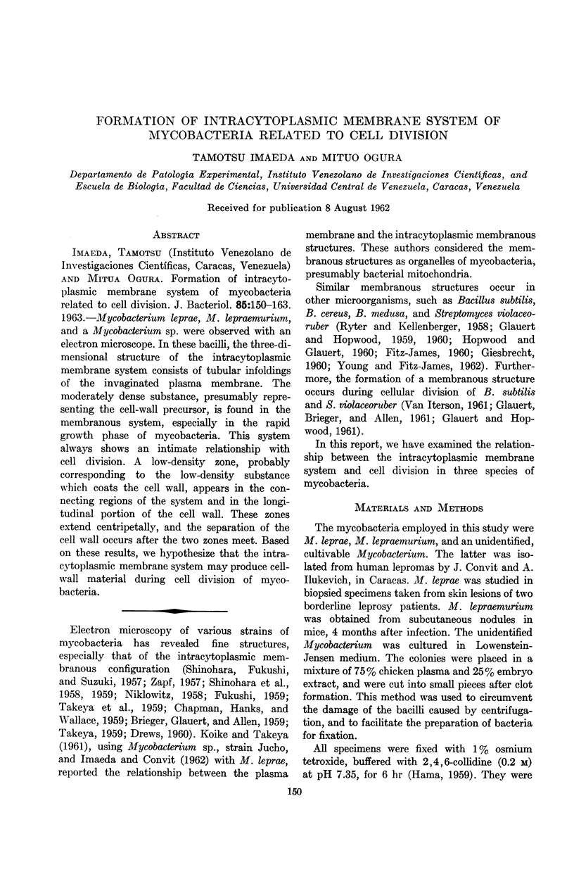
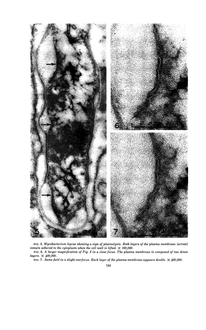
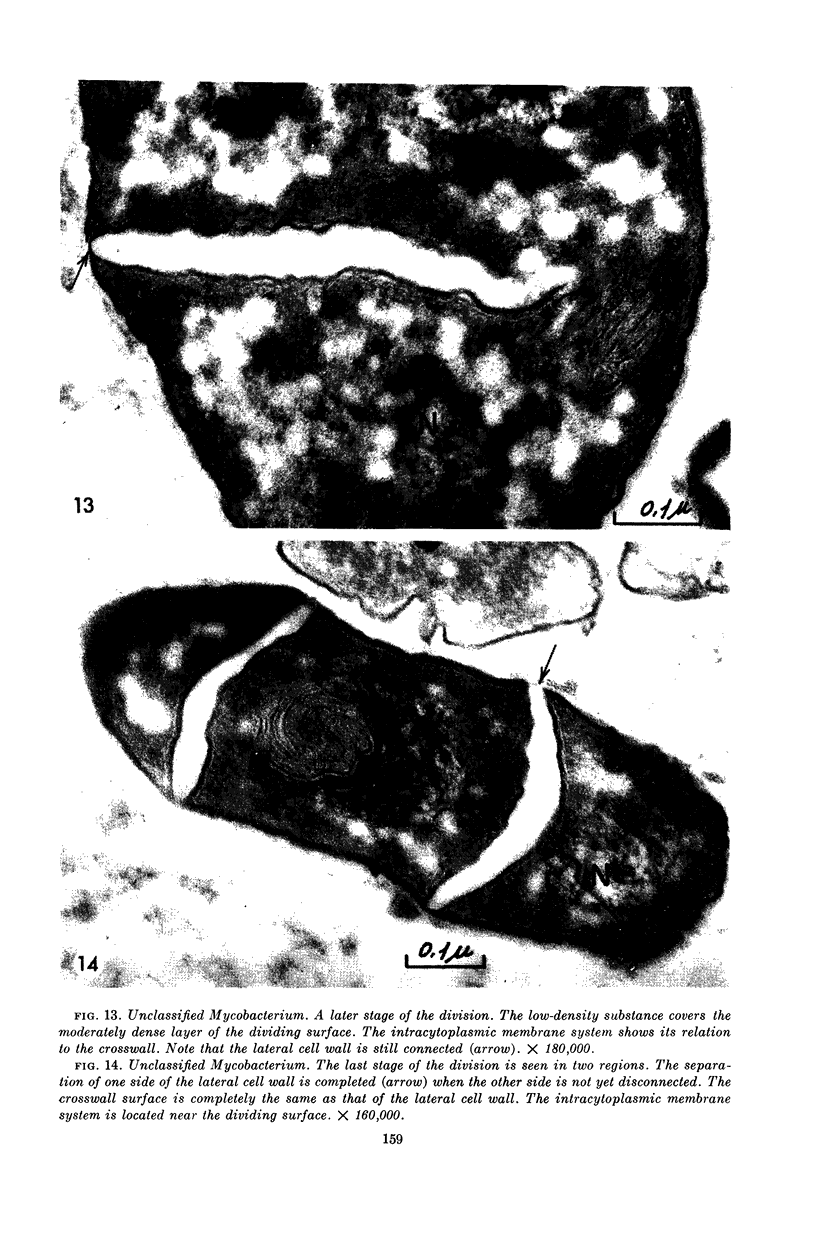
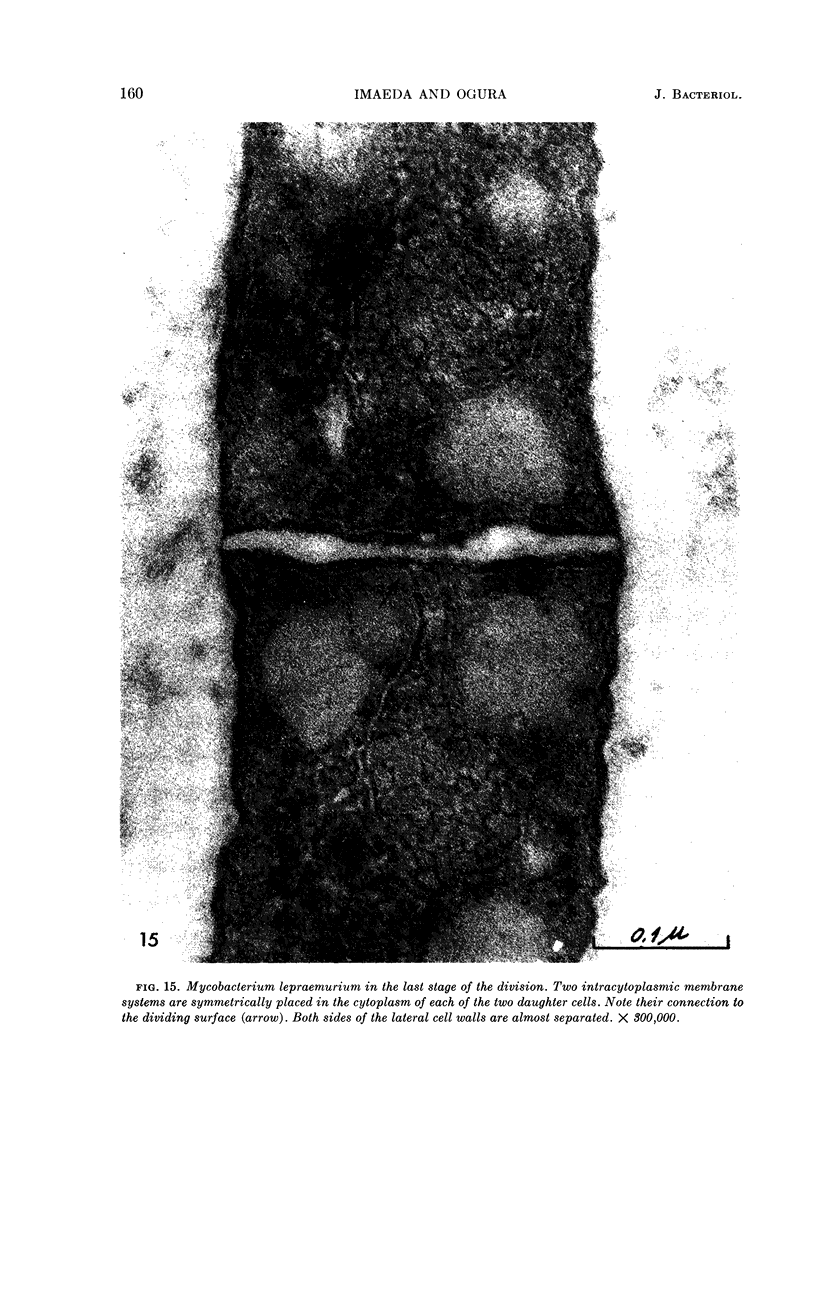
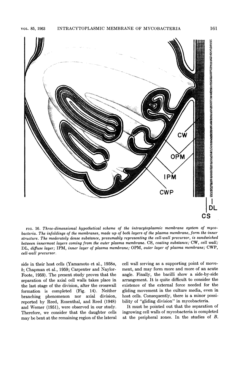
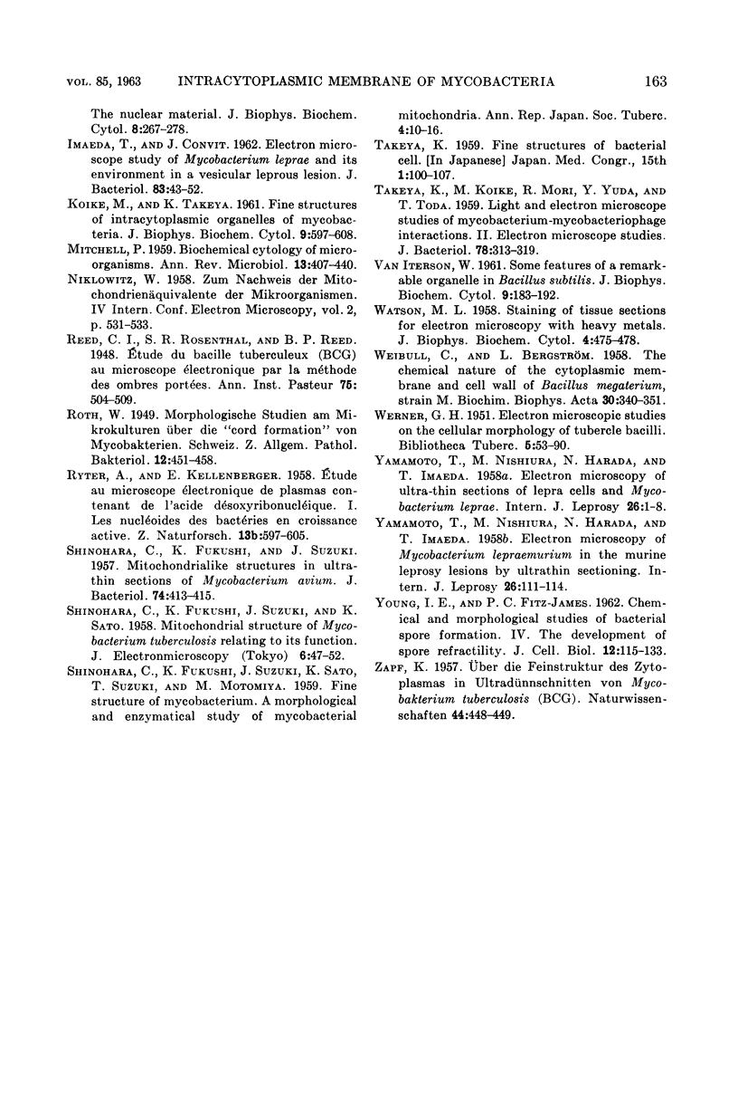
Abstract
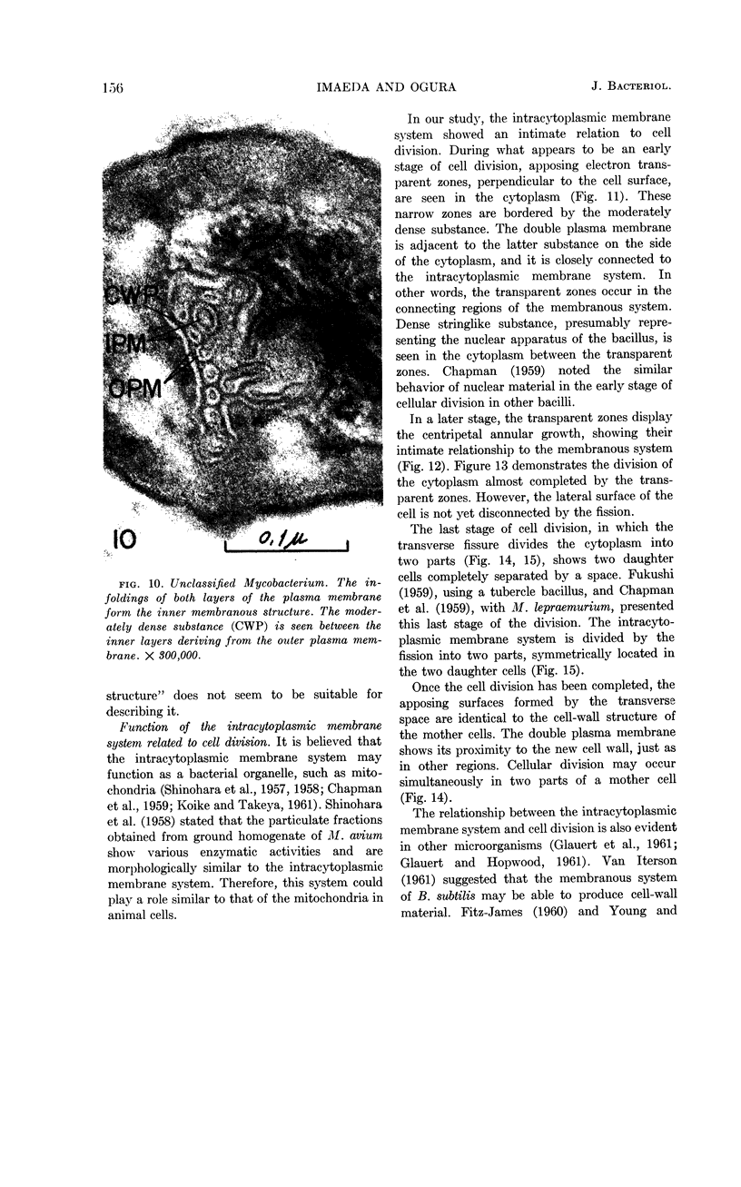
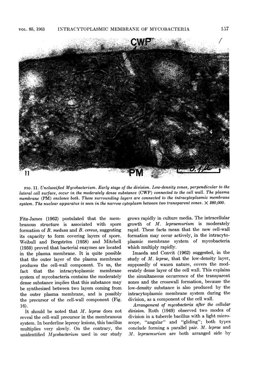
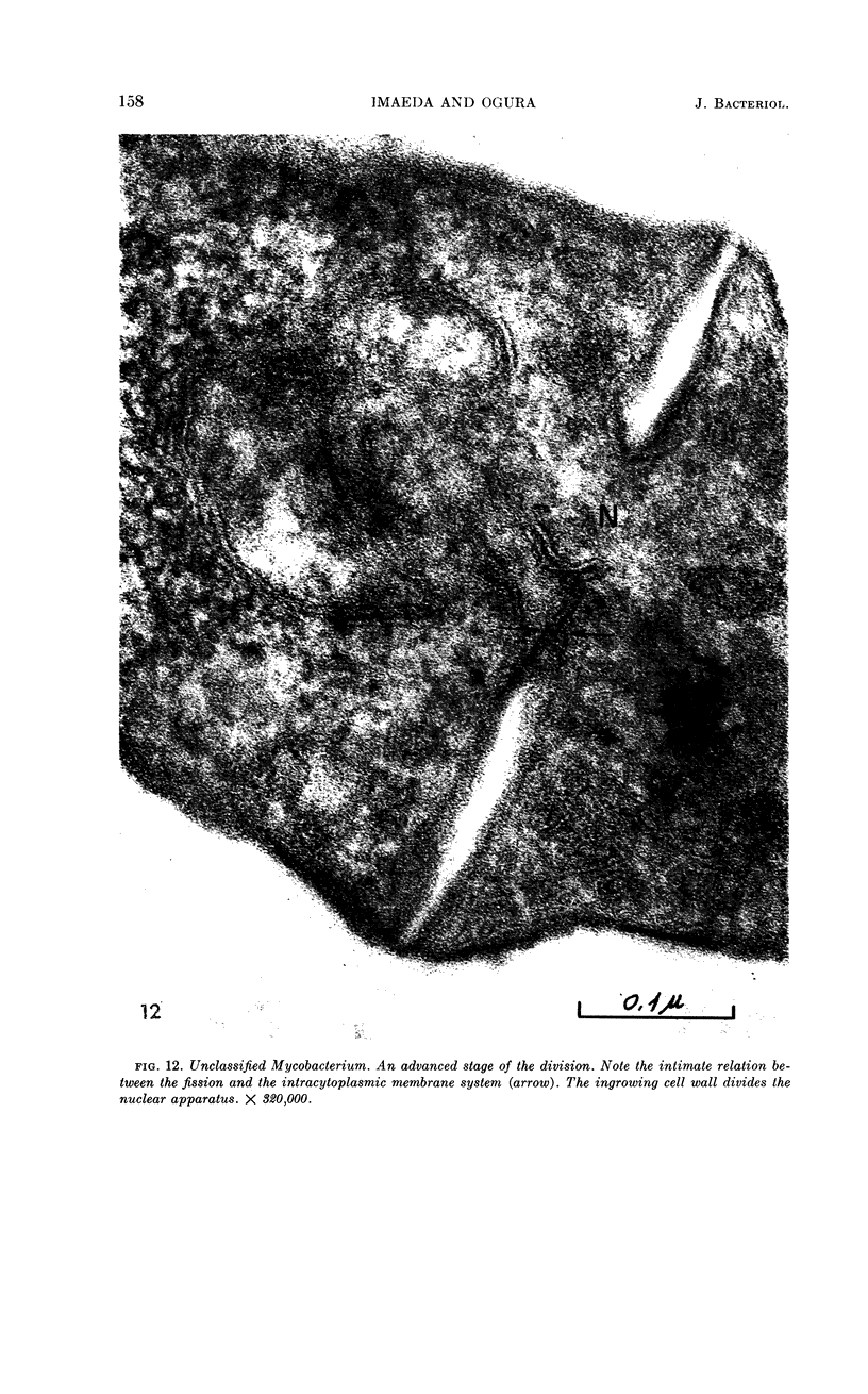
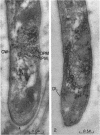
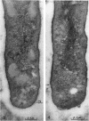
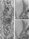
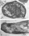
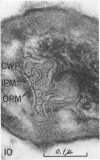
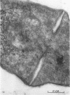
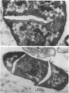
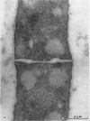
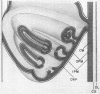
Imaeda, Tamotsu (Instituto Venezolano de Investigaciones Científicas, Caracas, Venezuela) and Mitua Ogura. Formation of intracytoplasmic membrane system of mycobacteria related to cell division. J. Bacteriol. 85:150–163. 1963.—Mycobacterium leprae, M. lepraemurium, and a Mycobacterium sp. were observed with an electron microscope. In these bacilli, the three-dimensional structure of the intracytoplasmic membrane system consists of tubular infoldings of the invaginated plasma membrane. The moderately dense substance, presumably representing the cell-wall precursor, is found in the membranous system, especially in the rapid growth phase of mycobacteria. This system always shows an intimate relationship with cell division. A low-density zone, probably corresponding to the low-density substance which coats the cell wall, appears in the connecting regions of the system and in the longitudinal portion of the cell wall. These zones extend centripetally, and the separation of the cell wall occurs after the two zones meet. Based on these results, we hypothesize that the intracytoplasmic membrane system may produce cell-wall material during cell division of mycobacteria.
Full text
PDF


Images in this article
Selected References
These references are in PubMed. This may not be the complete list of references from this article.
- BRIEGER E. M., GLAUERT A. M., ALLEN J. M. ytoplasmic structure in Mycobacterium leprae. Exp Cell Res. 1959 Oct;18:418–421. doi: 10.1016/0014-4827(59)90032-1. [DOI] [PubMed] [Google Scholar]
- CHAPMAN G. B. Electron microscope observations on the behavior of the bacterial cytoplasmic membrane during cellular division. J Biophys Biochem Cytol. 1959 Oct;6:221–224. doi: 10.1083/jcb.6.2.221. [DOI] [PMC free article] [PubMed] [Google Scholar]
- CHAPMAN G. B., HANKS J. H., WALLACE J. H. An electron microscope study of the disposition and fine structure of Mycobacterium lepraemurium in mouse spleen. J Bacteriol. 1959 Feb;77(2):205–211. doi: 10.1128/jb.77.2.205-211.1959. [DOI] [PMC free article] [PubMed] [Google Scholar]
- CHAPMAN G. B., HILLIER J. Electron microscopy of ultra-thin sections of bacteria I. Cellular division in Bacillus cereus. J Bacteriol. 1953 Sep;66(3):362–373. doi: 10.1128/jb.66.3.362-373.1953. [DOI] [PMC free article] [PubMed] [Google Scholar]
- Conti S. F., Gettner M. E. ELECTRON MICROSCOPY OF CELLULAR DIVISION IN ESCHERICHIA COLI. J Bacteriol. 1962 Mar;83(3):544–550. doi: 10.1128/jb.83.3.544-550.1962. [DOI] [PMC free article] [PubMed] [Google Scholar]
- DREWS G. [Electron microscopic studies on Mycobacterium phlei. (Structure and formation of metachromatic granula)]. Arch Mikrobiol. 1960;35:53–62. [PubMed] [Google Scholar]
- FITZ-JAMES P. C. Participation of the cytoplasmic membrane in the growth and spore fromation of bacilli. J Biophys Biochem Cytol. 1960 Oct;8:507–528. doi: 10.1083/jcb.8.2.507. [DOI] [PMC free article] [PubMed] [Google Scholar]
- GLAUERT A. M., BRIEGER E. M., ALLEN J. M. The fine structure of vegetative cells of Bacillus subtilis. Exp Cell Res. 1961 Jan;22:73–85. doi: 10.1016/0014-4827(61)90087-8. [DOI] [PubMed] [Google Scholar]
- GLAUERT A. M., HOPWOOD D. A. A membranous component of the cytoplasm in Streptomyces coelicolor. J Biophys Biochem Cytol. 1959 Dec;6:515–516. doi: 10.1083/jcb.6.3.515. [DOI] [PMC free article] [PubMed] [Google Scholar]
- GLAUERT A. M., HOPWOOD D. A. The fine structure of Streptomyces coelicolor. I. The cytoplasmic membrane system. J Biophys Biochem Cytol. 1960 Jun;7:479–488. doi: 10.1083/jcb.7.3.479. [DOI] [PMC free article] [PubMed] [Google Scholar]
- GLAUERT A. M., HOPWOOD D. A. The fine structure of Streptomyces violaceoruber (S. coelicolor). III. The walls of the mycelium and spores. J Biophys Biochem Cytol. 1961 Aug;10:505–516. doi: 10.1083/jcb.10.4.505. [DOI] [PMC free article] [PubMed] [Google Scholar]
- Imaeda T., Convit J. ELECTRON MICROSCOPE STUDY OF MYCOBACTERIUM LEPRAE AND ITS ENVIRONMENT IN A VESICULAR LEPROUS LESION. J Bacteriol. 1962 Jan;83(1):43–52. doi: 10.1128/jb.83.1.43-52.1962. [DOI] [PMC free article] [PubMed] [Google Scholar]
- KOIKE M., TAKEYA K. Fine structures of intracytoplasmic organelles of mycobacteria. J Biophys Biochem Cytol. 1961 Mar;9:597–608. doi: 10.1083/jcb.9.3.597. [DOI] [PMC free article] [PubMed] [Google Scholar]
- ROTH W. Morphologische Studien an Mikrokulturen über die cord-formation von Mycobakterien. Schweiz Z Pathol Bakteriol. 1949;12(5):451–458. [PubMed] [Google Scholar]
- RYTER A., KELLENBERGER E., BIRCHANDERSEN A., MAALOE O. Etude au microscope électronique de plasmas contenant de l'acide désoxyribonucliéique. I. Les nucléoides des bactéries en croissance active. Z Naturforsch B. 1958 Sep;13B(9):597–605. [PubMed] [Google Scholar]
- SHINOHARA C., FUKUSHI K., SUZUKI J. Mitochondria-like structures in ultrathin sections of Mycobacterium avium. J Bacteriol. 1957 Sep;74(3):413–415. doi: 10.1128/jb.74.3.413-415.1957. [DOI] [PMC free article] [PubMed] [Google Scholar]
- TAKEYA K., KOIKE M., MORI R., YUDA Y., TODA T. Light and electron microscope studies of Mycobacterium-mycobacteriophage interactions. II. Electron microscope studies. J Bacteriol. 1959 Sep;78:313–319. doi: 10.1128/jb.78.3.313-319.1959. [DOI] [PMC free article] [PubMed] [Google Scholar]
- VAN ITERSON W. Some features of a remarkable organelle in Bacillus subtilis. J Biophys Biochem Cytol. 1961 Jan;9:183–192. doi: 10.1083/jcb.9.1.183. [DOI] [PMC free article] [PubMed] [Google Scholar]
- WATSON M. L. Staining of tissue sections for electron microscopy with heavy metals. J Biophys Biochem Cytol. 1958 Jul 25;4(4):475–478. doi: 10.1083/jcb.4.4.475. [DOI] [PMC free article] [PubMed] [Google Scholar]
- WEIBULL C., BERGSTROM L. The chemical nature of the cytoplasmic membrane and cell wall of Bacillus megaterium, strain M. Biochim Biophys Acta. 1958 Nov;30(2):340–351. doi: 10.1016/0006-3002(58)90059-3. [DOI] [PubMed] [Google Scholar]
- WERNER G. H. Electron-microscopic studies on the cellular morphology of tubercle bacilli. Bibl Tuberc. 1951;5:53–90. [PubMed] [Google Scholar]
- YAMAMOTO T., NISHIURA M., HARADA N., IMAEDA T. Electron microscopy of Mycobacterium leprae murium in ultra-thin sections of murine leprosy lesions. Int J Lepr. 1958 Apr-Jun;26(2):111–114. [PubMed] [Google Scholar]
- YAMAMOTO T., NISHIURA M., HARADA N., IMAEDA T. Electron microscopy of ultra-thin sections of lepra cells and Mycobacterium leprae. Int J Lepr. 1958 Jan-Mar;26(1):1–8. [PubMed] [Google Scholar]
- YOUNG I. E., JAMES P. C. Chemical and morphological studies of bacterial spore formation. IV. The development of spore refractility. J Cell Biol. 1962 Jan;12:115–133. doi: 10.1083/jcb.12.1.115. [DOI] [PMC free article] [PubMed] [Google Scholar]