Abstract
Photoinhibition and production of reactive oxygen species were studied in tobacco plants overexpressing the plastid terminal oxidase (PTOX). In high light, these plants was more susceptible to photoinhibition than wild-type plants. Also oxygen-evolving activity of isolated thylakoid membranes from the PTOX-overexpressing plants was more strongly inhibited in high light than in thylakoids from wild-type plants. In contrast in low light, in the PTOX overexpressor, the thylakoids were protected against photoinhibition while in wild type they were significantly damaged. The production of superoxide and hydroxyl radicals was shown by EPR spin-trapping techniques in the different samples. Superoxide and hydroxyl radical production was stimulated in the overexpressor. Two-thirds of the superoxide production was maintained in the presence of DNP-INT, an inhibitor of the cytochrome b6f complex. No increase of the SOD content was observed in the overexpressor compared with the wild type. We propose that superoxide is produced by PTOX in a side reaction and that PTOX can only act as a safety valve under stress conditions when the generated superoxide is detoxified by an efficient antioxidant system.
Introduction
The plastid terminal oxidase (PTOX2 or IMMUTANS) is a plastid-located plastoquinol:oxygen oxidoreductase (1–3). It is distantly related to the alternative oxidase (AOX) of the mitochondrial inner membrane. The active site of both oxidases, PTOX and AOX, comprises a non-heme di-iron center (4, 5). PTOX is a minor component (∼1% of PSII levels in Arabidopsis thaliana) of the thylakoid membrane and is located in the stroma lamellae (6, 7). PTOX plays an important role in carotenoid biosynthesis and seems to be involved in phytoene desaturation reactions (8–13).
The physiological importance of the role of PTOX as plastoquinol oxidase in alternative photosynthetic electron transport pathways is unclear. Evidence that PTOX acts as a plastoquinol oxidase was shown in tobacco plants, which constitutively expressed the A. thaliana PTOX gene (14). It has been suggested that PTOX may serve to keep the photosynthetic electron transport chain relatively oxidized. Exposure of plants to excess light may result in over-reduction of the plastoquinol pool and may lead to photoinhibition (15). However, no major role for PTOX in oxidizing the PQ pool was found in chlorophyll fluorescence assays when thylakoids from wt A. thaliana and the immutans mutant lacking PTOX were compared (16).
Recently, several groups reported that the PTOX level increased under natural stress conditions in several species specialized to harsh environmental conditions. This was the case in Ranunculus glacialis, an alpine plant, when it was acclimated to high light and low temperature (17); in the halophyte Thellungiella halophila when it was exposed to salt stress (18); and in Brassica fruticulosa when it was exposed to elevated temperature and high light (19). Although these findings support the hypothesis that PTOX may serve as a safety valve under stress conditions, they are in direct conflict with the data of Rosso et al. (20). These authors have shown that overexpression of PTOX in A. thaliana did not result in an increased capacity to keep the plastoquinone pool oxidized and did not provide any significant photoprotection. A more detailed study of photoinhibition and the generation of reactive oxygen species (ROS) under photoinhibitory illumination seems to be required to answer the question if and under which conditions PTOX can contribute to photoprotection. Using wt and PTOX+ plants that overexpress PTOX (14), we investigated the susceptibility to light by measuring the loss of variable chlorophyll fluorescence in leaves and oxygen evolution in isolated thylakoids. Furthermore, we followed the light-induced generation of ROS by spin trapping EPR spectroscopy. We suggest, based on Western blots, that PTOX can only act as a safety valve and protect against photoinhibition when the level of SOD is adjusted to the actual level of PTOX.
EXPERIMENTAL PROCEDURES
Chemicals
All chemicals were of the highest grade from commercial suppliers. The spin trap α-(4-pyridyl-1-oxide)-N-tert-butyl nitrone (4-POBN) was obtained from Sigma. The nitrone 5-diethoxyphosphoryl-5-methyl-1-pyrroline N-oxide (DEPMPO) was synthesized according to Ref. 21. Commercial antibodies directed against D1, Cu/Zn-SOD, and Fe-SOD were obtained from Agrisera (Sweden).
Plant Material
Tobacco wild-type plants (Nicotiana tabacum var. petit Havana) and the plants overexpressing PTOX (14) were grown for 3 months on soil in a growth cabinet (24 °C day/18 °C night) under an irradiance of 150 or 450 μmol quanta m−2 s−1.
Escherichia coli Expressing PTOX
E. coli cells expressing Arabidopsis PTOX (22) were grown in M9/glycerol medium until D600 = 0.3. Isopropyl thio-β-d-galactoside was then added (final concentration 40 μm) to induce the expression of the recombinant gene during 12 h. The control strain was grown in parallel. E. coli membranes were prepared according to Ref. 22.
Chloroplast Preparation
Intact chloroplasts were prepared according to Ref. 23. MnCl2 was omitted from the medium because it interfered with EPR measurements. Intact chloroplasts (20–50 μg of Chl ml−1) were shocked for 20 s in 5 mm MgCl2, 25 mm HEPES, pH 7.5. Then an equal volume of 0.6 m sorbitol, 5 mm MgCl2, 25 mm HEPES, pH 7.5, was added. The intactness of the chloroplasts was determined as the ratio of light-driven reduction of the membrane impermeable K3[Fe(CN)6] measured with intact and osmotically shocked chloroplasts. The intactness of the chloroplasts was 70–80%.
Photoinhibition Treatment Leaves
The leaves were first kept at low light (8 μmol quanta m−2 s−1) for 4 h with the petioles in lincomycin solution (1 g/liter). The photoinhibitory illumination was done in a growth cabinet with controlled temperature (22 °C), and the temperature of the illuminated leaves was around 28 °C. The petioles were kept in the lincomycin solution during the whole illumination period. During the illumination, samples were taken from the leaves for measurements of fluorescence.
Thylakoids
Prior to the photoinhibitory treatment, the chloroplasts were shocked. Thylakoids were illuminated with white light (120 or 1500 μmol quanta m−2 s−1), stirred, and kept at 20–22 °C.
Fluorescence Measurements
The initial (Fo) and maximum (Fm) fluorescence levels were measured with a pulse amplitude modulated fluorometer (PAM 101; Heinz Walz, Effeltrich, Germany), using a saturating flash (7000 μmol quanta m−2 s−1; duration, 1 s) for Fm. The variable fluorescence (Fv = Fm − Fo) was calculated. The samples were dark adapted for 30 min before each measurement to allow most of the reversible light-induced fluorescence quenching to relax.
Measurements of Electron Transport Activity
Light-saturated electron transport activity was measured as the rate of oxygen evolution in shocked chloroplasts using 1 mm K3[Fe(CN)6] (ferricyanide) or 2,6-dichloro-1,4-benzoquinone (DCBQ) as electron acceptor and 0.5 mm NH4Cl as uncoupler using an oxygen electrode.
EPR Measurements
Spin-trapping assays with 4-POBN were carried out using freshly shocked chloroplasts at a concentration of 10 μg of Chl ml−1. Samples were illuminated for 5 min with white light (120 or 1500 μmol quanta m−2 s−1) in the presence of 50 mm 4-POBN, 4% ethanol, 50 μm Fe-EDTA, and buffer (25 mm Hepes, pH 7.5, 5 mm MgCl2, 0.3 m sorbitol).
When E. coli membranes were used, sonicated membranes (0.4 mg protein ml−1) were incubated for 5 min in 20 mm Tris/HCl pH 7.5, 10 mm KCl, 5 mm MgCl2 with 5 mm succinate as electron donor in the presence of 50 mm 4-POBN, 4% ethanol, 50 μm Fe-EDTA.
Spin-trapping assays with DEPMPO were carried out with thoroughly washed thylakoids (washing buffer contained 25 mm HEPES, pH 7.5, and 5 mm MgCl2) at a concentration of 50 μg of Chl ml−1. Samples were illuminated for 5 min with white light (1500 μmol quanta m−2 s−1) in the presence of 50 mm DEPMPO, 1 mm DTPA and buffer (25 mm Hepes, pH 7.5, 5 mm MgCl2).
EPR spectra were recorded at room temperature in a standard quartz flat cell using an ESP-300 X-band (9.73 GHz) spectrometer (Bruker, Rheinstetten, Germany). The following parameters were used: microwave frequency 9.73 GHz, modulation frequency 100 kHz, modulation amplitude: 1G, microwave power: 63 milliwatt in DEPMPO assays, or 6.3 milliwatt in 4-POBN assays, receiver gain: 2 × 10⁴, time constant: 40.96 ms; number of scans: 4.
SDS-PAGE and Western Blotting
SDS-PAGE was carried out in 12% polyacrylamide gel. Western blotting was performed using nitrocellulose membrane and a Multiphor II Novablot unit (Amersham Biosciences). For detection, the enhanced chemoluminescence (ECL) system (Amersham Biosciences) was used according to the manufacturer's protocol.
RESULTS
Influence of High Amounts of PTOX on Photoinhibition
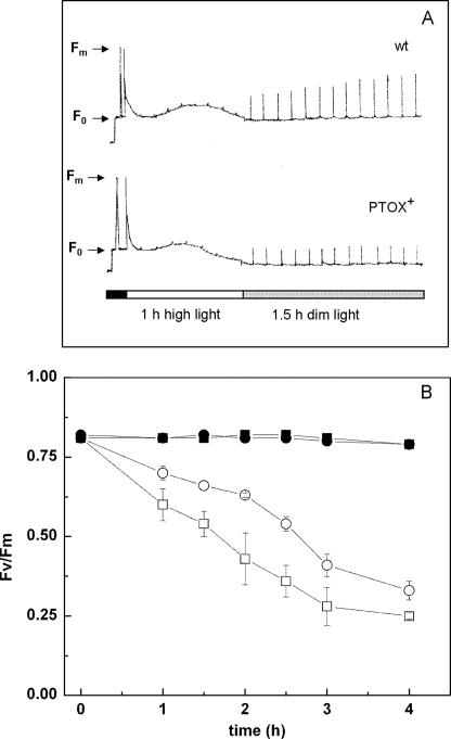
In this study, tobacco plants (PTOX+) overexpressing the plastid terminal oxidase from A. thaliana (14) were used. PTOX+ and wt plants were grown at 450 μmol quanta m−2 s−1. When attached leaves were exposed to photoinhibitory light (1500 μmol quanta m−2 s−1) a similar loss of variable fluorescence was observed for both, wt and PTOX+ plants. The recovery of variable fluorescence in low light (8 μmol quanta m−2 s−1) was significantly faster in the wild type than in PTOX+ plants (Fig. 1A). To investigate whether the PTOX+ plants were more susceptible to photoinhibition, leaves of wt and PTOX+ were incubated for 4 h in lincomycin to block the synthesis of D1 and thereby the repair of damaged PSII centers. Leaves were illuminated for up to 4 h with 850 μmol quanta m−2 s−1 and the ratio of Fv/Fm, a measure of the maximum quantum yield of photosynthesis, was determined. Prior to high light exposure, Fv/Fm was 0.81 for wt and PTOX+, consistent with measurements on a wide range of unstressed higher plants (24). During high light exposure, the loss of variable fluorescence was considerably higher in PTOX+ than in wt (Fig. 1B). When plants grown at 150 μmol quanta m−2 s−1 were illuminated with 400 μmol quanta m−2 s−1, a much lower loss of Fv/Fm was observed and no significant difference between PTOX+ and wt was found (data not shown).
FIGURE 1.
Effect of high light on chlorophyll fluorescence in wt and PTOX+ leaves. A, fluorescence measurements on attached leaves of wild-type and PTOX+ plants. Plants were dark-adapted for 30 min prior to the measurements. Segments of the leaves were illuminated for 1 h with white light (1500 μmol quanta m−2 s−1) and then transferred to dim light (8 μmol quanta m−2 s−1) for 1.5 h. B, detached leaves were incubated with lincomycin. Circles, wt; squares, PTOX+ leaves, filled symbols, incubated at low light (8 μmol quanta m−2 s−1), open symbols, incubated at high light (850 μmol quanta m−2 s−1). Error bars represent S.D. (nine independent experiments).
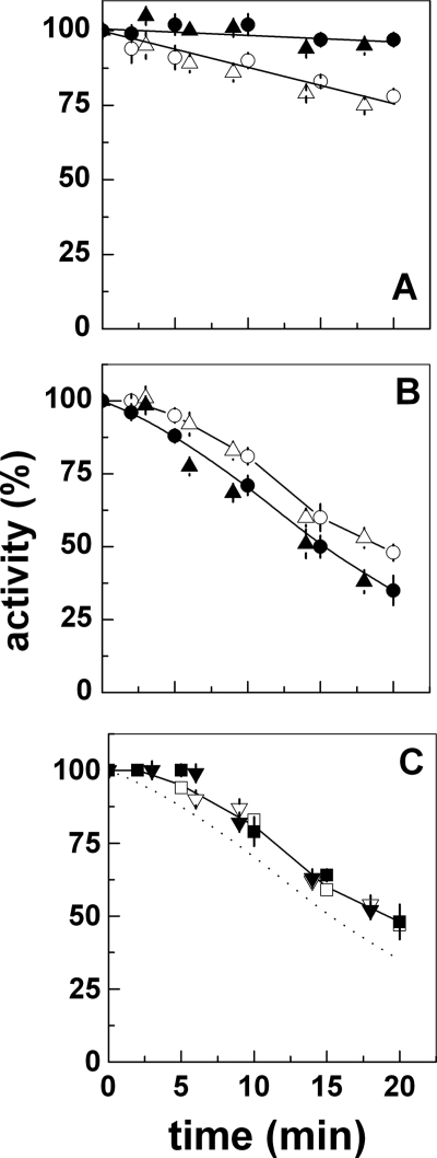
In addition to measurements of chlorophyll fluorescence in leaves, photoinhibition was measured as loss of the activity of the electron transfer chain in isolated thylakoids from wt and PTOX+ (Fig. 2). Thylakoids from PTOX+ illuminated with low light (120 μmol quanta m−2 s−1) showed almost no loss of activity while in wt thylakoids 20% of oxygen evolution was lost after 20 min of illumination. However, when thylakoids were illuminated with high light (1500 μmol quanta m−2 s−1) just the opposite effect was observed (Fig. 2B). Thylakoids from PTOX+ were more susceptible to illumination with high light intensities than those from wt (Fig. 2A). The loss of electron transport activity was about 10% higher than in wt thylakoids. To distinguish between damage of the total electron transport chain and damage of PSII, the activity was measured by using ferricyanide as electron acceptor to determine the activity of the total linear electron transport and by using DCBQ as electron acceptor for PSII. The data show that the most susceptible part of the electron transport chain is PSII. Addition of SOD rescued the electron transfer activity of PTOX+ thylakoids to a level comparable with the wt (Fig. 2C). The addition of SOD had no effect on the light-induced damage of photosynthetic electron transport in wt thylakoids (Fig. 2C). These photoinhibition experiments were performed in the absence of an uncoupler. No external electron acceptor was present during the photoinhibition treatment leaving oxygen as the only available electron acceptor.
FIGURE 2.
Photoinhibition assays on wt and PTOX+ thylakoids in low and high light. Freshly shocked thylakoids were incubated: (A) at low light (120 μmol quanta m−2 s−1), wt with ferricyanide (○), and DCBQ (▵), and PTOX+ with ferricyanide (●), and DCBQ (▴); B, same as A but at high light (1500 μmol quanta m−2 s−1); C, high light (1500 μmol quanta m−2 s−1) in the presence of SOD, wt with ferricyanide (□), and DCBQ (▿), and PTOX+ with ferricyanide (■), and DCBQ (▾). 50 μg SOD/ml were added prior to the photoinhibitory illumination. The dotted line is the same as the lower line in panel B, and the difference between it and the solid line shows the protective effect of SOD. The maximum activity was between 270–290 μmol of O2 mg Chl−1 h−1 for both wt and PTOX+ thylakoids in the presence of ferricyanide and 400–425 μmol O2 mg Chl−1 h−1 in the presence of DCBQ as electron acceptor. Error bars represent S.D. (3–4 independent experiments).
Influence of High Amounts of PTOX on ROS Production
ROS, including superoxide (O2˙̄), hydrogen peroxide (H2O2), and hydroxyl radicals (HO•), can be generated by the reduction of oxygen in photosynthetic electron transfer in a number of different reactions: O2 can act as terminal acceptor in the so-called Mehler reaction at the acceptor side of PSI, it can be reduced by plastosemiquinones in the membrane (25), it can be reduced at the acceptor side of PSII (26–30), and it is the electron acceptor of the plastid terminal oxidase, which uses two plastoquinol molecules as electron donor (4, 5, 31). We investigated the light-induced formation of ROS by EPR spectroscopy using either ethanol/4-POBN or DEPMPO as the spin traps. Performing 4-POBN spin trapping in the presence of ethanol is a general procedure to indirectly prove the formation of HO• through the detection of the secondary 4-POBN/α-hydroxyethyl spin adduct (32). The cyclic nitrone DEPMPO was used because it forms characteristic, non-exchangeable adducts with OH• and O2˙̄, and the EPR signal patterns are easily distinguishable (21).
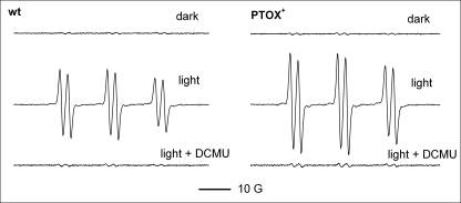
Whereas little or no signal was observed when samples were maintained in the dark in the presence of ethanol/4-POBN, illumination of thylakoids resulted in strong EPR signals as sextets of lines (aN = 15.61 G; aHβ = 2.55 G) characteristic of 4-POBN/α-hydroxyethyl aminoxyl (Fig. 3). Representative spectra are shown in Fig. 3. PTOX+ thylakoids produced a signal that was ∼50% larger than the signal obtained from wt thylakoids after 5 min of illumination with 1500 μmol quanta m−2 s−1 white light (Table 1). Addition of DCMU, an inhibitor of electron transfer in PSII, almost completely inhibited spin adduct formation. Taken altogether, the data of Fig. 3 show that OH• radicals were generated by photosynthetic electron transfer reactions and not by excitation of chlorophylls in disordered reaction centers or antenna systems.
FIGURE 3.
Light-induced hydroxyl radical formation in wt and PTOX+ thylakoids. Generation of hydroxyl radicals is shown by indirect spin trapping with 4-POBN/ethanol. Typical EPR spectra of the 4-POBN/α-hydroxyethyl adduct are shown. Samples were illuminated for 5 min at high light (1500 μmol quanta m−2 s−1). Where indicated, 10 μm DCMU was added prior to the illumination.
TABLE 1.
ROS production in wt and PTOX+ thylakoids
Thylakoids were illuminated for 5 min at high light (1500 μmol quanta m−2 s−1) in the presence of the spin traps 4-POBN (50 mm)/ethanol or DEPMPO (50 mm). Where indicated 10 μm DCMU, 10 μm DNP-INT, or 50 μg of SOD/ml were added prior to the illumination. The double integral of the total signal obtained with wt thylakoids after illumination was set to unity. If not stated otherwise four spectra of different preparations were used to calculate the mean and the standard deviation.
| EPR signal size |
||
|---|---|---|
| wt | PTOX+ | |
| 4-POBN/EtOH | ||
| Light | 1.00 ± 0.10 | 1.56 ± 0.15 |
| Light + DCMU | 0.03 ± 0.01 | 0.04 ± 0.01 |
| Dark | 0.02 ± 0.01 | 0.02 ± 0.01 |
| DEPMPO | ||
| Light | 1.00 ± 0.15 (n = 4) | 1.55 ± 0.23 (n = 9) |
| Light + DNP-INT | 0.31 ± 0.10 (n = 9) | 0.63 ± 0.09 (n = 5) |
| Light + DCMU | Not detectable | Not detectable |
| Light + SOD | 0.07 ± 0.02 (n = 4) | Not detectable |
| Dark | Not detectable | Not detectable |
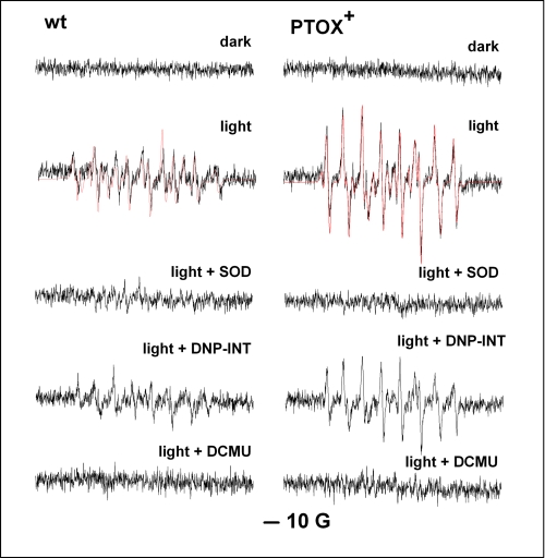
In our system, HO• could result from a metal ion-assisted Fenton mechanism involving H2O2, the disproportionation product of O2˙̄. To investigate whether O2˙̄ was the primary radical species formed experiments were carried out in the presence of DEPMPO instead of 4-POBN/ethanol (Fig. 4). Again no EPR signal was detected in the dark while a complex signal was observed upon illumination. A satisfactory simulation of the signals in Fig. 4 was obtained assuming a mixture of DEPMPO/O2˙̄ (DEPMPO-OOH; the asymmetrical signal of which could be simulated by the 1:1 mixture of two species having the coupling constants: aN = 13.1 (13.2); aP = 50.7 (49.9) and aHβ = 11.7 (10.5) G), DEPMPO/HO• (DEPMPO-OH: aN = 13.0; aP = 47.4 and aHβ = 15.2 G) and a carbon-centered/DEPMPO spin adduct (DEPMPO-R; aN = 14.4; aP = 45.5 and aHβ = 21.3 G). In all spectra, DEPMPO-OOH was the dominant species (∼70%) while the remaining DEPMPO-OH and DEPMPO-R appeared in approximately 1:1 mixtures.
FIGURE 4.
Light-induced superoxide formation in wt and PTOX+ thylakoids. Typical EPR spectra of the DEPMPO-OOH adduct are shown. Thylakoid samples were illuminated as described in Fig. 4 in the presence of 50 mm DEPMPO. Typical EPR spectra are mixtures of superoxide (∼70%), hydroxyl and carbon-centered DEPMPO adducts (red lines, simulated spectra). DEPMPO-OOH is generated as a relatively stable radical after DEPMPO reacts with O2˙̄. Samples were illuminated for 5 min at high light (1500 μmol quanta m−2 s−1). Where indicated, 50 μg SOD/ml, 10 μm DNP-INT, or 10 μm DCMU was added prior to the illumination.
PTOX+ thylakoids produced an EPR signal which was ∼60% larger than the signal obtained from wt thylakoids after 5 min illumination with 1500 μmol quanta m−2 s−1 white light (Fig. 4 and Table 1). In the presence of SOD, the signal in PTOX+ thylakoids was completely suppressed while in wt thylakoids a small signal was still observed (Fig. 4). These data indicate that O2˙̄, which was primarily generated by illumination, partially decomposed into HO•, which could in turn undergo hydrogen abstraction from a series of cellular targets to form carbon-centered radicals. The higher level of O2˙̄ production in the overexpressor suggests that PTOX is responsible for the additional O2˙̄ generation. However, the Mehler reaction will also contribute to O2˙̄ generation in thylakoids. To measure electron transport to oxygen in the absence of that reaction, the cytochrome b6f inhibitor DNP-INT, a specific inhibitor of the Qo binding site (33), was added prior to the illumination. In the presence of DNP-INT, the EPR signal size in DEPMPO experiments was reduced by ∼30% in PTOX+ thylakoids. In wt thylakoids, the signal size decreased by about 60% in comparison to the signal size measured in the absence of the inhibitor. The data obtained in the presence of DNP-INT show that the Mehler reaction counts for about two-thirds of the O2˙̄ generation in wt thylakoids. Unfortunately, DEPMPO could only detect O2˙̄ in thoroughly washed thylakoids while 4-POBN could be used with freshly shocked intact chloroplasts. Although DEPMPO-OOH does not significantly self-decompose into DEPMPO-OH (21), at least two reductive mechanisms occurring in cells can alter the DEPMPO-OOH EPR signal, i.e. conversion into DEPMPO-OH by certain enzymes such as glutathione peroxidase (21, 34) or formation of EPR-silent hydroxylamines such as with ascorbate (35, 36). Intense washing did not damage the thylakoids as can be seen by the complete inhibition of the signal in the presence of DCMU.
To test whether the presence of PTOX leads in general to an increase in ROS generation, PTOX was expressed in E. coli (Fig. 5), and ROS generation was measured by the spin trap EtOH/POBN in the presence of succinate as electron donor to complex II of the respiratory chain. In the presence of 1 mm KCN a significant level of ROS was formed in control membranes while this ROS generation was reduced by about 50% in PTOX-containing membranes (data not shown). This shows that PTOX functions as an alternative oxidase in this system. PTOX-containing membranes generated a small, but significant, amount of ROS while ROS production in control membranes was hardly detectable (Table 2). Decyl-plastoquinone was added to obtain higher rates of electron flow to PTOX (22). Under these conditions, ROS generation was detectable in control membranes and to a 1.6-fold higher level in PTOX-containing membranes. This result clearly shows that PTOX forms ROS when the quinone pool is over-reduced.
FIGURE 5.
Expression of PTOX in E. coli. The immunoblot was decorated with polyclonal antibodies directed against PTOX. 250 μl of culture was taken as a sample at given times after induction by isopropyl-1-thio-β-d-galactopyranoside (0–4 h and overnight, o.n.), centrifuged, and the pellet was resuspended in 50 μl of SDS loading buffer. 10 μl were loaded.
TABLE 2.
ROS production in control and PTOX-containing E. coli membranes
Membranes (0.4 mg protein ml−1) were incubated for 5 min with 5 mm succinate and, when indicated, 0.2 mm decyl-plastoquinone in the presence of the spin trap 4-POBN (50 mm)/ethanol. The double integral of the total signal obtained with control membranes in the presence of decyl-plastoquinone was set to unity. Five spectra were used to calculate the mean and the standard deviation.
| EPR signal size |
||
|---|---|---|
| Control membranes | PTOX-containing membranes | |
| No decyl-PQ | Not detectable | 0.6 ± 0.05 |
| +decyl-PQ | 1.0 ± 0.1 | 1.6 ± 0.1 |
SOD Protein Level in PTOX+ and Wt
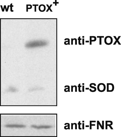
The question arises why PTOX+ thylakoids were more susceptible to photoinhibition in strong light while they were protected in low light compared with wt thylakoids. One possibility is that the antioxidant system in PTOX+ cannot cope with the heavier burden of O2˙̄ generation in high light conditions while it is sufficient to detoxify the amount of O2˙̄ under low light illumination. The antioxidant system of the chloroplast consists of SODs and several forms of ascorbate peroxidases. We focused here on SODs, which detoxify O2˙̄. The presence of a Cu/Zn-SOD and an Fe-SOD in chloroplasts of A. thaliana has been shown (37). In tobacco, one Cu/Zn-SOD is present in the chloroplast and there is evidence for a Fe-SOD on the transcript level (Data Bank Uniprot/Swiss-Prot). To show amounts of SOD, we performed immunoblots of the proteins of intact chloroplasts of wt and PTOX+ using polyclonal antibodies directed against PTOX, plastid Cu/Zn-SOD, and Fe-SOD (Fig. 6). Wt and PTOX+ chloroplasts contained about the same amount of Cu/Zn-SOD as shown by the reaction with the specific anti-Cu/Zn-SOD antibody. No protein was detected with the anti-FeSOD antibody.
FIGURE 6.
PTOX and Cu/Zn-SOD protein levels in wt and PTOX+ chloroplasts. The immunoblot was decorated with polyclonal antibodies directed against PTOX and Cu/Zn-SOD. FNR antibodies were used for a loading control. 10 μg of chlorophyll were loaded.
DISCUSSION
In the literature it has been shown that PTOX is involved in plastoquinol oxidation (14), and it has been suggested that it plays an important role as a stress-induced safety valve (15, 38) and helps the plants to adapt to stress conditions like salt stress (18), extreme temperatures and high light (17, 19, 39). However, PTOX overexpression did not protect PSII against photoinhibition in either tobacco (14) or Arabidopsis plants (20). It is likely that PTOX has a minimal impact on electron transport between PSII and PSI in mature leaves (3, 16, 20) but that it plays an important role in keeping the PQ pool oxidized during chloroplast biogenesis and assembly of the photosynthetic apparatus (20).
In the present study, the susceptibility to light was studied in wt and PTOX+ tobacco plants. In high light, PTOX+ plants were more damaged than the wt (Figs. 1 and 2) and higher levels of O2˙̄ (Fig. 4) and other ROS (H2O2 and OH•, Fig. 3) were produced in these plants than in wt. In low light, thylakoids from PTOX+ plants were protected against photoinhibition. In low light, in the presence of oxygen as the only available electron acceptor, the reduction state of the PQ pool will be high in wt plants. Under these conditions, the acceptor side of PSII becomes easily reduced and charge recombination reactions occur between the reduced semiquinones at the acceptor side of PSII and oxidized states of the donor side. Charge recombination reactions in PSII can lead to the formation of triplet chlorophyll in the reaction center which reacts with O2 to 1O2. 1O2 is likely to be the main responsible species for photoinhibition in low light (40–42). In PTOX+ plants, the PQ pool will be less reduced and, therefore, 1O2-triggered damage of PSII will be lower. The protein level of SOD will be sufficient to detoxify O2˙̄ under low light illumination.
In high light, O2˙̄ produced by the Mehler reaction and other alternative pathways plays an important role in photoinhibition in addition to 1O2. The photoinhibition treatment was performed in the absence of an uncoupler so that the presence of a high proton gradient (photosynthetic control) limits oxygen reduction at the level of PSI. We attribute most of the O2˙̄ production measured in thylakoids to PTOX activity since O2˙̄ generation was largely increased in PTOX+ plants and since it was only partially inhibited by DNP-INT (Fig. 4). Overexpression of PTOX does clearly not protect the photosynthetic electron transport chain from ROS generation but is just doing the opposite. The question arises which process causes the increase in O2˙̄ production in PTOX+ thylakoids and plants. One possibility is that the overexpressed protein is either not correctly assembled or not well integrated into the membrane and produces therefore more O2˙̄. This seems to be rather unlikely, because Joët et al. (14) have shown that the PQ pool was much faster oxidized in PTOX+ plants by measuring chlorophyll fluorescence curves during dark to light transitions.
A second possibility is that the plastoquinol oxidation by PTOX itself produces O2˙̄ in a side reaction. PTOX is a plastoquinol oxidase that catalyzes the four-electron reduction of oxygen to water. PTOX is distantly related to the AOX of the mitochondrial inner membrane. The active site of both oxidases, PTOX and AOX, comprises a non-heme di-iron center. The molecular mechanism of the reduction of oxygen to water by these oxidases is not known. In analogy to a side reaction of the methane monooxygenase, which contains a non-heme di-iron center and is capable of fully reducing oxygen to water, it was proposed that peroxide intermediates are formed in AOX and PTOX (4, 43). Alternatively it has been proposed that instead of a peroxide intermediate a diferryl center or a divalent ferric-ferryl or differic iron site together with a protein-derived radical is formed in analogy to the reaction intermediates of the R2 subunit of the class I ribonucleotide reductase (see (43) for a discussion of the different mechanisms). The reaction intermediates of oxidases with non-heme di-iron centers are likely to be sensitive to oxidation. This may lead to the generation of ROS like superoxide. In methane monooxygenase and also in Δ9-desaturase, reactivity toward oxygen is controlled to prevent potential side reactions which lead to ROS formation (4). In both enzymes, special mechanisms are employed to ensure the presence of both substrates before the reaction starts. It is an open question if the alternative oxidases, AOX and PTOX, use a similar strategy to ensure that the second quinol is already bound before oxygen binds and thereby allow the timely delivery of the two last electrons needed for the full reduction of oxygen to water. If this is the case, a coupling protein might be needed to avoid O2˙̄ generation like in the methane monooxygenase. In such a case, it might be that the expression of such an enzyme is not up-regulated in the PTOX overexpressor.
Alternatively, it could also be envisaged that a tight coupling between the level of SOD and PTOX is needed for an efficient detoxification of the produced O2˙̄. As can be seen in Fig. 6, the amount of Cu/Zn-SOD is the same in chloroplasts from PTOX+ and wt. Under low light conditions, the amount of SOD seems to be sufficient to detoxify the produced O2˙̄ while in high light the SOD is limiting and the O2˙̄ may cause the observed higher damage to PSII in PTOX+ thylakoids and leaves (Figs. 1 and 2). In a tobacco mutant lacking complex I of the respiratory chain a higher amount of AOX was found and, at the same time, the level of Mn-SOD (localized in the mitochondria) was increased (44). Plants completely lacking PTOX are more susceptible to photoinhibition (12), while plants which are exposed to an environmental stress that promotes photoinhibition have increased protein levels of PTOX (17–19). Further investigations are needed to show whether there is a correlation between PTOX and SOD levels in alpine plants at high light and cold temperatures (17), in halophytes under salt stress (18) and in oat and B. fruticulosa at high light and elevated temperatures (19, 39). Furthermore, a putative functional relationship between PTOX and SOD has to be established in plants which have increased protein levels of both, PTOX and SOD. If this is the case, PTOX could act as a safety valve even under high light conditions in which the PQ pool is highly reduced when the O2˙̄ level is controlled by SODs.
Acknowledgments
We thank Bernard Genty, CEA Cadarache, for providing us with seeds of PTOX+. We would like to thank Sun Un, CEA Saclay, and Peter Streb, Université Paris-Sud, for stimulating discussions. We are grateful to Marcel Kuntz, Université Grenoble, who provided the E. coli strain expressing PTOX and antibodies against PTOX and to R. J. Berzborn, Ruhr-Universität Bochum, who provided the antibodies against FNR.
Footnotes
- PTOX
- plastid alternative oxidase
- AOX
- alternative oxidase
- wt
- wild type
- ROS
- reactive oxygen species
- DEPMPO
- 5-diethoxyphosphoryl-5-methyl-1-pyrroline N-oxide
- SOD
- superoxide dismutase
- DNP-INT
- 2′iodo-6-isopropyl-3-methyl-2′,4,4′-trinitrodiphenylether.
REFERENCES
- 1.Berthold D. A., Stenmark P. (2003) Annu. Rev. Plant Biol. 54, 497–517 [DOI] [PubMed] [Google Scholar]
- 2.Kuntz M. (2004) Planta 218, 896–899 [DOI] [PubMed] [Google Scholar]
- 3.Rumeau D., Peltier G., Cournac L. (2007) Plant Cell Environ. 30, 1041–1051 [DOI] [PubMed] [Google Scholar]
- 4.Berthold D. A., Andersson M. E., Nordlund P. (2000) Biochim. Biophys. Acta 1460, 241–254 [DOI] [PubMed] [Google Scholar]
- 5.Fu A., Park S., Rodermel S. (2005) J. Biol. Chem. 280, 42489–42496 [DOI] [PubMed] [Google Scholar]
- 6.Andersson M. E., Nordlund P. (1999) FEBS Lett. 449, 17–22 [DOI] [PubMed] [Google Scholar]
- 7.Lennon A. M., Prommeenate P., Nixon P. J. (2003) Planta 218, 254–260 [DOI] [PubMed] [Google Scholar]
- 8.Wetzel C. M., Jiang C. Z., Meehan L. J., Voytas D. F., Rodermel S. R. (1994) Plant J. 6, 161–175 [DOI] [PubMed] [Google Scholar]
- 9.Carol P., Stevenson D., Bisanz C., Breitenbach J., Sandmann G., Mache R., Coupland G., Kuntz M. (1999) Plant Cell 11, 57–68 [DOI] [PMC free article] [PubMed] [Google Scholar]
- 10.Wu D., Wright D. A., Wetzel C., Voytas D. F., Rodermel S. (1999) Plant Cell 11, 43–55 [DOI] [PMC free article] [PubMed] [Google Scholar]
- 11.Carol P., Kuntz M. (2001) Trends Plant Sci. 6, 31–36 [DOI] [PubMed] [Google Scholar]
- 12.Shahbazi M., Gilbert M., Labouré A. M., Kuntz M. (2007) Plant Physiol. 145, 691–702 [DOI] [PMC free article] [PubMed] [Google Scholar]
- 13.Aluru M. R., Rodermel S. R. (2004) Physiol. Plant. 120, 4–11 [DOI] [PubMed] [Google Scholar]
- 14.Joët T., Genty B., Josse E. M., Kuntz M., Cournac L., Peltier G. (2002) J. Biol. Chem. 277, 31623–31630 [DOI] [PubMed] [Google Scholar]
- 15.Niyogi K. K. (2000) Curr. Opin. Plant Biol. 3, 455–460 [DOI] [PubMed] [Google Scholar]
- 16.Nixon P. J., Rich P. R. (2006) The Structure and Function of Plastids (Wise R. R., Hoober J. K. eds) pp. 237–251, Springer, The Netherlands [Google Scholar]
- 17.Streb P., Josse E. M., Gallouët E., Baptist F., Kuntz M., Cornic G. (2005) Plant Cell Environ. 28, 1123–1135 [Google Scholar]
- 18.Stepien P., Johnson G. N. (2009) Plant Physiol. 149, 1154–1165 [DOI] [PMC free article] [PubMed] [Google Scholar]
- 19.Díaz M., De Haro V., Muñoz R., Quiles M. J. (2007) Plant Cell Environ. 30, 1578–1585 [DOI] [PubMed] [Google Scholar]
- 20.Rosso D., Ivanov A. G., Fu A., Geisler-Lee J., Hendrickson L., Geisler M., Stewart G., Krol M., Hurry V., Rodermel S. R., Maxwell D. P., Hüner N. P. (2006) Plant Physiol. 142, 574–585 [DOI] [PMC free article] [PubMed] [Google Scholar]
- 21.Frejaville C., Karoui H., Tuccio B., Le Moigne F., Culcasi M., Pietri S., Lauricella R., Tordo P. (1995) J. Med. Chem. 38, 258–265 [DOI] [PubMed] [Google Scholar]
- 22.Josse E. M., Alcaraz J. P., Labouré A. M., Kuntz M. (2003) Eur. J. Biochem. 270, 3787–3794 [DOI] [PubMed] [Google Scholar]
- 23.Laasch H. (1987) Planta 171, 220–226 [DOI] [PubMed] [Google Scholar]
- 24.Bjorkman O., Demmig B. (1987) Planta 170, 489–504 [DOI] [PubMed] [Google Scholar]
- 25.Ivanov B., Mubarakshina M., Khorobrykh S. (2007) FEBS Lett. 581, 1342–1346 [DOI] [PubMed] [Google Scholar]
- 26.Ananyev G., Renger G., Wacker U., Klimov V. (1994) Photosynth. Res. 41, 327–338 [DOI] [PubMed] [Google Scholar]
- 27.Pospísil P., Arató A., Krieger-Liszkay A., Rutherford A. W. (2004) Biochemistry 43, 6783–6792 [DOI] [PubMed] [Google Scholar]
- 28.Arató A., Bondarava N., Krieger-Liszkay A. (2004) Biochim. Biophys. Acta 1608, 171–180 [DOI] [PubMed] [Google Scholar]
- 29.Kruk J., Strzalka K. (1999) Photosynth. Res. 62, 273–279 [Google Scholar]
- 30.Kruk J., Strzalka K. (2001) J. Biol. Chem. 276, 86–91 [DOI] [PubMed] [Google Scholar]
- 31.Cournac L., Josse E. M., Joët T., Rumeau D., Redding K., Kuntz M., Peltier G. (2000) Philos. Trans. R. Soc. Lond. B Biol. Sci. 355, 1447–1454 [DOI] [PMC free article] [PubMed] [Google Scholar]
- 32.Ramos C. L., Pou S., Britigan B. E., Cohen M. S., Rosen G. M. (1992) J. Biol. Chem. 267, 8307–8312 [PubMed] [Google Scholar]
- 33.Trebst A., Wietoska H., Draber W., Knops H. J. (1978) Z. Naturforsch., C: J. Biosci. 33, 919–927 [Google Scholar]
- 34.Culcasi M., Rockenbauer A., Mercier A., Clément J. L., Pietri S. (2006) Free Radic. Biol. Med. 40, 1524–1538 [DOI] [PubMed] [Google Scholar]
- 35.Berliner J., Khramtsov V., Fujii H., Clanton T. L. (2001) Free Radic. Biol. Med. 30, 489–499 [DOI] [PubMed] [Google Scholar]
- 36.Karoui H., Rockenbauer A., Pietri S., Tordo P. (2002) Chem. Commun. (Camb) 24, 3030–3031 [DOI] [PubMed] [Google Scholar]
- 37.Kliebenstein D. J., Monde R. A. (1998) Plant Physiol. 118, 637–650 [DOI] [PMC free article] [PubMed] [Google Scholar]
- 38.Peltier G., Cournac L. (2002) Annu. Rev. Plant Biol. 53, 523–550 [DOI] [PubMed] [Google Scholar]
- 39.Quiles M. J. (2006) Plant Cell Environ. 29, 1463–1470 [DOI] [PubMed] [Google Scholar]
- 40.Keren N., Gong H., Ohad I. (1995) J. Biol. Chem. 270, 806–814 [DOI] [PubMed] [Google Scholar]
- 41.Keren N., Berg A., van Kan P. J., Levanon H., Ohad I. (1997) Proc. Natl. Acad. Sci. U.S.A. 94, 1579–1584 [DOI] [PMC free article] [PubMed] [Google Scholar]
- 42.Krieger-Liszkay A. (2005) J. Exp. Bot. 56, 337–346 [DOI] [PubMed] [Google Scholar]
- 43.Affourtit C., Albury M. S., Crichton P. G., Moore A. L. (2002) FEBS Lett. 510, 121–126 [DOI] [PubMed] [Google Scholar]
- 44.Dutilleul C., Garmier M., Noctor G., Mathieu C., Chétrit P., Foyer C. H., de Paepe R. (2003) Plant Cell 15, 1212–1226 [DOI] [PMC free article] [PubMed] [Google Scholar]


