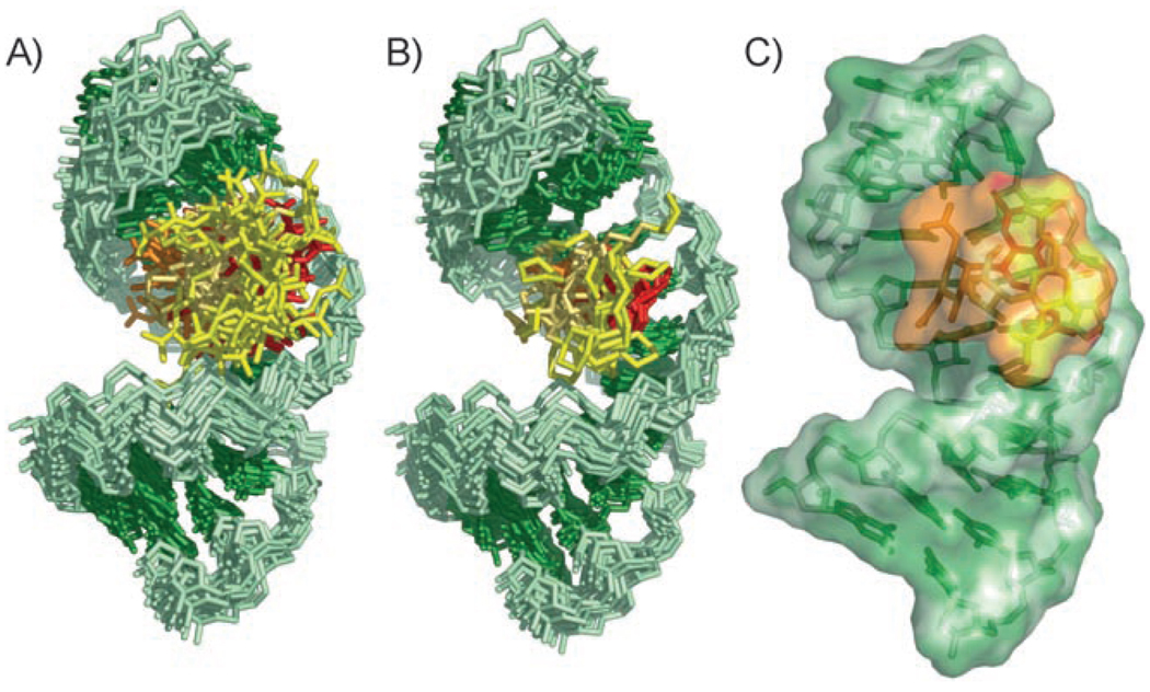
Figure 6.
Structure derived by NMR spectroscopy of the HIV-1 frameshift-site stem-loop RNA complexed with GNB. The RNA is green, and rings I, II, III, and IV of GNB are colored orange, red, pale orange, and yellow, respectively. The view is into the major groove of the GNB-binding site of the RNA. A) Superimposition of the 20 lowest-energy structures over all heavy atoms. B) Superimposition of the 20 lowest-energy structures over all heavy atoms of the RNA and only the rings of GNB. The unrestrained guanidinium groups are omitted for clarity. C) Lowest-energy structure of the complex between the frameshift-site stem loop and GNB.

