Abstract
After developing in the thymus, recent thymic emigrants (RTEs) enter the lymphoid periphery and undergo a maturation process as they transition into the mature naïve (MN) T cell compartment. This maturation presumably shapes RTEs into a pool of T cells best fit to function robustly in the periphery without causing autoimmunity; however, the mechanism and consequences of this maturation process remain unknown. Using a transgenic mouse system that specifically labels RTEs, we tested the influence of MHC molecules, key drivers of intrathymic T cell selection and naive peripheral T cell homeostasis, in shaping the RTE pool in the lymphoid periphery. We found that the TCRs expressed by RTEs are skewed to longer CDR3 regions compared to those of MN T cells, suggesting that MHC does streamline the TCR repertoire of T cells as they transition from the RTE to the MN T cell stage. This conclusion is borne out in studies in which the representation of individual TCRs was followed as a function of time since thymic egress. Surprisingly, we found that MHC is dispensable for the phenotypic and functional maturation of RTEs.
This is an author-produced version of a manuscript accepted for publication in The Journal of Immunology (The JI). The American Association of Immunologists, Inc. (AAI), publisher of The JI, holds the copyright to this manuscript. This version of the manuscript has not yet been copyedited or subjected to editorial proofreading by The JI; hence, it may differ from the final version published in The JI (online and in print). AAI (The JI) is not liable for errors or omissions in this author-produced version of the manuscript or in any version derived from it by the U.S. National Institutes of Health or any other third party. The final, citable version of record can be found at www.jimmunol.org
Keywords: T cells, Cell Differentiation, Repertoire Development, Recent Thymic Emigrants, Homeostasis
Introduction
Recent thymic emigrants (RTEs) are those naïve T cells that have newly entered the lymphoid periphery following development in the thymus. RTEs constantly replenish T cell diversity in the periphery, and their contribution is particularly important in neonates and lymphopenic adults. While RTEs were originally thought to have completed development in the thymus and to enter the lymphoid periphery ready to scan for and distinguish foreign antigen from self, recent evidence has emerged that RTEs undergo a post-thymic maturation period in the lymphoid periphery (1–5). RTEs display reduced proliferation and cytokine secretion in response to stimulation, and a phenotype that is IL-7RαloTCRhiCD3hiCD28loCD24hiQa2loCD45RBlo relative to mature naïve (MN) T cells (1).
While RTEs differ phenotypically from MN T cells, they lack an unambiguous cell surface marker, creating limitations and technical challenges in studying this subset of T cells. However, a murine transgenic (Tg) model system (6) has facilitated the study of RTEs by labeling these cells, allowing for easy identification and separation. These mice contain a transgene encoding GFP under the control of the RAG2 promoter (RAG2p-GFP Tg mice), which is expressed in the thymus and specifically labels RTEs as GFP+ in peripheral T cells (1). Because no new GFP is made in the periphery, this label is brightest in the youngest RTEs (7), and decays over time until it can no longer be detected on T cells that have been out in the lymphoid periphery for more than 3 weeks (1).
Although knowledge of the distinctions between RTEs and MN T cells is amassing, little is known about the mechanisms that govern the maturation process RTEs undergo in the lymphoid periphery. We have previously shown that RTE maturation requires thymic egress and exposure to secondary lymphoid organs (4). MHC molecules are a well-known regulator of naïve T cell homeostasis, transmitting a basal survival signal through the TCR important for the long-term survival of naive T cells (reviewed in 8). Given that RTEs are part of the naïve T cell pool, it is reasonable to hypothesize that MHC molecules, expressed by a variety of cell types within secondary lymphoid organs, deliver signals that shape the population of RTEs. Consistent with this idea is the observation that NKT cells undergo TCR-dependent maturation in the lymphoid periphery driven by the non-classical MHC molecule CD1d (9, 10). MHC molecules may also shape the population of RTEs by narrowing the repertoire, as T cells develop with a range of affinity for self MHC+peptide, and those T cells that leave the thymus with too high (potentially autoreactive), or too low (non-productive) self-reactivity may be winnowed out of the population of RTEs before joining the fully immunocompetent MN T cell pool. Age-dependent shaping of the TCR repertoire in the lymphoid periphery is suggested by the fact that the CD4:CD8 ratio of RTEs is much higher than that of MN T cells (1, 3), and the observation that thymectomized female mice gradually lose reactivity to the male antigen H-Y (11). Here, we show that MHC shapes the population of RTEs by modulating the TCR repertoire of cells that become MN T cells, but does not drive phenotypic maturation of RTEs.
Materials and Methods
Mice
CD4−/−, classical MHC II−/− (I-Abβ−/−), and complete MHC II−/− (lacking the entire MHC II locus) C57BL/6 (B6) mice were purchased from The Jackson Laboratory (Bar Harbor, ME). B6 mice have a disrupted I-Eα gene, but retain expression of I-Eβ (12). Tg B6 mice expressing I-Aβ under control of the K14 promoter (K14p-I-Aβ Tg mice, reference (13) were a gift from M. Bevan (University of Washington). These mice are also I-Aβ−/− and thus lack classical MHC II molecule expression in the lymphoid periphery but express MHC class II on cortical thymic epithelial cells. RAG2p-GFP Tg mice (6) were originally a gift from M. Nussenzweig (The Rockefeller University) and were backcrossed in our lab at least 10 generations onto the B6 background. Also used were chicken ovalbumin-specific MHC class I restricted CD45.1+ OT-I (14) and MHC class II restricted CD45.1+ OT-II (15) and lymphocytic choriomeningitis virus-specific MHC class I restricted CD45.1+ P14 (16) and MHC class II restricted CD45.2+ SMARTA (17) TCR Tg B6 mice. Each of these TCR Tg lines of mice was crossed onto the RAG2p-GFP Tg background to allow identification of RTEs, and GFP brightness of RTEs from polyclonal and TCR Tg mice were similar.
To make radiation chimeras, ~5×10⁶ T cell-depleted bone marrow cells were injected i.v. into lethally irradiated (1000 rads) RAG2p-GFP Tg recipient mice, which were maintained on water containing neomycin sulfate (Mediatech, Inc., Manassas, VA) and Polymyxin B (Invitrogen, Carlsbad, CA) from 1 day (d) before to 14 d after irradiation. TCR Tg and polyclonal T cell populations in competitive bone marrow chimeras were identified using antibodies against a combination of congenic markers and transgene-encoded TCRs. Injected polyclonal T cell marrow expressed the same congenic marker as the recipient, to allow identification of polyclonal RTEs derived from both donor and host bone marrow. Reconstituted chimeras were analyzed ≥ 8 weeks later, while all other mice were used at 6–12 weeks of age. Adoptive transfers were performed by i.v. injection of the indicated number of cells into unmanipulated hosts. All experiments were performed in compliance with the University of Washington Institutional Animal Care and Use Committee.
Cell preparation, staining, enrichment, sorting, and stimulation
Single cell suspensions of thymus, spleen, and lymph nodes were prepared and stained as described in (4) with Abs against the following molecules: CD4 (clone RM4-5), CD8 (53–6.7), CD44 (Pgp-1), CD62L (MEL-14), CD24 (M1/69), Qa2 (1-1-2), CD45RB (16A), CD45.1 (A-20), CD45.2 (104), I-Ab (M5/114.15.2), and TCR subunits Vα2 (B20.1), Vα3.2 (RR3-16), Vα8 (B21.14), Vβ2 (B20.6), Vβ3 (KJ25), Vβ4 (KT4-10), Vβ5 (MR9-4), Vβ6 (RR4-7), Vβ7 (TR310), Vβ8.1/8.2 (KJ16), Vβ9 (MR10-2), Vβ10 (B21.5), Vβ11 (RR3-15), Vβ13 (MR12-3), and Vβ14 (14-2), all from eBioscience (San Diego, CA) or BD Biosciences (San Jose, CA). Biotinylated Abs were detected with PE- or allophycocyanin-conjugated streptavidin (eBioscience). Events were collected on a FACSCanto (BD Biosciences) and data were analyzed on FlowJo (Treestar, Ashland, OR) after excluding doublets from live-gated samples. Fluorescence-Minus-One samples (18) were run when appropriate. Sorting to obtain peripheral naïve (CD62Lhi) CD4 or CD8 RTEs (GFP+) or MN T cells (GFP−) or pre-RTEs (CD4 or CD8 single positive (SP) CD62LhiGFP+ thymocytes) from RAG2p-GFP Tg mice was done as described (4). Briefly, untouched T cells were enriched with an EasySep kit (StemCell Technologies, Vancouver, BC) and sorted on a FACSAria (BD Biosciences) to >97% purity, except pre-RTEs, which were contaminated with a larger population of double-negative thymocytes that were gated out upon analysis.
Where indicated, freshly sorted CD4+CD62Lhi RTEs and MNs, or RTEs transferred to classical MHC II−/− and CD4−/− mice 9 d previously were CFSE labeled and stimulated as described (4). Briefly, 1.25 × 10⁵ freshly sorted CFSE-labeled congenic RTEs or MNs were stimulated for 3 d with 30 ng/ml anti-CD3 and 1 μg/ml anti-CD28 in the presence of 2 × 10⁶ irradiated T-depleted splenocytes in 24-well plates. Alternatively, 2 × 10⁶ CD8 T-cell depleted spleens and lymph node cells from classical MHC II−/− and CD4−/− recipients were CFSE-labeled and subjected to the same stimulation conditions for 3 d.
Spectratyping
TCR CDR3 spectratyping was performed as previously described (19, 20), with modifications. Total RNA was extracted using the RNeasy mini kit (Qiagen, Valencia, CA) and RNA from equivalent numbers of sorted CD4 RTEs and MN T cells and CD8 RTEs and MN T cells was then converted to cDNA using oligo(dT) or random hexamer primers with SuperScript III reverse transcriptase (Invitrogen). The TCR CDR3 region was amplified by a first step PCR using a Vβ- and Cβ-specific primer pair. Amplified CDR3 products were then labeled with a second step PCR using an internal FAM-labeled primer specific for Cβ or Jβ (Invitrogen); the latter reaction amplifies the CDR3 region encoded by a specific Vβ-Jβ recombination. Primer sequences and PCR conditions were based on (20). FAM-labeled runoff products were resolved on a DNA sequencer by the Genomics Facility at the Fred Hutchinson Cancer Research Center (Seattle, WA). Peak heights were calculated from the sequencer data using Genescan software (Applied Biosystems, Foster City, CA). For each sample, peak heights were then expressed as a percentage of total peak heights within that sample.
Statistical analysis
Statistical significance between two groups was determined using a 2-tailed Student’s t-test with equal variance, except where indicated, a paired 2-tailed Student’s t-test was used. Differences with p < .05 were considered significant.
Results
RTEs and MN T cells have globally similar TCR repertoires
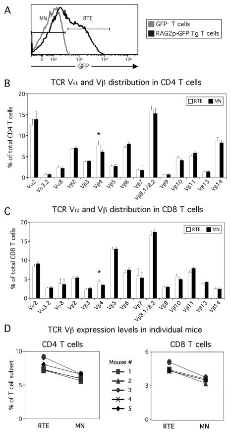
To determine the influence of MHC on RTEs, we first considered the effect of MHC in shaping the TCR repertoire. We hypothesized that, as happens during thymic positive and negative selection, peripheral MHC and self-peptide might transmit survival signals to cells with useful TCR specificities, while causing elimination of cells with too high or too low self-reactivity. For a broad overview of TCR diversity, we assessed the expressed TCR Vα and Vβ repertoire using available V-region-specific antibodies. We identified GFP+ RTEs and GFP− MN T cells in RAG2p-GFP Tg mice (Fig. 1A). For both CD4 (Fig. 1B) and CD8 (Fig. 1C) T cells, we found that RTEs and MN T cells had broadly similar TCR Vα/β usage. However, statistical analysis revealed some subtle differences, including Vβ4 usage frequency. The RTE subset of both CD4 and CD8 T cells from each mouse we analyzed had higher Vβ4 usage relative to their MN counterparts, suggesting that some T cells expressing this V region may be deleted soon after entry into the lymphoid periphery (Fig. 1D).
Figure 1.
The TCR repertoires of RTEs and MN T cells are globally similar. A, Naïve (CD44lo/mid) T cells from RAG2p-GFP Tg mice (black line) were gated as RTEs and MN T cells on the basis of GFP level, using GFP− T cells from B6 mice (gray line) to determine the cutoff for RTEs. B, C, RTE and MN T cells were subdivided on the basis of TCR Vα and Vβ expression for bothCD4 (B) and CD8 (C) T cells. Data are averaged from 6 independent experiments analyzing a total of 5–12 mice per TCR Vα/β subunit, with error bars representing SD. *, p < .005. D, RTE and MN expression levels of TCR Vβ4 are shown for individual mice. p = .002 and p = .0001 for CD4 RTEs vs. MN T cells and CD8 RTEs vs. MN T cells, respectively, using a paired 2-tailed Student’s t-test.
A closer look at TCR usage reveals repertoire differences between RTEs and MN T cells
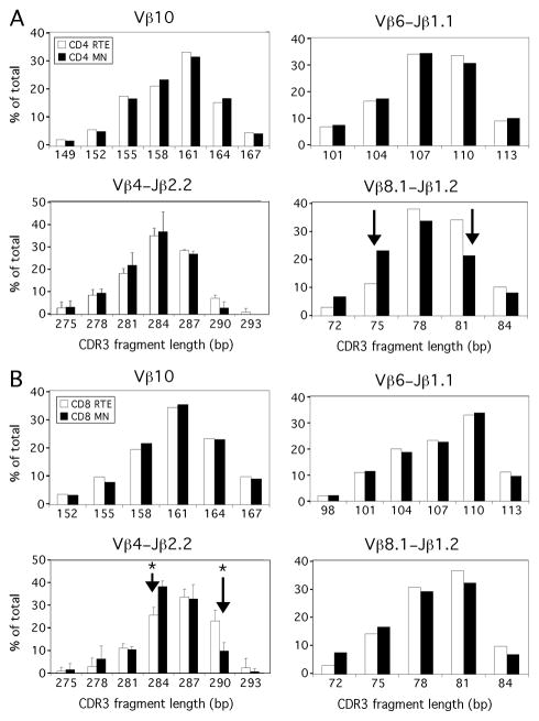
For a higher-resolution TCR repertoire analysis, we compared TCR Vβ CDR3 lengths in TCRs expressed by CD4 and CD8 RTEs and MN T cells using CDR3 length spectratyping (20). Using this method, each Vβ-Jβ combination shows an approximately Gaussian distribution of 6–10 CDR3 lengths in 3 base pair increments (representing in-frame rearrangements). In analyses within a given Vβ, containing rearrangements to all 12 Jβs, RTEs resembled MN T cells in both the CD4 and CD8 compartments for Vβ10 (Fig. 2A, B upper left panels) and the other Vβs assessed (Vβ4, 5, 6, 8.1, 8.2, and 11, data not shown). When we narrowed our focus to CDR3 diversity within particular Vβ-Jβ rearrangements, we found that most of the 48 segments analyzed (all 12 Jβs paired with Vβ4, 6, 8.1, 8.2) showed RTEs to be comparable to MN T cells within both the CD4 and CD8 compartments, including Vβ6-Jβ1.1 (Fig. 2A, B, upper right panels). However, some notable differences were detected, as CD8 RTEs and MN T cells differed in distribution of Vβ4-Jβ2.2 CDR3 lengths, and CD4 RTEs differed from CD4 MN T cells for Vβ8.1-Jβ1.2 (Fig. 2A, B, lower panels). In each of these cases, TCRs expressed by RTEs are skewed towards longer CDR3 regions.
Figure 2.
The TCR repertoires of RTEs and MN T cells are subtly distinct. TCR CDR3 length spectratyping was performed on cDNA from sorted populations of naïve (CD44lo/mid CD62Lhi) CD4 and CD8 RTEs and MN T cells. Representative analyses of the indicated TCR Vβ or Vβ-Jβ recombination are shown for both CD4 (A) and CD8 (B) T cells. Arrows indicate ≥ 10% CDR3 length differences between RTEs and MN T cells, approximately 2-fold the SD. Data are representative of 3 replicates from a pool of 5 mice. Error bars indicate SD from the mean of values from 3 replicates. *, p < .02.
These TCR distributions differences were statistically significant for Vβ4-Jβ2.2 rearrangements in CD8 T cells, but did not reach that threshold for Vβ8.1-Jβ1.2 rearrangements in CD4 T cells. This, in addition to high sample to sample variation, encouraged us to use a different system to determine whether the frequency of cells expressing a given TCR is modulated during the transition from RTE to MN compartment.
The frequency of T cells expressing monoclonal TCRs is modulated at the RTE stage
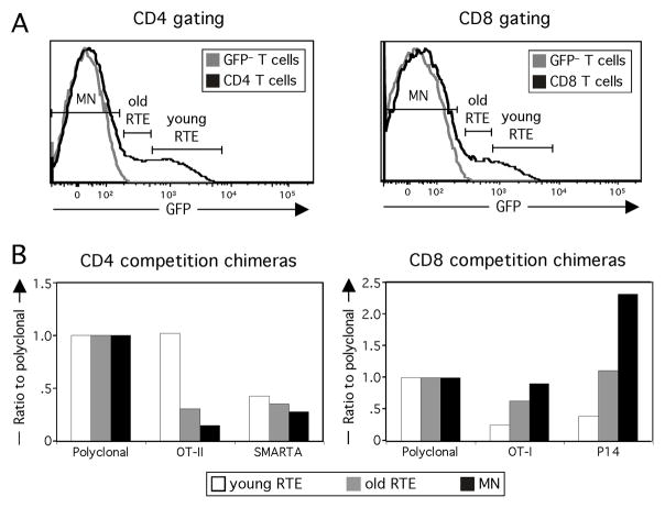
To determine whether repertoire shaping could be detected at the level of an individual TCR, we set up competitive bone marrow chimeras, in which hosts were reconstituted with a mixture of polyclonal and TCR Tg bone marrow, all on the RAG2p-GFP Tg background to allow identification of RTEs and MN T cells within each population of cells. In CD4 competition chimeras, polyclonal T cells were competed with OT-II and SMARTA TCR Tg T cells and in CD8 competition chimeras, polyclonal T cells were competed with OT-I and P14 TCR Tg T cells to determine if their relative frequencies changed as a function of cellular age. T cells from each population of cells were categorized by residence time in the periphery on the basis of GFP brightness (7) into young RTEs, old RTEs, and MN T cells (Fig. 3A), and the frequency of each TCR Tg population relative to polyclonal T cells was then calculated. In all 12 chimeras analyzed, the frequency of OT-II Tg relative to polyclonal CD4 T cells in the periphery was highest in young RTEs, dropping at each stage to a nadir in MN T cells (Fig. 3B, left panel). The difference in relative frequency between young RTE and MN OT-II Tg T cell was statistically significant (p < .001). Given that T cells expressing the OT-II TCR are likely eliminated in the periphery by a superantigen that deletes Vβ5+ T cells (21, 22), this finding confirms that age-dependent modulation of TCR frequency is detectable in our competitive bone marrow chimeras. There was not a striking modulation of the frequency of SMARTA Tg T cells, although two thirds of chimeras analyzed showed a small decrease in frequency from the young RTE to the MN T cell stage, suggesting that subtle modulation might be occurring (p = .069, comparison of young RTE and MN SMARTA TCR Tg T cell relative frequencies). In all 12 CD8 competition chimeras, both OT-I Tg and even more prominently, P14 Tg T cells, increased in frequency over time in the periphery relative to polyclonal CD8 T cells (Fig. 3B, right panel). These differences were statistically significant (p < .001). This molding of the TCR repertoire was seen in chimeras reconstituted with 1:1:1 or 5:1:1 polyclonal:TCR Tg:TCR Tg bone marrow (data not shown). These data suggest that cells expressing these transgene-encoded TCRs are selectively surviving and/or proliferating relative to polyclonal CD8s as a whole. Given that the naïve TCR Tg T cells divided little in the periphery, on the basis of their low BrdU incorporation and low frequency of CD44hi effector/memory cells (data not shown), it is likely that most of the increase in frequency of cells expressing these TCRs is due to enhanced survival. As seen in Fig. 3B, the TCR Tg populations generally seeded the periphery at a frequency lower than their original 1:1:1 proportion in the donor bone marrow. This finding is expected, given that selection of cells expressing uniform TCRs in a competitive situation is limited by thymic expression of positively selecting ligands (23–25).
Figure 3.
The frequency of individual TCRs is modulated in RTEs. Lethally irradiated mice were reconstituted with a mixture of bone marrow from polyclonal and 2 TCR Tg donors. A, T cells from each population of cells were divided into 3 groups for both CD4 (left panel) and CD8 (right panel) chimeras: GFPhi (young) and GFPlo (old) RTEs, and GFP− MN T cells. B, The frequencies of OT-II and SMARTA Tg (left panel) and OT-I and P14 Tg (right panel) T cells within young RTE, old RTE, and MN T cell populations was normalized to the polyclonal level at that stage. Data are representative of 3 independent sets of chimeras for a total of 6–12 individual mice.
RTEs mature upon transfer to a classical MHC class II-deficient environment
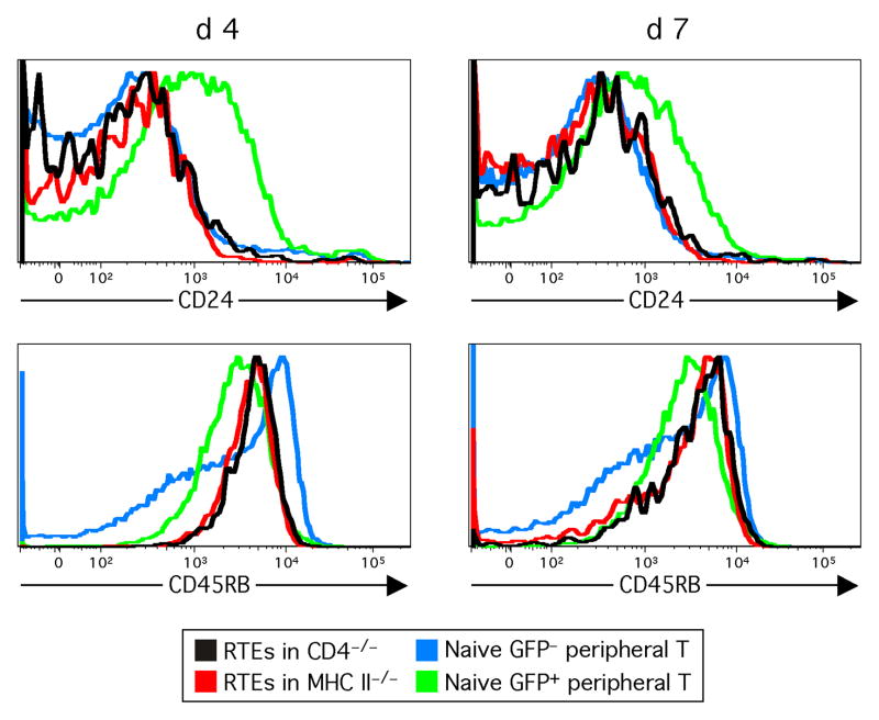
To determine whether MHC imparts maturation signals to RTEs, we placed RTEs in an environment lacking MHC. Sorted CD4 RTEs were transferred to classical MHC II-deficient mice (I-Aβ−/− I-Eα−/− B6 mice) or control CD4−/− B6 mice, which are similarly CD4 T cell lymphopenic, but express MHC class II. RTE maturation in experimental and control recipients was tracked by determining expression levels of CD24, downregulated during RTE maturation, and Qa2 and CD45RB, upregulated during RTE maturation (1, 3), and as seen in overlays of the blue and green histograms in Fig. 4. CD4 RTEs have matured to a comparable degree on d 4 and d 7 following transfer to MHC-deficient and MHC-replete environments (Fig. 4 and data not shown). Thus, the absence of classical MHC class II molecules does not prevent CD4 RTE maturation. Furthermore, while CD24 appeared to be fully downregulated by d 4 post-transfer, upregulation of CD45RB expression continued from d 4 to d 7 in MHC class II-deficient mice (Fig. 4), suggesting that CD4 RTE maturation in the absence of MHC class II progresses normally.
Figure 4.
RTEs mature normally following transfer to a classical MHC II-deficient environment. At the indicated time points post-transfer, CD24 and CD45RB expression by sorted CD4 RTEs transferred into I-Aβ-deficient (RTEs in MHC II−/−) or CD4- deficient (RTEs in CD4−/−) mice were determined. Splenocytes from an unmanipulated RAG2p-GFP Tg mouse were analyzed on the same day for marker expression by CD4+CD44lo/mid RTEs (Naive GFP+ peripheral T) and MN (Naïve GFP− peripheral T) cells. Data are representative of 4 independent experiments.
RTEs mature normally in situ in a classical MHC class II-deficient environment
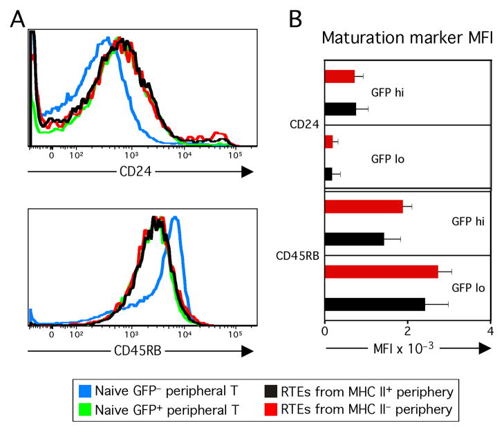
We next analyzed RTE maturation in the absence of exposure to a MHC II+ periphery and without the complication of adoptive transfer. RAG2p-GFP Tg and K14p-I-Aβ Tg mice were crossed to generate RTE reporter mice that lack classical MHC class II expression in the lymphoid periphery but express MHC II on cortical thymic epithelial cells, allowing for positive selection of CD4 T cells (13). CD4 RTEs from MHC class II-expressing mice and from peripheral MHC II-deficient mice exhibited comparable maturation as assessed by CD24 and CD45RB levels, confirming that RTEs can mature normally in a MHC-deficient periphery (Fig. 5A). Note that RTEs analyzed in situ in both of these mice are phenotypically identical to unmanipulated RTEs from RAG2p-GFP Tg mice. Here, the cells are not transferred, and are instead analyzed in situ, and should have an identical phenotype to the unmanipulated control. Furthermore, from the GFPhi young RTE to the GFPlo old RTE stage, “undisturbed” RTEs in the MHC-deficient periphery showed the normal pattern of progressive maturation (Fig. 5B).
Figure 5.
RTEs mature normally in situ in a classical MHC II-deficient lymphoid periphery. A, B, CD24 and CD45RB expression levels by CD4+GFP+ RTEs from RAG2p-GFP Tg × K14-I-Aβ Tg mice (RTEs from MHC II− periphery) or from a MHC II+ periphery are shown. Splenocytes from an unmanipulated RAG2p-GFP Tg mouse were analyzed on the same day for marker expression by CD4+CD44lo/mid RTEs (Naive GFP+ peripheral T) and MN (Naive GFP− peripheral T) cells. Representative data are shown in A, and data in B are averaged median fluorescence intensity of RTEs split into young GFPhi and old GFPlo populations from 3 independent experiments analyzing a total of 6–10 mice per group, with error bars representing SD. Differences were not statistically significant (p > .05).
RTEs mature normally upon transfer to a complete MHC class-II deficient environment
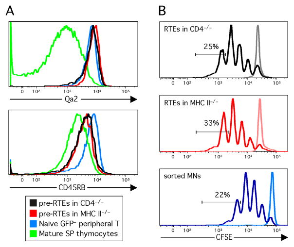
While I-A−/− mice lack expression of native MHC II molecules, these mice do express very low levels of hybrid I-Aα/I-Eβ MHC class II molecules (26, 27). To eliminate hybrid MHC class II expression, we used as adoptive hosts B6 mice in which the entire MHC class II locus is deleted (28). To obtain donor T cells that were never exposed to MHC class II in the periphery, the most developmentally mature subset of CD4 SP thymocytes (pre-RTEs) were sorted from RAG2p-GFP Tg mice and transferred to full MHC II-deficient or control CD4−/− mice. CD4 pre-RTEs (Fig. 6A) and transferred RTEs (data not shown) matured normally in the absence of MHC class II. Both transferred populations exhibited phenotypic maturation (i.e. increased marker expression relative to that by mature SP thymocytes). Furthermore, there was no statistically significant defect in Qa2 and CD45RB expression levels on pre-RTEs from MHC-deficient mice as compared to those on CD4−/− controls (n = 4 per group).
Figure 6.
CD4 pre-RTEs mature normally following transfer to mice expressing no MHC II molecules. A, Sorted CD4+CD8−GFP+CD62Lhi thymocytes (pre-RTEs) were transferred into complete MHC II−/− or CD4−/− mice and Qa2 and CD45RB expression levels were determined on cells 8 d following transfer. Thymocytes and splenocytes from an unmanipulated RAG2p-GFP Tg mouse were analyzed on the same day for marker expression by CD4+CD8−GFP+CD62Lhi thymocytes (Mature SP thymocytes) and CD4+CD44lo/mid MN (Naïve GFP− peripheral T) cells. Data are representative of 2 experiments, analyzing a total of 4 mice per condition. B, Sorted congenic CD4+CD62Lhi RTEs were transferred to classical MHC II−/− or CD4−/− mice, and at 9 d following transfer, were CFSE-labeled and stimulated in vitro for 3 d with anti-CD3 and anti-CD28. Freshly sorted CD4+CD62Lhi MN T cells were concomitantly CFSE-labeled and stimulated. Labeled histograms depict CFSE dilution of stimulated (darker lines) or unstimulated (lighter lines) samples. Indicated are the percents of cells that have divided 4 or more times. Data are representative of 2 recipients per condition.
To determine if RTEs undergo functional maturation in an MHC-deficient environment, we compared the proliferative capacity of CD4 RTEs allowed to age upon transfer to classical MHC II−/− and CD4−/− mice. Cells were CFSE-labeled and stimulated in vitro with anti-CD3 and anti-CD28 9 d following transfer. RTEs showed a comparable rate of division in both recipients (Fig. 6B), suggesting that MHC is not required for functional maturation. Furthermore, the transferred RTEs appeared to be fully mature by this measure, as their level of division was comparable to that of stimulated, freshly sorted CD4 MN T cells (Fig. 6B). This proliferative capacity is greater than the level of division shown by RTEs that have not aged in an adoptive host (1, 3). Compared to CD4 MN T cells, fewer than half the number of such “fresh” CD4 RTEs have undergone 4 or more rounds of division during the 3 d in vitro stimulation period (data not shown).
Discussion
The work presented here shows that the TCR repertoires of RTEs and MN T cells differ. Because MN T cells are derived from the subset of RTEs that survive for longer than 3 weeks in the periphery, the most likely interpretation is that RTEs cells with some TCR specificities do not survive to the MN T cell stage. This TCR repertoire shaping is likely driven in part by tonic signals transmitted to naïve T cells by MHC molecules, signals known to be important for cell survival (reviewed in 8). Naive T cells with too low an affinity for self-MHC+peptide may not receive sufficient survival signals to persist in the lymphoid periphery. This may in part explain why OT-I and P14 TCR Tg RTEs persist better than polyclonal CD8 RTEs, in which some T cells expressing TCRs with very low self-affinity may not persist. The 3 weeks T cells remain RTEs is a time period in which interaction with self-MHC molecules might be expected to shape the population of RTEs, given that naïve T cells prevented from recognizing MHC begin to die within this time span (29–31). To directly show that TCR repertoire changes in RTEs require MHC would necessitate adoptive transfers into MHC-deficient recipients; however, interpretation of these technically challenging experiments would be significantly confounded by the fact that T cells would begin to die in the absence of MHC.
In addition to poor survival, naïve T cells with very low self-affinity also are unable to undergo undergo lymphopenia-induced proliferation (LIP) (8). This may partially explain why OT-II TCR Tg RTEs, which do not undergo LIP (32), compete poorly with polyclonal T cells. Preferential LIP could also contribute to the observed increase in OT-I and P14, but this contribution is likely to be small, given that these TCR Tg cells remained largely CD44lo, and cells undergoing extensive LIP would be expected to become CD44hi (8).
On the other hand, T cells expressing TCRs with too high an affinity for self-MHC+peptide in the lymphoid periphery can be anergized and/or deleted. Indeed, probable deletion via superantigen is seen in OT-II TCR Tg RTEs in our TCR Tg competition chimera system (Fig. 3). Deletion of autoreactive polyclonal RTEs may account for some of the observed increase in OT-I and P14 T cells in the MN T cell compartment. Consistent with a role for MHC in shaping RTEs is the observation that CD8 RTEs appear to have specific gut homing properties. This exposure to the antigen-rich gut microenvironment is speculated to be important for inducing T cell tolerance to intestinal microflora (33).
Modulation of the TCR repertoire is subtle in RTEs, and is not apparent at the level of differences in TCR Vα or Vβ proportions between RTEs and MN T cells, aside from the relative loss of Vβ4 expression by MN T cells compared to RTEs in both CD4 and CD8 subsets (Fig. 1). However, repertoire shifts are apparent by TCR CDR3 length distribution. Interestingly, the TCR CDR3 repertoire differences showed that RTEs are skewed to expressing TCRs with longer CDR3 regions (Fig. 2). A longer CDR3 may extend further out into space, thereby allowing for a higher degree of contact and potentially greater affinity for a specific MHC+peptide ligand, while a shorter CDR3 may not contact peptide well, but instead interact mainly with the MHC (34–36). This idea is consistent with the observation that negative selection appears to shorten the mean CDR3 length in thymocytes (37, 38). These data suggest that potentially autoreactive RTEs expressing TCRs with longer CDR3 regions are deleted upon encounter with ligand in the lymphoid periphery. It is not known which self-ligands may be influencing the stage-specific expression of TCR Vβ4, but viral superantigens are an unlikely source (39).
Our data also surprisingly suggest that while MHC shapes the RTE population, it does not drive the phenotypic or functional maturation of RTEs in the lymphoid periphery. The simplest experiment, transferring RTEs to I-Aβ−/− recipients, suggests that maturation occurs normally in the absence of MHC (Fig. 4). However, harvesting donor RTEs from the lymphoid periphery allows some exposure to peripheral MHC, thus opening the possibility that this brief encounter drives RTE maturation even after transfer to MHC-deficient recipients. Our in situ maturation model alleviates those concerns, as RTEs from this environment are not subject to adoptive transfer and are never exposed to MHC in the periphery (Fig. 5). An additional caveat from these experiments is the possibility that the low level of hybrid MHC II molecule expression in I-Aβ−/− mice (26, 27) could influence maturation. However, adoptive transfers of pre-RTEs into mice in which the entire MHC II locus was deleted showed that maturation progressed normally in this environment (Fig. 6), while maturation of RTEs cannot be completed without thymic egress (4). In addition, the adoptive transfer experiments circumvent the T cell lymphopenia seen in the in situ maturation model. This lymphopenia, equally noticeable in MN and RTE CD4 T cell subsets (data not shown), may be due both to altered T cell development in the thymus and lower T cell survival in the periphery. In the adoptive transfer experiments, the same numbers of RTEs were recovered from the spleens of both MHC II−/− and CD4−/− hosts (7–8 % of input at d 7 post-transfer), and were comparable to the numbers of transferred MN T cells that were recovered. This suggests that the bulk of transferred CD4 RTEs survive in the absence of MHC class II for the duration of the experiment, and that maturation is not due to preferential survival of a mature subset of RTEs, in concordance with previous findings (4). A role for MHC I in driving CD8 RTE maturation was not directly tested due to the technical challenge posed by the expression of MHC I by T cells themselves. However, given that the maturation process appears to be largely identical in CD4 and CD8 RTEs (1), it is likely that the same factor(s) modulate these processes.
Taken together, our data suggest that RTEs are receiving multiple signals in the lymphoid periphery. One signal comes from an unknown factor(s) that drives RTEs to fully mature in the lymphoid periphery. An additional signal comes from MHC, that, while not influencing RTE maturation, appears to shape the RTE TCR repertoire. This shaping process likely preserves RTEs with useful TCR specificities for incorporation into the MN T cell pool, but eliminates RTEs with TCR specificities that are potentially autoreactive or that may possess an affinity for self that is too low to be functional. While 99% of developing thymocytes never exit the thymus, those cells that are allowed to escape to seed the lymphoid periphery are still subject to additional selective forces. Selection of immunocompetent but self-tolerant T cells occurs at multiple levels and exacts a great price.
Non-standard abbreviations
- Tg
Transgenic
- RTE
recent thymic emigrant
- MN
mature naïve
- RAG2p-GFP Tg
mice transgenic for GFP under control of the RAG2 promoter
- SP
single positive
- LIP
lymphopenia-induced proliferation
- C57BL/6
B6
- day
d
Footnotes
Disclosures
The authors declare no competing financial interests. The content is solely the responsibility of the authors and does not necessarily represent the official views of either the NIH or the NCI.
Funding This work was supported by grants from the National Cancer Institute (T32CA0095) to E.G.H. and the National Institutes of Health (R01 AI 064318) to P.J.F.
References
- 1.Boursalian TE, Golub J, Soper DM, Cooper CJ, Fink PJ. Continued maturation of thymic emigrants in the periphery. Nat Immunol. 2004;5:418–425. doi: 10.1038/ni1049. [DOI] [PubMed] [Google Scholar]
- 2.Haines CJ, Giffon TD, Lu LS, Lu X, Tessier-Lavigne M, Ross DT, Lewis DB. Human CD4+ T cell recent thymic emigrants are identified by protein tyrosine kinase 7 and have reduced immune function. J Exp Med. 2009;206:275–285. doi: 10.1084/jem.20080996. [DOI] [PMC free article] [PubMed] [Google Scholar]
- 3.Hale JS, Boursalian TE, Turk GL, Fink PJ. Thymic output in aged mice. Proc Natl Acad Sci USA. 2006;103:8447–8452. doi: 10.1073/pnas.0601040103. [DOI] [PMC free article] [PubMed] [Google Scholar]
- 4.Houston EG, Jr, Nechanitzky R, Fink PJ. Cutting edge: Contact with secondary lymphoid organs drives postthymic T cell maturation. J Immunol. 2008;181:5213–5217. doi: 10.4049/jimmunol.181.8.5213. [DOI] [PMC free article] [PubMed] [Google Scholar]
- 5.Opiela SJ, Koru-Sengul T, Adkins B. Murine neonatal recent thymic emigrants are phenotypically and functionally distinct from adult recent thymic emigrants. Blood. 2009;113:5635–5643. doi: 10.1182/blood-2008-08-173658. [DOI] [PMC free article] [PubMed] [Google Scholar]
- 6.Yu W, Nagaoka H, Jankovic M, Misulovin Z, Suh H, Rolink A, Melchers F, Meffre E, Nussenzweig MC. Continued RAG expression in late stages of B cell development and no apparent re-induction after immunization. Nature. 1999;400:682–687. doi: 10.1038/23287. [DOI] [PubMed] [Google Scholar]
- 7.McCaughtry TM, Wilken MS, Hogquist KA. Thymic emigration revisited. J Exp Med. 2007;204:2513–2520. doi: 10.1084/jem.20070601. [DOI] [PMC free article] [PubMed] [Google Scholar]
- 8.Surh CD, Sprent J. Homeostasis of naive and memory T cells. Immunity. 2008;29:848–862. doi: 10.1016/j.immuni.2008.11.002. [DOI] [PubMed] [Google Scholar]
- 9.McNab FW, Berzins SP, Pellicci DG, Kyparissoudis K, Field K, Smyth MJ, Godfrey DI. The influence of CD1d in postselection NKT cell maturation and homeostasis. J Immunol. 2005;175:3762–3768. doi: 10.4049/jimmunol.175.6.3762. [DOI] [PubMed] [Google Scholar]
- 10.McNab FW, Pellicci DG, Field K, Besra G, Smyth MJ, Godfrey DI, Berzins SP. Peripheral NK1.1- NKT cells are mature and functionally distinct from their thymic counterparts. J Immunol. 2007;179:6630–6637. doi: 10.4049/jimmunol.179.10.6630. [DOI] [PubMed] [Google Scholar]
- 11.Di Rosa F, Ramaswamy S, Ridge JP, Matzinger P. On the lifespan of virgin T lymphocytes. J Immunol. 1999;163:1253–1257. [PubMed] [Google Scholar]
- 12.Grusby MJ, Johnson RS, Papaioannou VE, Glimcher LH. Depletion of CD4+ T cells in major histocompatibility complex class II-deficient mice. Science. 1991;253:1417–1420. doi: 10.1126/science.1910207. [DOI] [PubMed] [Google Scholar]
- 13.Laufer TM, DeKoning J, Markowitz JS, Lo D, Glimcher LH. Unopposed positive selection and autoreactivity in mice expressing class II MHC only on thymic cortex. Nature. 1996;383:81–85. doi: 10.1038/383081a0. [DOI] [PubMed] [Google Scholar]
- 14.Hogquist KA, Jameson SC, Heath WR, Howard JL, Bevan MJ, Carbone FR. T cell receptor antagonist peptides induce positive selection. Cell. 1994;76:17–27. doi: 10.1016/0092-8674(94)90169-4. [DOI] [PubMed] [Google Scholar]
- 15.Barnden MJ, Allison J, Heath WR, Carbone FR. Defective TCR expression in transgenic mice constructed using cDNA-based alpha- and beta-chain genes under the control of heterologous regulatory elements. Immunol Cell Biol. 1998;76:34–40. doi: 10.1046/j.1440-1711.1998.00709.x. [DOI] [PubMed] [Google Scholar]
- 16.Pircher H, Burki K, Lang R, Hengartner H, Zinkernagel RM. Tolerance induction in double specific T-cell receptor transgenic mice varies with antigen. Nature. 1989;342:559–561. doi: 10.1038/342559a0. [DOI] [PubMed] [Google Scholar]
- 17.Oxenius A, Bachmann MF, Zinkernagel RM, Hengartner H. Virus-specific MHC-class II-restricted TCR-transgenic mice: effects on humoral and cellular immune responses after viral infection. Eur J Immunol. 1998;28:390–400. doi: 10.1002/(SICI)1521-4141(199801)28:01<390::AID-IMMU390>3.0.CO;2-O. [DOI] [PubMed] [Google Scholar]
- 18.Herzenberg LA, Tung J, Moore WA, Herzenberg LA, Parks DR. Interpreting flow cytometry data: a guide for the perplexed. Nat Immunol. 2006;7:681–685. doi: 10.1038/ni0706-681. [DOI] [PubMed] [Google Scholar]
- 19.Hendricks DW, Fink PJ. Uneven colonization of the lymphoid periphery by T cells that undergo early TCR a rearrangements. J Immunol. 2009;182:4267–4274. doi: 10.4049/jimmunol.0804180. [DOI] [PMC free article] [PubMed] [Google Scholar]
- 20.Pannetier C, Cochet M, Carche S, Casrouge A, Zoller M, Kourilsky P. The sizes of the CDR3 hypervariable regions of the murine T cell receptor b chains vary as a function of the recombined germ-line segments. Proc Natl Acad Sci USA. 1993;90:4319–4323. doi: 10.1073/pnas.90.9.4319. [DOI] [PMC free article] [PubMed] [Google Scholar]
- 21.Fink PJ, Fang CA, Turk GL. The induction of peripheral tolerance by the chronic activation and deletion of CD4+Vβ5+ cells. J Immunol. 1994;152:4270–4281. [PubMed] [Google Scholar]
- 22.Fink PJ, Swan K, Turk G, Moore MW, Carbone FR. Both intrathymic and peripheral selection modulate the differential expression of Vβ5 among CD4+ and CD8+ T cells. J Exp Med. 1992;176:1733–1738. doi: 10.1084/jem.176.6.1733. [DOI] [PMC free article] [PubMed] [Google Scholar]
- 23.Dillon SR, Fink PJ. Thymic selection events mediated by the pre-TCR do not depend upon a limiting ligand. Int Immunol. 1995;7:1363–1373. doi: 10.1093/intimm/7.8.1363. [DOI] [PubMed] [Google Scholar]
- 24.Huesmann M, Scott B, Kisielow P, von Boehmer H. Kinetics and efficacy of positive selection in the thymus of normal and T cell receptor transgenic mice. Cell. 1991;66:533–540. doi: 10.1016/0092-8674(81)90016-7. [DOI] [PubMed] [Google Scholar]
- 25.Merkenschlager M, Benoist C, Mathis D. Evidence for a single-niche model of positive selection. Proc Natl Acad Sci USA. 1994;91:11694–11698. doi: 10.1073/pnas.91.24.11694. [DOI] [PMC free article] [PubMed] [Google Scholar]
- 26.Dorfman JR, Stefanova I, Yasutomo K, Germain RN. CD4+ T cell survival is not directly linked to self-MHC-induced TCR signaling. Nat Immunol. 2000;1:329–335. doi: 10.1038/79783. [DOI] [PubMed] [Google Scholar]
- 27.Martin B, Bourgeois C, Dautigny N, Lucas B. On the role of MHC class II molecules in the survival and lymphopenia-induced proliferation of peripheral CD4+ T cells. Proc Natl Acad Sci USA. 2003;100:6021–6026. doi: 10.1073/pnas.1037754100. [DOI] [PMC free article] [PubMed] [Google Scholar]
- 28.Madsen L, Labrecque N, Engberg J, Dierich A, Svejgaard A, Benoist C, Mathis D, Fugger L. Mice lacking all conventional MHC class II genes. Proc Natl Acad Sci USA. 1999;96:10338–10343. doi: 10.1073/pnas.96.18.10338. [DOI] [PMC free article] [PubMed] [Google Scholar]
- 29.Hataye J, Moon JJ, Khoruts A, Reilly C, Jenkins MK. Naive and memory CD4+ T cell survival controlled by clonal abundance. Science. 2006;312:114–116. doi: 10.1126/science.1124228. [DOI] [PubMed] [Google Scholar]
- 30.Labrecque N, Whitfield LS, Obst R, Waltzinger C, Benoist C, Mathis D. How much TCR does a T cell need? Immunity. 2001;15:71–82. doi: 10.1016/s1074-7613(01)00170-4. [DOI] [PubMed] [Google Scholar]
- 31.Polic B, Kunkel D, Scheffold A, Rajewsky K. How ab T cells deal with induced TCR a ablation. Proc Natl Acad Sci USA. 2001;98:8744–8749. doi: 10.1073/pnas.141218898. [DOI] [PMC free article] [PubMed] [Google Scholar]
- 32.Ernst B, Lee DS, Chang JM, Sprent J, Surh CD. The peptide ligands mediating positive selection in the thymus control T cell survival and homeostatic proliferation in the periphery. Immunity. 1999;11:173–181. doi: 10.1016/s1074-7613(00)80092-8. [DOI] [PubMed] [Google Scholar]
- 33.Staton TL, Habtezion A, Winslow MM, Sato T, Love PE, Butcher EC. CD8+ recent thymic emigrants home to and efficiently repopulate the small intestine epithelium. Nat Immunol. 2006;7:482–488. doi: 10.1038/ni1319. [DOI] [PubMed] [Google Scholar]
- 34.Fazilleau N, Cabaniols JP, Lemaitre F, Motta I, Kourilsky P, Kanellopoulos JM. Vα and Vβ public repertoires are highly conserved in terminal deoxynucleotidyl transferase deficient mice. J Immunol. 2005;174:345–355. doi: 10.4049/jimmunol.174.1.345. [DOI] [PubMed] [Google Scholar]
- 35.Gavin MA, Bevan MJ. Increased peptide promiscuity provides a rationale for the lack of N regions in the neonatal T cell repertoire. Immunity. 1995;3:793–800. doi: 10.1016/1074-7613(95)90068-3. [DOI] [PubMed] [Google Scholar]
- 36.Gilfillan S, Waltzinger C, Benoist C, Mathis D. More efficient positive selection of thymocytes in mice lacking terminal deoxynucleotidyl transferase. Int Immunol. 1994;6:1681–1686. doi: 10.1093/intimm/6.11.1681. [DOI] [PubMed] [Google Scholar]
- 37.Nishio J, Suzuki M, Nanki T, Miyasaka N, Kohsaka H. Development of TCRB CDR3 length repertoire of human T lymphocytes. Int Immunol. 2004;16:423–431. doi: 10.1093/intimm/dxh046. [DOI] [PubMed] [Google Scholar]
- 38.Matsutani T, Ohmori T, Ogata M, Soga H, Kasahara S, Yoshioka T, Suzuki R, Itoh T. Comparison of CDR3 length among thymocyte subpopulations: impacts of MHC and BV segment on the CDR3 shortening. Mol Immunol. 2007;44:2378–2387. doi: 10.1016/j.molimm.2006.10.026. [DOI] [PubMed] [Google Scholar]
- 39.Luther SA, Acha-Orbea H. Mouse mammary tumor virus: immunological interplays between virus and host. Adv Immunol. 1997;65:139–243. [PubMed] [Google Scholar]


