Abstract
Global histone modifications and their putative relevance to short and long term cellular programming have drawn substantial interest in the study of chromatin. Here we describe the use of reverse-phase liquid chromatography coupled to Linear Ion Trap-Fourier Transform Mass Spectrometry (RPLC–LTQ-FTMS) to quickly profile post-translationally modified isoforms and variants for core histone proteins from as few as 5×10⁴ cells at isotopic resolution. Such LC-MS profiling greatly facilitated detection of histones from HeLa S3 or 293T cells experiencing shRNA- or siRNA-knockdown of histone deacetylase (HDAC) 1, 2, 3 or 1 and 2 together. In no case was significant global histone hyperacetylation relative to control cells observed, suggesting possible compensation of deacetylation activity by partially redundant enzymes in the 18-member HDAC family. This contrasts sharply with yeast where genetic deletion of HDAC rpd3 causes massive hyperacetylation. Treatment of cells with TSA and class I selective HDAC inhibitors had similar ability to induce global histone hyperactylation, though to different extents in HeLa S3 vs. 293T cells.
Keywords: acetylation, histone, HDACs, LC-FTMS
Introduction
Dynamic organization of chromatin is of great importance in the regulation of eukaryotic gene expression [1]. The local conformation of chromatin influences the access of chromatin-associated proteins to DNA. Specific modifications of the histone tails modulate interaction affinities of chromatin-associated proteins and transcription factors. Different modifications on the same or different histone tails may be interdependent, but clearly generate hundreds of distinct combinations of protein forms [2, 3] suggesting a “histone code” that significantly extends the information potential of the genetic code [4]. In particular, histone acetylation is associated with transcriptional activation [5]. Additionally, acetylation of core histones has been correlated with other genome functions, including chromatin assembly, DNA repair and recombination [6]. The dynamic equilibrium of histone acetylation levels is achieved by two kinds of enzymes, histone acetyltransferases (HATs), such as p300, CBP and P/CAF, and histone deacetylases (HDACs). Five HDACs have been identified from yeast and eighteen human HDACs are classified into four classes based on their homology to yeast HDACs and phylogenetic analysis [7]. HDACs often function in concert with other proteins to form multiprotein complexes (e.g., NuRD [8], N-CoR [9], SMRT [10], and Sin3 [8]) recruited to specific loci to remove acetyl groups from histones and in turn suppress gene transcription. Overexpressed or sustained HDAC activity, occurring in some cancer cells [11, 12], results in deacetylation of the histone tails (a.k.a. histone hypoacetylation). Hence histone deacetylation by HDAC may be a mechanism for silencing some tumor suppressor genes, such as p53, and cyclin-dependent kinase inhibitors, such as p21, responsible for cell differentiation or proliferation.
Most frequently, histone modifications are analyzed using highly specific antibodies directed against specific modifications at particular amino acids. However, despite being highly sensitive, these methods are not informative about the global state of chromatin in cells and can give false readings from neighboring modifications affecting antibody binding [13, 14, 15]. Recently, the coupling of high performance liquid chromatography and mass spectrometry (LC-MS) has enabled the detection of multiple post-translational modifications occurring on histone H2A and H4 [16, 17]. More detailed information associated to the location of a specific modification, if required, can be obtained by tandem mass spectrometry (MS/MS). Both top-down [18, 19, 20, 21, 22] and bottom-up [23, 24, 25] MS approaches have been successful in generating a comprehensive map of isolated histone PTMs and hundreds of specific combinations of PTMs on the most heavily modified histone, H3 [3].
Here, we use top-down mass spectrometry to study the global profile of human histones, by an online LC-MS method using >102 better mass resolving power than prior LC-MS histone assays [16, 17]. This method provides a global view of core histones from <105 cells after treatment with inhibitors or HDAC knockdown. Broad spectrum HDAC inhibitors caused global hyperacetylation on all core histones whereas knocking down one or two of class I HDACs did not affect global histone acetylation levels significantly. These data contrast with genetic disruption of their homologous HDAC in yeast (rpd3) which caused significant hyperacetylation on histone H3 and H4. Inhibition of HATs also caused detectable changes in the level of global acetylation. This LC-MS method is now suitable for screening of HDAC/HAT inhibitors and efficiently provides a gene-specific snapshot of each histone in the context of global chromatin.
Experimental procedures
Cell culture and Reagents
Hela S3 cells and 293T cells were maintained in Dulbecco’s modified Eagle’s medium (DMEM) with 4.5 g/L glucose containing 10% fetal bovine serum at 37°C with 5% CO2. Reagents for siRNA were ordered from Dharmacon and prepared according to the vendor’s instructions. Transfection with control siRNA, HDAC1-specific or HDAC2-specific siRNA (SMARTPool) was performed by using DharmaFECT (Dharmacon). Sodium butyrate, TSA, apicidin and valproic acid and Anacardic acid were purchased from Sigma.
Lentivirus-delivered RNAi
shRNAs for human HDAC3 constructed in pLKO.1-Puro vectors were purchased from Sigma (Mission shRNA) and contained 5 constructs with different target sequences, all of which were packaged for viral production and infection, and tested for target knockdown. shRNA with scrambled sequence was used for a negative control. For viral packaging, pLKOshRNA, pCMV-dR8.91 and pCMV-VSV-G were co-transfected into 293T cells using Fugene 6 (Roche). Media containing viruses were collected 48 h after transfection. Hela S3 cells were infected with the viruses in the presence of polybrene (8 mg/mL) for 24 h, and then subjected to selection by 2 μg/mL puromycin for 5 days prior to further analysis.
Treatment with HDAC and HAT inhibitors
HeLa S3 cells and 293T cells were treated by 10 mM sodium butyrate and 1 μM TSA for 4 h, respectively. 293T cells were treated by 2.5 μM apicidin or 5 mM valproic acid for 4 h. HAT inhibitor, anacardic acid was used to treat HeLa S3 cells and 293T cells for 24 h in a final concentration of 15 μM. All control cells were treated by the same volume of DMSO.
Histone purification
Mammalian histones were purified as described below. Briefly, cells were scraped from plates in nucleus isolation buffer (15 mM Tris-HCl; pH 7.5, 60 mM KCl, 11 mM CaCl2, 5 mM NaCl, 5 mM MgCl2, 250 mM sucrose, 1 mM dithiothreitol, 5 nM microcystin-LR, 500 μM 4-(2-aminoethyl) benzenesulfonyl fluoride, 10 mM sodium butyrate and 0.3% NP-40). Nuclei were gently pelleted by centrifugation and washed with nucleus isolation buffer (same as mentioned above, but without NP-40). Histones were acid extracted with 0.4 N H2SO4 for 30 min. and precipitated with 20% trichloroacetic acid (TCA) overnight, followed by washes with cold acetone containing 0.1% HCl and then pure acetone. Before further separation by RPLC, the sample was oxidized by incubating at room temperature in 3% (v/v) formic acid and 3% H2O2 (v/v) for 4 h [26]. Yeast histones were purified and analyzed as described previously [27].
Western blotting
Protein concentrations of cell lysates were measured by the Bradford method. Approximately 20-30 μg of protein is loaded to 12% SDS-PAGE and transferred to a PVDF membrane. The membranes were probed with primary antibodies against HDAC1 (1:3000), HDAC2 (1:3000) (Upstate Biotechnology, Lake Placid, NY), HDAC3 (1:2000) and GAPDH (1:5000) (Santa Cruz) in 0.05% Tween 20/PBS and then with an HRP-labeled secondary antibody (1:3000; Santa Cruz). Proteins were visualized by chemiluminescence with ECL Western blotting detection reagents (Pierce).
Online LC-MS
All LC-MS experiments were performed on an LTQ-FT mass spectrometer operating at 7 Tesla (Thermo Fisher Scientific, San Jose, CA). Histones were separated by reverse-phase HPLC (Alltech Vydac TP C18 column, 1.0 mm i.d., 250 mm, Nicholasville, KY) at a constant flow rate of 50 μL/min. Mobile phases were (A) 0.05% trifluoroacetic acid (TFA) in water and (B) 0.05% trifluoroacetic acid in acetonitrile. Using 0.05% TFA increased separation power and reduced the presence of TFA adducts. Phase B increased from 0 to 30% in 20 min. and from 30% to 60% in 100 min. Between each run, the column was washed by 3 gradients from 0% B to 100% B and then equilibrated at 0% B for 25 min. To minimize adduction of phosphate anions and ion-pairing reagents, a setting of 20 V was used for mild ion activation in the ESI source. The LTQ-FT method consisted of two scan events. The initial event performed a full scan in the ion trap. The second segment was used for a broadband high-resolution scan in the trapped ion cell of the Fourier-Transform (FTMS) portion of the instrument with AGC setting at 1,000,000 ions and resolution of 100,000 at m/z 400. The monoisotopic mass of each peak was calculated by using the Xtract algorithm for mass assignment of FT-ICR MS data.
Results
HPLC separation and MS detection of intact histones
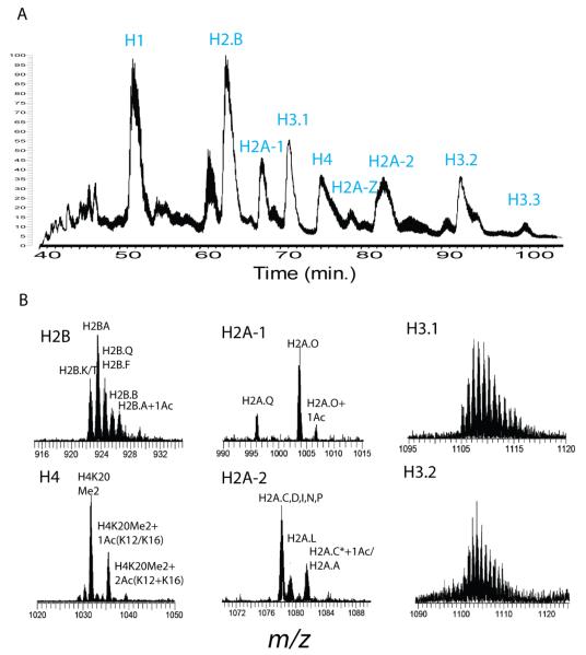
Top-down mass spectrometry has been successfully applied to the identification and characterization of histone forms with pre-purified histone fractions with large sample requirements [18, 19, 20, 21, 22]. Oxidation of samples before RPLC separation converted methionine to methionine sulfone and helped to resolve each histone type into an individual chromatographic peak (Figure 1A). All the FTMS spectra during the elution time of each chromatographic peak were summed together to generate the averaged broadband spectrum of each histone (Figure 1B). Combined with previous data acquired from extensive MS/MS for each peak in the spectra [18, 19, 20, 22], we were able to distinguish different modified histone forms or variants of H2A, H2B and H4 from HeLa S3 cells using intact protein masses alone (Figure 1B). Since histone H3 has a more complicated spectrum, which form each peak represents is not completely elucidated though much is known about the composition of intact H3 [3, 22]. The relative ratios obtained from intact histone forms of at least moderate abundance (i.e., specific histone forms present at >10% relative abundance in global chromatin) did not change appreciably (i.e., less than 10% variation) from 3 repeated samplings of untreated cells (e.g., Figure 2 B), consistent with our prior work on histone H4 [28].
Figure 1.
LC-FTMS profile of core histones from HeLa S3 cells. (A) Total Ion Chromatogram (TIC) of core histones from ~107 HeLa S3 cells. (B) Spectra of each core histone. Each isotopic distribution represents different modified forms or histone variants.
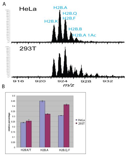
Figure 2.
Deviation of different H2B variants from HeLa cells and 293T cells. (A) Spectrum of histone H2B from HeLa cells and 293T cells. (B) Relative percentage of different H2B variants from different samplings.
Use of FTMS provides ~100,000 resolving power to clearly distinguish histone forms from one to another, including each histone isotopic distribution; for example, the most abundant form of human histone H4 obtained from our LC-FTMS method has been shown to have an N-terminal acetylation and two methylations on K20 previously [29]. The experimental monoisotopic mass (11331.42 Da) matched well with the theoretical neutral mass (11331.37 Da) with less than 5 ppm error (Supplementary Figure 1) and these performance metrics were retained even at low sample amount (vide infra).
Human epithelial carcinoma cell line HeLa S3 and embryonic 293 T cells derived from human kidney showed similar histone profiles of H2A, H3 and H4 (Figures 1 and Supplementary Figure 4). Histone H2B from these two cell lines showed significant deviation in expression of different isoforms (Figure 2). The most abundant H2B isoform in HeLa S3 cells is H2B.A (13830.50 Da). However, in 293T cells the most abundant form is either H2B.Q or H2B.F or a mixture which have the same mass (13844.51 Da). To calculate the relative abundance of these different H2B isoforms, H2B.E/T, H2B.A and H2BQ, F, we measured the relative MS intensity of the most abundant isotopic peak of each form from 3 different samples on different days. The relative percentage of each peak was calculated and plotted in Figure 2B. Most of the H2B forms observed here differ only by one or two amino acids. The differences including alanine changing to serine or threonine which could be potential phosphorylation sites are difficult to be observed by bottom-up approach due to its relative low sequence coverage. Since the specific functions of different histone variants in cells are still not fully understood, LC-MS profiling of histones with high resolution can shed a light on finding histone variants expression variation from different cellular events.
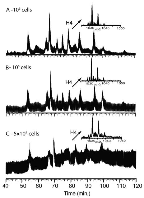
Populations of 293T cells were trypsinzed and counted via a hemocytometer. Histones were purified from 106, 105 or 5×10⁴ starting cell counts and analyzed by LC-FTMS. Figure 3 displays the total ion chromatograms and spectra of histone H4 from each run. While higher counts did transfer to spectra with a higher signal to noise ratio, 5×10⁴ cells were sufficient to generate histone profiles showing modified forms and variants of H2A, H2B and H4 (data not shown). Hence this LC-FTMS method was able to record histone PTM profiles from a number of cells readily workable in diverse applications; this provides the possibility to study histone profiles from clinical samples and even whole tissues, as histone extraction works clearly in such cases [30]. Further improvements in sensitivity can easily be achieved by targeting narrow m/z ranges for particular histones but handling <<104 cells is challenging without an altogether different type of sample preparation.
Figure 3.
TIC and partial FT mass spectrum of histone H4 obtained from 106 (A), 105 (B) and 5×10⁴ (C) 293T cells.
Knockdown of selected HDACs
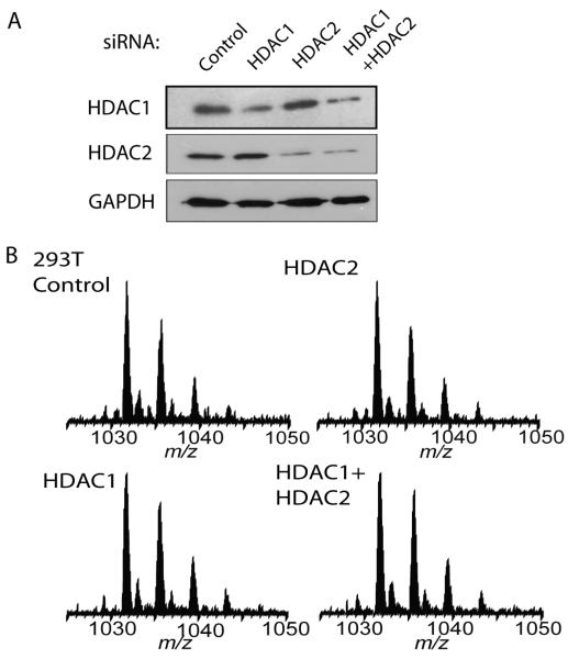
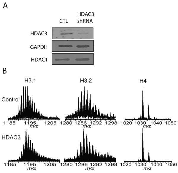
With an ability to produce clear PTM profiles from few cells, HDAC1 and HDAC2 were knocked down separately by specific siRNA in 293T cells at ~50% and ~70% respectively (Figure 4A). The global histone acetylation levels were not affected significantly (Figure 4B). Since HDAC1 and HDAC2 are usually found to interact with each other in a protein complex [31, 32], both of them were knocked down simultaneously in 293T cells; no evidence for significant hyperacetylation of global histone was observed by LC-FTMS (Figure 4 and Supplementary Figure 2). HDAC3 was knocked down by ~70% using shRNA in HeLa S3 cells (Figure 5A) and also global histone acetylation as profiled here was not significantly affected (Figure 5B and data not shown).
Figure 4.
Knockdown of HDAC1 and HDAC2 does not affect global histone acetylation of 293T cells. (A) Western blotting showing knocking down of HDAC1, HDAC2 only or both in 293T cells. (B) Spectra of histone H4 from control cells and HDAC knock down cells.
Figure 5.
Knock down of HDAC3 by shRNA does not affect global histone acetylation of HeLa S3 cells. (A) Western blotting showing knock down of HDAC3 in HeLa S3 cells. (B) Spectra of histone H3.1, H3.2 and H4 from control cells and HDAC3 knock down cells.
Profiling the effects of HDAC and HAT inhibition
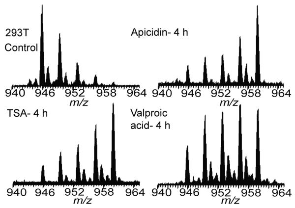
Upon treatment of HeLa S3 cells and 293T cells using either 10 mM sodium butyrate or 1 μM Trichostatin (TSA) for 4 h, all core histones displayed hyperacetylation profiles (Supplementary Figure 3 and 4). TSA and sodium butyrate are both HDAC inhibitors with a broad spectrum of activity against class I and II HDACs, but not the SIR2 family [33]. When class I and II HDACs were inhibited, HATs act to hyperacetylate core histones. Two HDAC class-I-selective inhibitors, Valproic acid and apicidin [34], caused comparable level of histone hyperacetylation as the TSA treatment of 293T cells (Figure 6 and data not shown).
Figure 6.
Inhibitors that show specificity during in vitro assays (AP and VPA) induce similar hyperacetylation as the broad spectrum inhibitor, TSA.
When HAT activity was inhibited by anacardic acid which is a potent, non-competitive inhibitor of p300 and PCAF [35], a decrease of histone acetylation level was observed on H2A and H2B (Supplementary Figure 5). The results indicate under normal conditions global histone acetylation is controlled by both the activity of HDACs and HATs.
Cell lines differ in response to HDAC inhibitors
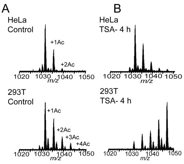
Comparing histone acetylation levels on H4 from populations of asynchronous control HeLa S3 and 293T cells, the data show somewhat greater levels of baseline acetylation in 293T cells where up to 4 internal acetylations can be observed from asynchronous cells (Figure 7A, bottom left). This may correlate with global HDAC activity in different cell lines. When we inhibited HDAC activities by using 1 μM TSA, increases in acetylation levels were observed in both cell lines. However, H4 from 293T cells showed far more hyperacetylation upon the same TSA treatment (Figure 7B). Assuming TSA has the same permeability into the two cell lines, it suggests that global HDAC activity is lower or global HAT activity is higher in 293T cells vs. HeLa. In order to rule out the possibility that two cell lines have different permeabilities for TSA, we also compared the data using other non-specific HDAC inhibitors, for instance, sodium butyrate, and the same response was observed (data not shown). This difference of cell line in response to HDACi may be caused by upregulation of HDAC protein expression or increased global HDAC activity in some cancer cells.
Figure 7.
Histone H4 from 293T cells has a higher acetylation level than that from HeLa cells. (A). Spectra of histone H4 from asynchronized HeLa cells and 293T cells. (B) Spectra of histone H4 from TSA treated HeLa cells and 293T cell.
Discussion
Histones have multiple gene family members and variants, all of which potentially serve specific biological functions. Most of the protein forms in each gene family are highly conserved. In some cases, they differ by as little as a single amino acid change. It is very difficult to distinguish these isoforms using antibody based approaches or bottom-up mass spectrometry. Profiling with LC-MS from the top-down provides a global perspective on chromatin and a method to distinguish very closely related histone species. For example, it has been shown that H2A variants expression ratios differ between chronic lymphocytic leukemia patients and normal volunteers [17]. In our approach, highly accurate intact mass measurement form online profiling and extensive offline fragmentation provides high confidence for protein identification. This ‘intact mass tag’ concept has also been used in other studies. [36, 37]
Histone acetyltransferases and deacetylases can be targeted to promoters to activate or repress gene transcription. In yeast, HATs and HDACs not only function in a loci specific manner but also at a global level throughout the genome [38]. Top-down mass spectrometry has shown that knocking out rpd3, one of the HDAC genes in yeast, caused hyperacetylation on histone H3 and H4 (Supplementary Figure 6). In mammalian cells, HDAC1, HDAC2, HDAC3 and HDAC8 belong to class I which is homologous to yeast RPD3. In sharp contrast to yeast, knockdown of individual class I HDAC protein in human does not affect global histone acetylation levels. Though global histone acetylation is not changed, HDACs’ function of repressing tumor suppressor genes is abolished once they are depleted or knocked down [39, 40, 41]. It is likely that only a small number of histones at specific chromatin regions are hyperacetylated once one HDAC is knocked down. These subtle changes may not be detected by our LC-FTMS approach which targets histone modification at the global level. Specific HDACs are recruited to specific gene loci and all or some of the different HDACs may be able to compensate depleted HDACs to maintain global histone acetylation level. The top-down LC-MS approach used here detects changes in histone expression ratios that are greater than ~10%, which translates to millions of nucleosomes experiencing histone remodeling (i.e., histone variant substitution or changes in modifications).
Another possibility to explain the HDAC compensation effect observed here is that different HDACs have different non-histone substrate targets. It was shown that HDAC1 deacetylates p53 in vitro and in vivo and SIRT1 also binds to and deacetylates p53 [42, 43, 44, 45]. It is also possible that knockdown efficiency is not high enough to cause loss HDAC function at sufficiently high levels to alter bulk histone acetylation. A small portion of HDAC protein left in the cells may still have sufficient activity to maintain global histone acetylation level.
HDAC inhibitors were originally discovered based on their ability to inhibit growth of transformed cells. HDAC inhibitor molecules induce transformed cell arrest and even cell death with only a minor fraction of genes being deregulated [46]. Our observation that the inhibitors cause a dramatic increase in global histone acetylation suggests that global histone acetylation is not directly linked with global gene transcription. The upregulation of tumor suppressor gene is likely to be regulated not only by the histone acetylation caused by HDAC inhibitor treatment but also by another level of transcriptional regulation. The hyperacetylation caused by HDACi treatment could weaken DNA- histone interaction and consequently change chromosome structure and dynamics enough to ultimately compromise cell viability.
Our data from class I selective reagents, valproic acid and apicidin, suggest that inhibition of certain HDACs is sufficient to induce global histone hyperacetylation at levels comparable to broad spectrum HDACi reagents. Clearly, biochemical HDAC inhibition assays performed in vitro do not reflect the actual inhibition occurring in live cells. It is possible that these two compounds do not have their purported HDAC selectivity in whole cells. Together with the knockdown experiments, our data suggest that global human histone acetylation is regulated by multiple class I (and II) HDACs, not individual HDACs.
To date, there are five distinct classes of HDACi according to their chemical structures [47] and various HDACi from several chemical classes have already advanced into clinical trials. However, the mechanisms by which HDAC inhibitors induce cell cycle arrest and cell death are not well elucidated. Key advancement in the development of potent HDAC inhibitors, however, is dependent on appropriate assays that require rapid screening applications with high sensitivity, selectivity and cost-effectiveness. Traditional assays for the in vitro determination of HDAC activity are based on the incubation of the purified enzyme or cell nuclear extracts with acetate-radiolabeled histones from chicken reticulocytes or cultured cells previously fed with [3H]acetyl-CoA [48, 49]. Currently, non-isotopic in vitro assays with fluorescent substrates and purified enzymes have well approached the suitability of high throughput screening. However, the in vitro assay condition does not completely match with the cellular conditions. HDAC inhibitors which showed good specificities in in vitro assays caused hyperacetylation similar to broad spectrum inhibitors in our studies. Our LC-FTMS based histone profiling provides the sensitivity and resolution for a cell-based HDAC (or HAT) inhibitor screen. Based on the knockdown results, global histone acetylation levels may not detect single HDAC specific inhibitor screening. Rather, a kinetic assay [28] using cell lines with varying propensity for hyperacetylation could be used as a method to better screen for specific HDACi activity, which should yield lower acetylation levels vs. broad spectrum inhibitors. Further, the use of different cell lines with varying overall HDAC activity (as judged by bulk histone acetylation levels) could be compared to their metastatic potential in animal models with further study using the efficient platform reported here.
Supplementary Material
Supplementary Figure 1. Highly accurate determination of mass from histone H4.
Supplementary Figure 2. LC-FTMS profiling shows that simultaneous knockdown of HDAC1 and HDAC2 does not effect global histone acetylation.
Supplementary Figure 3. Spectra of hyperacetylated core histones from sodium butyrate treated 293T cells.
Supplementary Figure 4 Spectra of hyperacetylated core histones from TSA treated 293T cells.
Supplementary Figure 5 Spectra of histones from anacardic acid treated 293T cells. Decrease of acetylation level was observed on H2A and H2B.
Supplementary Figure 6 Knockout of rpd3 in yeast causes massive hyperacetylation on histone H3 and H4.
Acknowledgements
The authors thank Dr. Jie Chen for pCMV-dR8.91 and pCMV-VSV-G plasmids for shRNA knockdown experiments. This work was supported by generous grants from the Packard Foundation and the National Institutes of Health (GM 067193-06) to N.L.K.
Footnotes
Publisher's Disclaimer: This is a PDF file of an unedited manuscript that has been accepted for publication. As a service to our customers we are providing this early version of the manuscript. The manuscript will undergo copyediting, typesetting, and review of the resulting proof before it is published in its final citable form. Please note that during the production process errors may be discovered which could affect the content, and all legal disclaimers that apply to the journal pertain.
References
- [1].Jaenisch R, Bird A. Nat Genet. 2003;33(Suppl):245. doi: 10.1038/ng1089. [DOI] [PubMed] [Google Scholar]
- [2].Wu J, Grunstein M. Trends Biochem Sci. 2000;25:619. doi: 10.1016/s0968-0004(00)01718-7. [DOI] [PubMed] [Google Scholar]
- [3].Garcia BA, Pesavento JJ, Mizzen CA, Kelleher NL. Nat Methods. 2007;4:487. doi: 10.1038/nmeth1052. [DOI] [PubMed] [Google Scholar]
- [4].Jenuwein T, Allis CD. Science. 2001;293:1074. doi: 10.1126/science.1063127. [DOI] [PubMed] [Google Scholar]
- [5].Workman JL, Kingston RE. Annu Rev Biochem. 1998;67:545. doi: 10.1146/annurev.biochem.67.1.545. [DOI] [PubMed] [Google Scholar]
- [6].Vidanes GM, Bonilla CY, Toczyski DP. Cell. 2005;121:973. doi: 10.1016/j.cell.2005.06.013. [DOI] [PubMed] [Google Scholar]
- [7].Hildmann C, Riester D, Schwienhorst A. Appl Microbiol Biotechnol. 2007;75:487. doi: 10.1007/s00253-007-0911-2. [DOI] [PubMed] [Google Scholar]
- [8].Ahringer J. Trends Genet. 2000;16:351. doi: 10.1016/s0168-9525(00)02066-7. [DOI] [PubMed] [Google Scholar]
- [9].Ishizuka T, Lazar MA. Mol Cell Biol. 2003;23:5122. doi: 10.1128/MCB.23.15.5122-5131.2003. [DOI] [PMC free article] [PubMed] [Google Scholar]
- [10].You A, Tong JK, Grozinger CM, Schreiber SL. Proc Natl Acad Sci U S A. 2001;98:1454. doi: 10.1073/pnas.98.4.1454. [DOI] [PMC free article] [PubMed] [Google Scholar]
- [11].Kim DH, Kim M, Kwon HJ. J Biochem Mol Biol. 2003;36:110. doi: 10.5483/bmbrep.2003.36.1.110. [DOI] [PubMed] [Google Scholar]
- [12].Lin RJ, Nagy L, Inoue S, Shao W, Miller WH, Jr., Evans RM. Nature. 1998;391:811. doi: 10.1038/35895. [DOI] [PubMed] [Google Scholar]
- [13].Kouzarides T. Cell. 2007;128:693. doi: 10.1016/j.cell.2007.02.005. [DOI] [PubMed] [Google Scholar]
- [14].Lo WS, Trievel RC, Rojas JR, Duggan L, Hsu JY, Allis CD, Marmorstein R, Berger SL. Mol Cell. 2000;5:917. doi: 10.1016/s1097-2765(00)80257-9. [DOI] [PubMed] [Google Scholar]
- [15].Edmondson DG, Davie JK, Zhou J, Mirnikjoo B, Tatchell K, Dent SY. J Biol Chem. 2002;277:29496. doi: 10.1074/jbc.M200651200. [DOI] [PubMed] [Google Scholar]
- [16].Su X, Jacob NK, Amunugama R, Lucas DM, Knapp AR, Ren C, Davis ME, Marcucci G, Parthun MR, Byrd JC, Fishel R, Freitas MA. Journal of Chromatography B. 2007;850:440. doi: 10.1016/j.jchromb.2006.12.037. [DOI] [PMC free article] [PubMed] [Google Scholar]
- [17].Zhang L, Freitas MA, Wickham J, Parthun MR, Klisovic MI, Marcucci G, Byrd JC. J Am Soc Mass Spectrom. 2004;15:77. doi: 10.1016/j.jasms.2003.10.001. [DOI] [PubMed] [Google Scholar]
- [18].Pesavento JJ, Kim YB, Taylor GK, Kelleher NL. J Am Chem Soc. 2004;126:3386. doi: 10.1021/ja039748i. [DOI] [PMC free article] [PubMed] [Google Scholar]
- [19].Boyne MT, 2nd, Pesavento JJ, Mizzen CA, Kelleher NL. J Proteome Res. 2006;5:248. doi: 10.1021/pr050269n. [DOI] [PubMed] [Google Scholar]
- [20].Siuti N, Roth MJ, Mizzen CA, Kelleher NL, Pesavento JJ. J Proteome Res. 2006;5:233. doi: 10.1021/pr050268v. [DOI] [PubMed] [Google Scholar]
- [21].Medzihradszky KF, Zhang X, Chalkley RJ, Guan S, McFarland MA, Chalmers MJ, Marshall AG, Diaz RL, Allis CD, Burlingame AL. Mol Cell Proteomics. 2004;3:872. doi: 10.1074/mcp.M400041-MCP200. [DOI] [PubMed] [Google Scholar]
- [22].Thomas CE, Kelleher NL, Mizzen CA. J Proteome Res. 2006;5:240. doi: 10.1021/pr050266a. [DOI] [PubMed] [Google Scholar]
- [23].Bonenfant D, Coulot M, Towbin H, Schindler P, van Oostrum J. Mol Cell Proteomics. 2006;5:541. doi: 10.1074/mcp.M500288-MCP200. [DOI] [PubMed] [Google Scholar]
- [24].Zhang K, Tang H. Journal of Chromatography B. 2003;783:173. doi: 10.1016/s1570-0232(02)00631-1. [DOI] [PubMed] [Google Scholar]
- [25].Zhang L, Eugeni E, Parthun M, Freitas M. Chromosoma. 2003;112:77. doi: 10.1007/s00412-003-0244-6. [DOI] [PubMed] [Google Scholar]
- [26].Pesavento JJ, Garcia BA, Streeky JA, Kelleher NL, Mizzen CA. Mol Cell Proteomics. 2007;6:1510. doi: 10.1074/mcp.M600404-MCP200. [DOI] [PubMed] [Google Scholar]
- [27].Jiang L, Smith JN, Anderson SL, Ma P, Mizzen CA, Kelleher NL. J Biol Chem. 2007;282:27923. doi: 10.1074/jbc.M704194200. [DOI] [PubMed] [Google Scholar]
- [28].Pesavento JJ, Mizzen CA, Kelleher NL. Anal Chem. 2006;78:4271. doi: 10.1021/ac0600050. [DOI] [PubMed] [Google Scholar]
- [29].Pesavento JJ, Bullock CR, Leduc RD, Mizzen CA, Kelleher NL. J Biol Chem. 2008 doi: 10.1074/jbc.M709796200. [DOI] [PMC free article] [PubMed] [Google Scholar]
- [30].Garcia BA, Thomas CE, Kelleher NL, Mizzen CA. J Proteome Re. 2008;7:4225. doi: 10.1021/pr800044q. [DOI] [PMC free article] [PubMed] [Google Scholar]
- [31].Ahringer J. Trends in Genetics. 2000;16:351. doi: 10.1016/s0168-9525(00)02066-7. [DOI] [PubMed] [Google Scholar]
- [32].Grozinger CM, Schreiber SL. Chem Biol. 2002;9:3. doi: 10.1016/s1074-5521(02)00092-3. [DOI] [PubMed] [Google Scholar]
- [33].Young DA, Lakey RL, Pennington CJ, Jones D, Kevorkian L, Edwards DR, Cawston TE, Clark IM. Arthritis Res Ther. 2005;7:R503. doi: 10.1186/ar1702. [DOI] [PMC free article] [PubMed] [Google Scholar]
- [34].Khan N, Jeffers M, Kumar S, Hackett C, Boldog F, Khramtsov N, Qian X, Mills E, Berghs SC, Carey N, Finn PW, Collins LS, Tumber A, Ritchie JW, Jensen PB, Lichenstein HS, Sehested M. Biochem J. 2008;409:581. doi: 10.1042/BJ20070779. [DOI] [PubMed] [Google Scholar]
- [35].Sun Y, Jiang X, Chen S, Price BD. FEBS Lett. 2006;580:4353. doi: 10.1016/j.febslet.2006.06.092. [DOI] [PubMed] [Google Scholar]
- [36].Gomez SM, Nishio JN, Faull KF, Whitelegge JP. Mol Cell Proteomics. 2002;1:46. doi: 10.1074/mcp.m100007-mcp200. [DOI] [PubMed] [Google Scholar]
- [37].Richard DS, Gordon AA, Mary SL, Ljiljana P-T, Yufeng S, Thomas PC, Timothy DV, Harold RU. PROTEOMICS. 2002;2:513. [Google Scholar]
- [38].Kurdistani SK, Robyr D, Tavazoie S, Grunstein M. Nat Genet. 2002;31:248. doi: 10.1038/ng907. [DOI] [PubMed] [Google Scholar]
- [39].Hartman HB, Yu J, Alenghat T, Ishizuka T, Lazar MA. EMBO Rep. 2005;6:445. doi: 10.1038/sj.embor.7400391. [DOI] [PMC free article] [PubMed] [Google Scholar]
- [40].Lagger G, O’Carroll D, Rembold M, Khier H, Tischler J, Weitzer G, Schuettengruber B, Hauser C, Brunmeir R, Jenuwein T, Seiser C. Embo J. 2002;21:2672. doi: 10.1093/emboj/21.11.2672. [DOI] [PMC free article] [PubMed] [Google Scholar]
- [41].Huang W, Tan D, Wang X, Han S, Tan J, Zhao Y, Lu J, Huang B. Biochem Biophys Res Commun. 2006;339:165. doi: 10.1016/j.bbrc.2005.11.010. [DOI] [PubMed] [Google Scholar]
- [42].Juan LJ, Shia WJ, Chen MH, Yang WM, Seto E, Lin YS, Wu CW. J Biol Chem. 2000;275:20436. doi: 10.1074/jbc.M000202200. [DOI] [PubMed] [Google Scholar]
- [43].Luo J, Su F, Chen D, Shiloh A, Gu W. Nature. 2000;408:377. doi: 10.1038/35042612. [DOI] [PubMed] [Google Scholar]
- [44].Luo J, Nikolaev AY, Imai S, Chen D, Su F, Shiloh A, Guarente L, Gu W. Cell. 2001;107:137. doi: 10.1016/s0092-8674(01)00524-4. [DOI] [PubMed] [Google Scholar]
- [45].Vaziri H, Dessain SK, Eaton E. Ng, Imai SI, Frye RA, Pandita TK, Guarente L, Weinberg RA. Cell. 2001;107:149. doi: 10.1016/s0092-8674(01)00527-x. [DOI] [PubMed] [Google Scholar]
- [46].Van Lint C, Emiliani S, Verdin E. Gene Expr. 1996;5:245. [PMC free article] [PubMed] [Google Scholar]
- [47].Gallinari P, Di Marco S, Jones P, Pallaoro M, Steinkuhler C. Cell Res. 2007;17:195. doi: 10.1038/sj.cr.7310149. [DOI] [PubMed] [Google Scholar]
- [48].Kolle D, Brosch G, Lechner T, Lusser A, Loidl P. Methods. 1998;15:323. doi: 10.1006/meth.1998.0636. [DOI] [PubMed] [Google Scholar]
- [49].Kijima M, Yoshida M, Sugita K, Horinouchi S, Beppu T. J Biol Chem. 1993;268:22429. [PubMed] [Google Scholar]
Associated Data
This section collects any data citations, data availability statements, or supplementary materials included in this article.
Supplementary Materials
Supplementary Figure 1. Highly accurate determination of mass from histone H4.
Supplementary Figure 2. LC-FTMS profiling shows that simultaneous knockdown of HDAC1 and HDAC2 does not effect global histone acetylation.
Supplementary Figure 3. Spectra of hyperacetylated core histones from sodium butyrate treated 293T cells.
Supplementary Figure 4 Spectra of hyperacetylated core histones from TSA treated 293T cells.
Supplementary Figure 5 Spectra of histones from anacardic acid treated 293T cells. Decrease of acetylation level was observed on H2A and H2B.
Supplementary Figure 6 Knockout of rpd3 in yeast causes massive hyperacetylation on histone H3 and H4.


