

Abstract
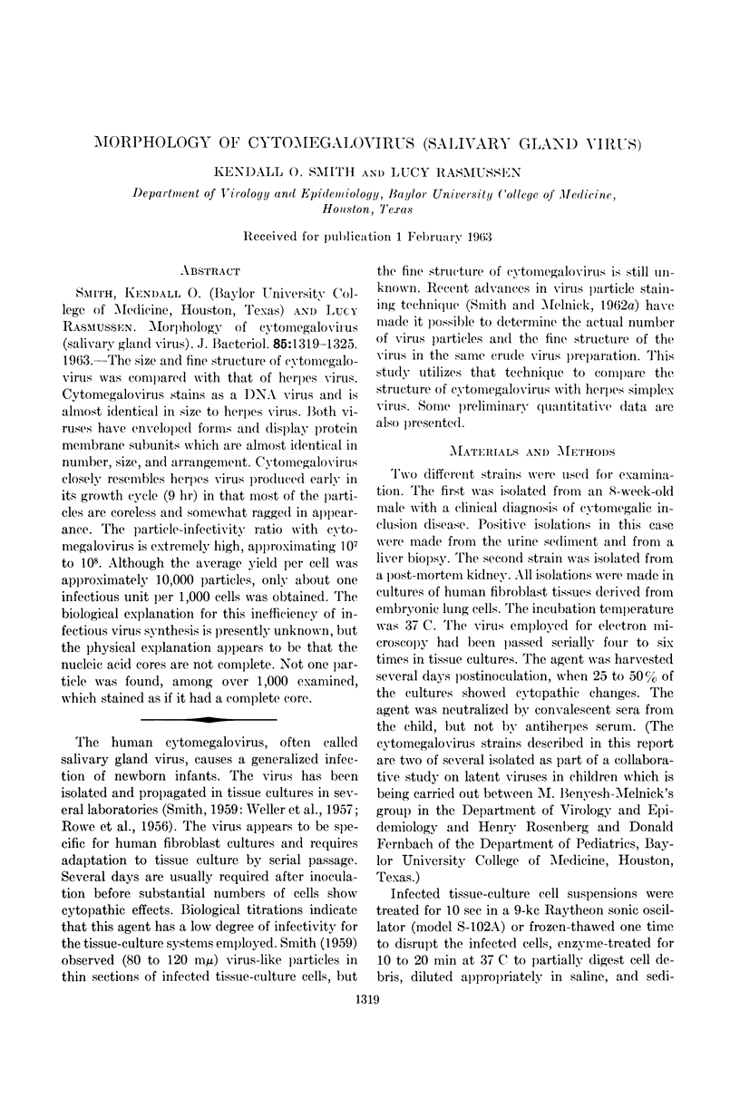
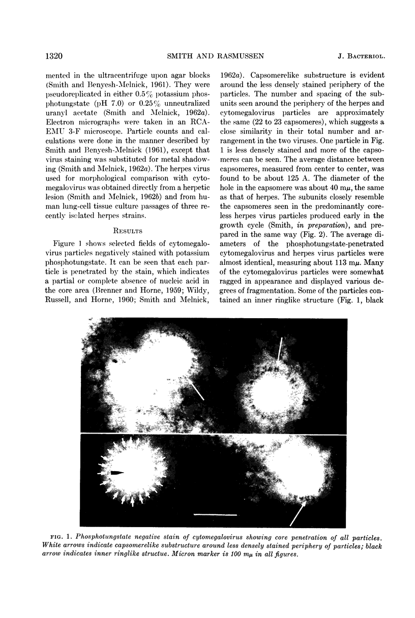
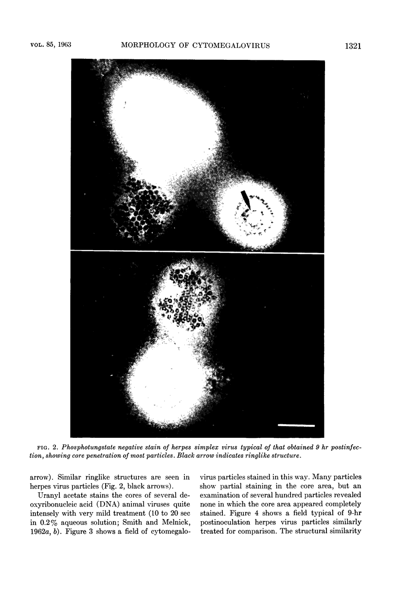
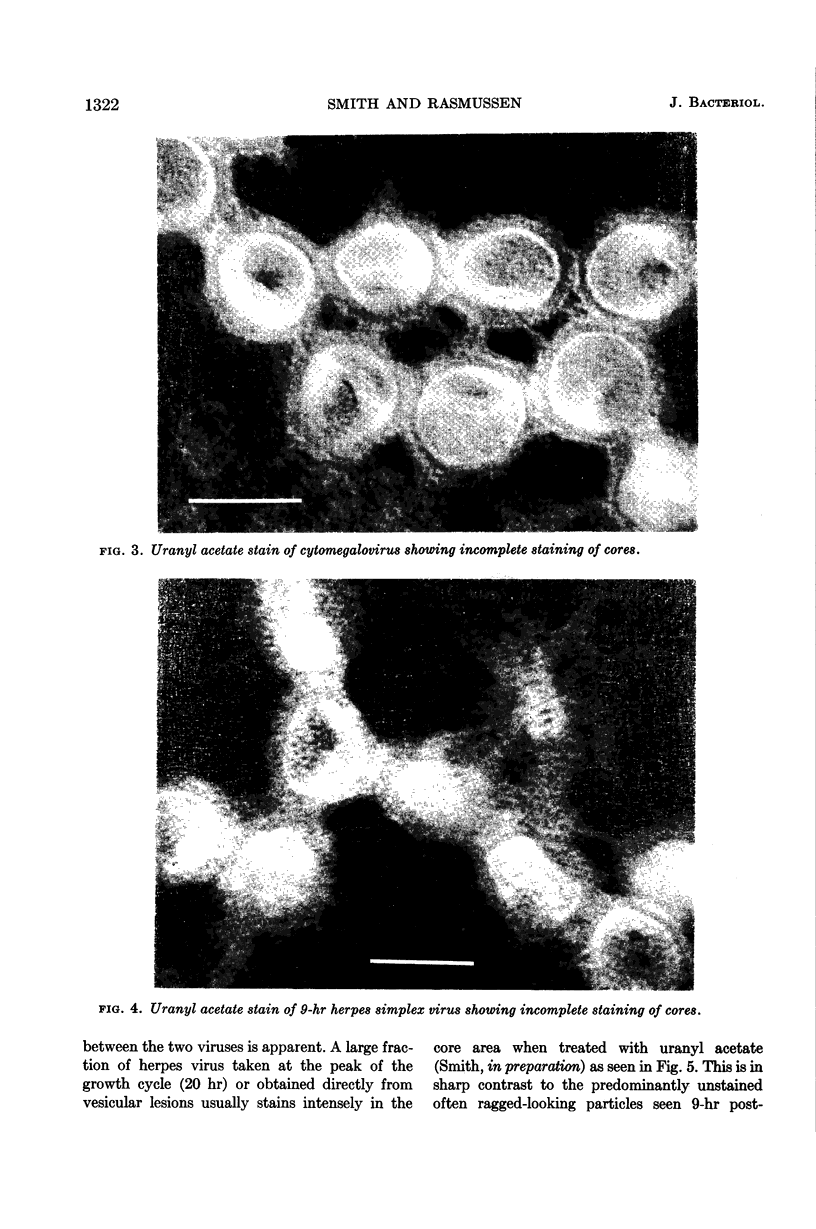
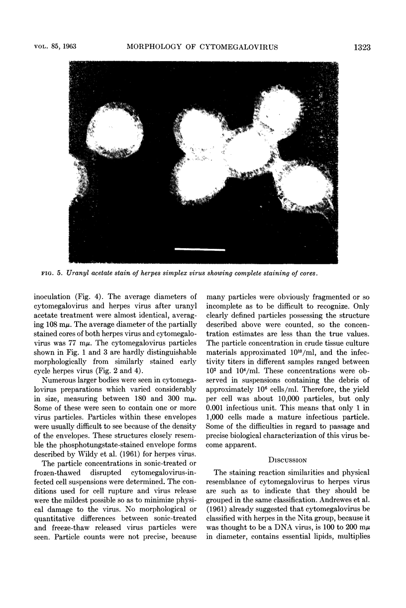
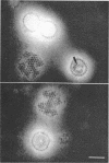
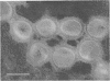
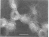
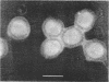
Smith, Kendall O. (Baylor University College of Medicine, Houston, Texas) and Lucy Rasmussen. Morphology of cytomegalovirus (salivary gland virus). J. Bacteriol. 85:1319–1325. 1963.—The size and fine structure of cytomegalovirus was compared with that of herpes virus. Cytomegalovirus stains as a DNA virus and is almost identical in size to herpes virus. Both viruses have enveloped forms and display protein membrane subunits which are almost identical in number, size, and arrangement. Cytomegalovirus closely resembles herpes virus produced early in its growth cycle (9 hr) in that most of the particles are coreless and somewhat ragged in appearance. The particle-infectivity ratio with cytomegalovirus is extremely high, approximating 107 to 108. Although the average yield per cell was approximately 10,000 particles, only about one infectious unit per 1,000 cells was obtained. The biological explanation for this inefficiency of infectious virus synthesis is presently unknown, but the physical explanation appears to be that the nucleic acid cores are not complete. Not one particle was found, among over 1,000 examined, which stained as if it had a complete core.
Full text
PDF
Images in this article
Selected References
These references are in PubMed. This may not be the complete list of references from this article.
- ANDREWES C. H., BURNET F. M., ENDERS J. F., GARD S., HIRST G. K., KAPLAN M. M., ZHDANOV V. M. Taxonomy of viruses infecting vertebrates: present knowledge and ignorance. Virology. 1961 Sep;15:52–55. doi: 10.1016/0042-6822(61)90076-9. [DOI] [PubMed] [Google Scholar]
- BRENNER S., HORNE R. W. A negative staining method for high resolution electron microscopy of viruses. Biochim Biophys Acta. 1959 Jul;34:103–110. doi: 10.1016/0006-3002(59)90237-9. [DOI] [PubMed] [Google Scholar]
- ROWE W. P., HARTLEY J. W., WATERMAN S., TURNER H. C., HUEBNER R. J. Cytopathogenic agent resembling human salivary gland virus recovered from tissue cultures of human adenoids. Proc Soc Exp Biol Med. 1956 Jun;92(2):418–424. [PubMed] [Google Scholar]
- SMITH K. O., MELNICK J. L. A method for staining virus particles and identifying their nucleic acid type in the electron microscope. Virology. 1962 Jul;17:480–490. doi: 10.1016/0042-6822(62)90143-5. [DOI] [PubMed] [Google Scholar]
- WILDY P., RUSSELL W. C., HORNE R. W. The morphology of herpes virus. Virology. 1960 Oct;12:204–222. doi: 10.1016/0042-6822(60)90195-1. [DOI] [PubMed] [Google Scholar]