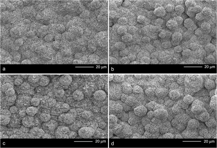
Figure 1.
Low magnification SEM images of the bone-like mineral surface following different DNA incorporation methods: a) Mineralized control b) Plasmid DNA coprecipitation c) DNA-Lipoplex adsorption and d) DNA-Lipoplex coprecipitation. Neither coprecipitation group demonstrated a difference in the size of the mineral nucleation sites as compared to the mineralized controls.

