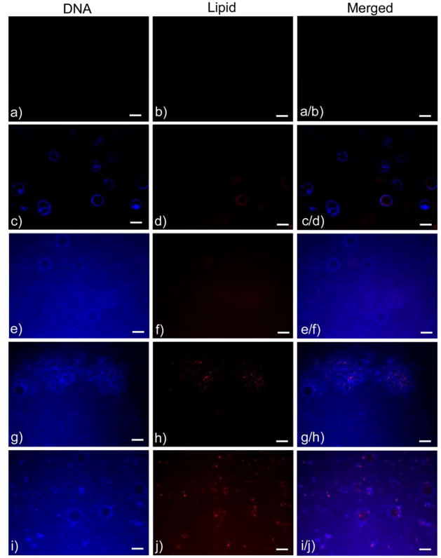
Figure 3.
Fluorescence images of DNA and lipid agent components from representative samples from each of the following groups: a–b) Mineralized controls, c–d) Plasmid DNA incorporated into PLGA, e–f) Plasmid DNA coprecipitated with mineral, g–h) Plasmid DNA-Lipoplex adsorbed to mineralized films, and i–j) Plasmid DNA-Lipoplex coprecipitated with mineral. Distribution of both the plasmid DNA and the lipid transfection agent on the bone-like mineral was demonstrated by the colocalization of the fluorescent staining (after thorough rinsing) in the adsorption and coprecipitation groups and the absence of staining in the mineralized controls. Scale bars represent 100 μm.

